KINS 158 Neuromotor Basis for Motor Control
 Call Kai
 Learn
 Practice Test
 Spaced Repetition
 Match
 Flashcards
 Knowt Play
1/23

There's no tags or description
Looks like no tags are added yet.
24 Terms
The human nervous system is divided into two major divisions. What are they and what is each responsible for?
Central NS - Brain, brainstem, cerebellum, and spinal cord
• Responsible for processing information and generating motor
commands
Peripheral NS - All neural structures outside the skull and vertebral column,
• Linking the CNS to the rest of the body
What does the term neuromotor system mean?
Refers specifically to the components of
the nervous system that coordinate and control voluntary,
purposeful movement.
What is the neuron responsible for?
Transmits information throughout the nervous system
Structure of a Neuron: Cell Body
The cell body contains the nucleus. What is the nucleus responsible for?
What is the role of the cell body?
Regulates homeostasis of neuron
Synthesizes a large quantity and variety of proteins used as neurotransmitters.
Structure of a Neuron: Dendrites
What are dendrites responsible for?
primarily responsible for receiving information from other neurons
Structure of a Neuron: Axon
What is the role of the axon?
What are the branches of the axon called?
They are covered by myelin. What is this?
What do the axon terminals do?
Responsible for sending information from the
neuron.
Collaterals
a cellular
membrane that speeds up the transmission of neural
signals along the axon
a signal transmission relay station for
neurotransmitters
Sensory Neurons
Transmit information from the ___ to the ___
Sensory neurons are Pseudounipolar. What does this mean?
Where is the cell body of sensory neurons located?
Transmit information from the periphery to the CNS
Appear to have a single projection from the cell body
Cell body of sensory neurons located at the dorsal root
ganglion of the spinal nerve
Motor Neurons
Transmit information from the ___ to ____ by activating ___
Alpha Motor Neurons - Where are they located and what do they do?
Gamma Motor Neurons - What do they do?
Transmit information from the CNS to muscles by activating muscle
contraction
Alpha Motor Neurons
• Located predominantly in spinal cord
• Axons synapse on skeletal muscle fibers to create the motor unit
Gamma Motor Neurons
• Activity regulated through alpha-gamma coactivation
• Regulates sensitivity of intrafusal fibers (muscle spindles) to maintain accurate level
of proprioception
Interneurons
What are these?
Transmits information between?
Specialized neurons that originate and terminate in the brain or spinal cord
• Axons from the brain and motor neurons
• Axons from the sensory nerves and the spinal nerves ascending to the
brain
What are the two ways cells communicate?
Action Potentials - Rapid electrical impulses that travel along
a neuron’s axon, allowing information to
move within a single neuron
Synaptic Transmission - process by which neurons communicate with
each other—or with muscles and glands—
through the release of neurotransmitters
across the synaptic cleft
Action Potential
A - Resting membrane potential
During resting membrane potential, what does the inside and outside of the cell look like?
How is the cell maintained at this state?
Inside the cell → High concentration of K⁺ (potassium).
• Outside the → High concentration of Na⁺ (sodium) and Cl⁻ (chloride).
Sodium-potassium pump (Na⁺/K⁺ pump) → pumps 3 NA⁺ out and 2 K⁺ in.
Leak channels: allow K⁺ to slowly leak out, further keeping the negative resting potential.
Action Potential
B - Depolarization
What 3 stages occur during depolarization?
Stimulus (e.g., neurotransmitter from another neuron) causes Na⁺ ligand-gated channels to open slightly → Na⁺ enters cell, makes inside less negative (small depolarization).
2. If membrane potential reaches threshold (~ -55 mV), an action potential fires.
3. Voltage-gated Na⁺ channels open, Na⁺ rushes in → rapid depolarization to +40 mV
Action Potential
C - Repolarization and Hyperpolarization
How does depolarization and hyperpolarization occur?
Voltage-gated K⁺ channels open, K⁺ flows out → repolarization.
extra K⁺ leaving, making the inside too negative (below –70 mV) → Hyperpolarization
Action Potential
D - Return to Resting Potential
Explain this stage
Na⁺/K⁺ pump restores ion balance.
Action Potential - Refractory Periods
What is the refractory period?
What is absolute refractory period?
What is relative refractory period?
Period when neuron cannot fire another action potential
Na⁺ channels unresponsive; neuron cannot fire again
Stronger stimulus required to trigger another potential
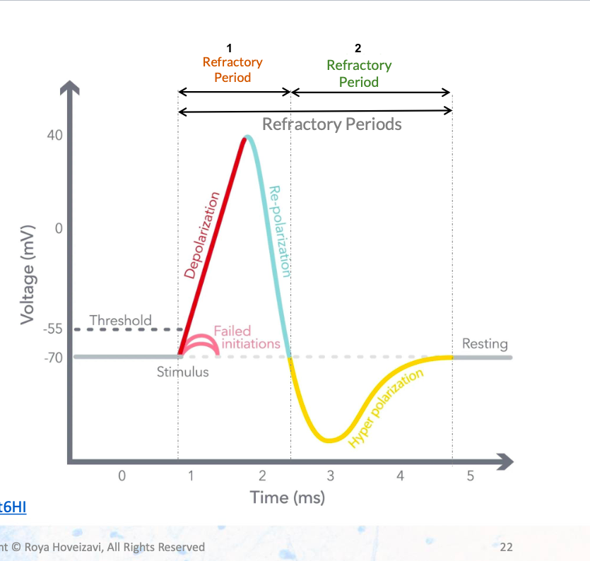
Absolute
Relative
Action Potential: Propagation of the Signal
Depolarization at one segment triggers…?
Myelin sheath (lipid-rich insulation) increases speed and efficiency by:
Depolarization at the next, spreading the signal along the axon
a. Preventing current leakage.
b. Allowing current to "jump" between Nodes of Ranvier (saltatory conduction). Nodes of Ranvier: Gaps in myelin/ rich in voltage-gated Na⁺ channels
Graded Potentials
What do graded potentials do?
What is EPSP?
What is IPSP?
change the membrane potential slightly (by a few millivolts) and can add up
(summate) over time and space.
Excitatory Postsynaptic Potential - a small depolarization of the postsynaptic
membrane that makes the neuron more likely to fire an action potential
Inhibitory Postsynaptic Potential - a small hyperpolarization that makes the neuron less likely to fire an action potential
Summation
Summation of graded potentials can trigger…?
What is temporal summation?
What is spatial summation?
The firing of an action potential
many potentials from one neuron trigger threshold point
potentials from many neurons trigger threshold point
What is the all-or-none law?
Once triggered, does the action potential speed up?
Neurons either fire or don’t — action potentials are not graded by stimulus intensity.
No. Once triggered, the action potential always has a constant amplitude and speed
Synapses
What is a synapse?
What is the most common type of synapse?
Are synapses electrical or chemical?
Specialized junctions where neurons communicate
Axodendritic synapse
Both
Chemical Synaptic Transmission
Explain the 5 stages
1. Action potential arrives at the axon terminal,
→ triggers the voltage gated Ca+ channels
2. Triggers synaptic vesicle mobilization
3. Vesicles fuse with the membrane (exocytosis) and
release neurotransmitters into the synaptic cleft.
4. Neurotransmitters bind to receptors on the
postsynaptic neuron → change its membrane
potential (excitation or inhibition).
5. Neurotransmitters are removed from the synaptic
cleft:
• Enzymatic breakdown (e.g., acetylcholinesterase).
• Reuptake into the presynaptic neuron (e.g., SSRIs
serotonin reuptake blocking).
The Motor Unit
One motor unit =
What is the Neuromuscular Junction?
the alpha motor neuron and all the muscle fibers it innervates
Connection site between alpha motor neuron and muscle fibers
Motor unit recruitment
Muscle force depends on?
More motor units activated = ?
Motor unit recruitment follows what principle?
Muscle force depends on how many muscle fibers are active
More motor units activated → greater force output.
Size Principle
• Recruitment starts with small, weak motor units.
• Progresses to large, powerful motor units as force demand increases