BIPN 105 Midterm 1
 Call Kai
 Learn
 Practice Test
 Spaced Repetition
 Match
 Flashcards
 Knowt Play
1/132

There's no tags or description
Looks like no tags are added yet.
133 Terms
Oscilloscope
Used to detect how electrical voltage changes over time
Axis usually volts / time
Measures voltage
Y axis gain (Range) → sensitivity
Large signal requires large range, thus sensitivity is low
Small signal requires small range, thus sensitivty is high
X axis gain (Timebase) → Sweep speed (time/div)
Sets how long oscilloscope will record for
Large signal (couple min) would sweep slowly to detect event
Small signal (couple ms) would sweep fast
Examples of Oscilloscope use
To detect Electromagnetic fields (EMFs), get closer to EMF noise increases
To measure electrical activity in a nerve, able to stimulate nerve past threshold thus generating AP (utilize preamplifier to see activity better before reaches oscilloscope)
To measure skeletal muscle contractions (utilize transducer which converts mechanical event to voltage)
Why must we calibrate the transducer
Through calibration equation must convert volts into appropriate units
Analog to digital mismatch
Computers take discrete samples whereas in real life its a series of continuous events
To reproduce more accurate results, it may be best to take as many samples to reproduce real life
Types of Filters
Band Pass
AC couple
DC Couple
Band pass filter
Band pass: only allows a certain band of frequencies to pass, removes unwanted frequencies found below and above signal we want to record (reduces noise)
(high pass/low cutoff, low pass/high cutoff)
AC couple filter
sets non /slowly changing parts of signal to 0, only allows fast changing parts go through unchanged (primarily used for fast events and to set baseline to 0)
DC couple filter
all parts of single unchanged (looks exact same as before, and used when turn off AC couple, primarily used for slow events)
Grounding
low resistance circuit, that passively spreading electricity goes into
Decreases amount of stimulus artifact (passive) that reaches recording electrodes
Permeable membrane
Solutes and solvent cross and at equilibrium both compartments are the same (same conc and same volume level)
Semi permeable membrane
ONLY solvent can cross
Diffusion
Movement of molecules from high to low concentration (both solute and solvent move)
Osmosis
Movement of solvent across semi-permeable membrane from high (low solute conc) to low solvent concentration (high solute conc)
only solvent moves
What generates osmotic pressure?
Solute particles generate osmotic pressure driving solvent towards it
What is Hydrostatic pressure
Physical push exerted by a fluid against a surface or container wall.
When hydrostatic pressure builds up on the concentrated side of a membrane, it pushes water back in the opposite direction.
Equilibrium occurs when the magnitude of the opposing hydrostatic pressure equals the osmotic pressure, stopping any net movement of water.
Osmotic pressure vs Hydrostatic pressure
Osmotic pressure pulling water
Hydrostatic pressure is pushing water out

What is this equation
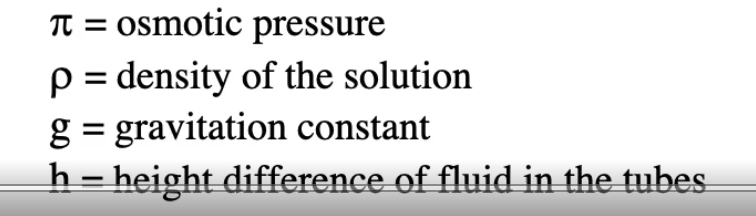

What is this equation?
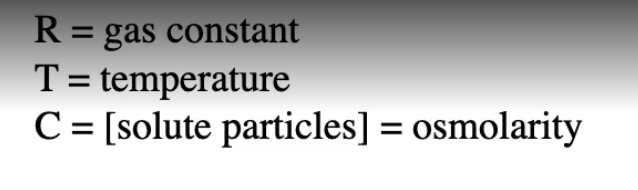
What does osmolarity = ?
conc of solute particles = osmolarity = S/V
S= # of particles
V = volume of distribution
Molarity
a measure of the concentration of a whole solute in a specific volume of solution
What is the difference between osmolarity and molarity
Osmolarity measures dissociated particles in solution, whereas molarity measures whole solutes
Not all solutes dissociate in solution, describe the differences
Non electrolyte and electrolyte solutes
Non-electrolyte solutes
Molecules that don’t dissociate into multiple particles
Thus, Molarity = osmolarity , 150mM sucrose = 150mOsm
EX: sucrose, urea, proteins, etc…
Electrolyte solutes
Molecules that dissociate into multiple particles
molarity dissociation # = osmolarity, 150 mM NACl * 2 = 300 mOsm
Terms to compare solutions/osmolarity
Isoosmotic, generate same osmotic pressure
Hypoosmotic, generates lower osmotic pressure (A is hypoosmotic to B)
Hyperosmotic, generates higher osmotic pressure (B is hyperosmotic to A)

Perm selective membrane
Solvents can cross, solutes have different permeabilities based on Permeability coefficient (P)
Moving down concentration gradient vs against concentration gradient
Conc gradient - high to low
Against gradient - low to high (utilizes energy
What is tonicity
Predicts water flow
Isotonic
no net water flow (water is crossing, but same amount is crossing generating no difference)
Hypotonic
A is hypotonic to B
Hypertonic
B is hypertonic to A (meaning B has higher conc of non permeable solutes)
Predict what will happen if a cell is put into solutions with different tonicities
Isotonic soln see no volume change
Hypotonic solution, water goes into cell, swells too much resulting in lysis
Hypertonic solution, water goes out of cell, cell shrinks
RBCs
no nucleus, few internal structures, little lipid bilayer bags of hemoglobin (Hb)
Cell membrane of RBCs are perm selective (Water and urea can easily cross, whereas other solutes cannot)
What is hemmatocrit test
Put blood solution in centrifuge, will find plasma (water & solutes), buffy coat (WBCs, platelets), RBC at bottom
% of RBCs in blood sample
Hct = vol of RBCs/ total volume
What would happen to hematocrit levels if blood is put into solutions with different tonicities?
Isotonic solution = no change in Hct
Hypotonic solution = increase in Hct (if lysis occurs decrease in Hct)
Hypertonic solution = decrease in Hct
TOTAL VOLUME DOESN”T CHANGE ONLY VOLUME OF RBCs
low HCT Anemia
Causes: Bleeding (lose RBCs), low iron (need iron to make HB (found within RBC) and for maturation of RBCs), pregnancy (expansion of plasma volume), RBC breakage (hemolytic anemias, immune attack on RBC), some type of kidney disease (decrease Erythropoietin “EPO” production, main hormone triggers production of RBC in bone marrow)
high HCT Polycythemia
causes: dehydration (decreased plasma volume), hereditary overproduction of RBC, low blood oxygen (high altitude, etc), blood packing, EPO injections
Derivation of Osmometer equation
1) V= W + B
V= total volume of RBC
W = volume of water in RBC (can be changed by osmotic forces)
b = “osmotically inactive volume”, can’t cross membranes such as proteins and solutes (can’t be changed by osmotic forces)
Rearrange to W = V-b
2) C = [solute] inside of an RBC
C= S/ W
S = # of particles
W = volume of water in RBC
S = W * C (S is constant since # of particles is same due to assumption solutes cant cross RBC membrane)
S= W C = Wo Co (normal state, isotonic blood solution)
Stick V-b in place for W
3) (V-b) C = (Vo- b) Co

Osmometer equation
Describes changes in RBC volume when put into different tonicity solutions
Anatomy of a neuron (nerve cell)
Dendrites, cell body, Hillock, Axon, Terminals
Dendrites
main input
Cell body
Has nucleus
Hillock
APs start
Axon
carries Action potentials towards other cells
Terminals
Axon branches to contact multiple cells
What happens if stimulus reaches threshold?
Will generate AP, AP will travel down axon and once it reaches the terminal, causes Ca2+ to rush into cell activating synaptic vesicles and releasing neurotransmitters into synaptic cleft
Why do neurons have membrane potential?
They have Sodium/Potassium ATPase pumps along the membrane that maintain ion concentration gradients across.
Typical cell membrane concentration
Find K+, anions, proteins inside cell
Find Na+, Ca2+, Cl- outside cell
Ions go down conc gradient unless utilize form of energy to go against conc gradient
Chemical and Electrical potential
K+ leaves the cell from high to low conc (Chemical potential), the cell will become more negative, which starts to pull K+ back into the cell (Electrical potential)
Nernst equation (Calculates equilibrium potential)
Tells you how much voltage need across membrane to balance chemical potential, no net flow of ion across membrane

Goldman equation (calculates membrane potential for all ions)
Why is it that at rest the permeability of K+ is larger than Na+?
Membrane potential at rest is much closer to K+ due to there being more K+ leak channels
Intracellular recording
Good for recording one neuron
Lec 3, 6:18
Define terms depolarize, repolarize, hyperpolarize
Depolarize: membrane potential past (Vrest)
Repolarize: membrane potential back to (Vrest)
Hyperpolarize: membrane potential below (Vrest)
Threshold potential
Vm at which an AP is initiated
APs are all or nothing
What is the purpose of Vt?
Serves as a noise filter and neurons use frequency modulation to code information (cant increase AP size instead increase frequency of AP)
AP phases
Depolariztion
Na+ channel activates
Large force drives Na+ into cell
Na+ influx depolarizes past threshold generating AP
Repolarization
Na+ channel inactivation (time dependent, very fast)
K+ channel activation
K+ flows out of cell
Hyperpolarization
Excess permeability/current of K+ (drives membrane potential below Vrest)
Return to Vrest
K+ channel deactivation (voltage dependent, not time dependent)
Na+ channel deactivation (voltage dependent, required to remove inactivation)
Leak channels are the only activated channels bring back to Vrest
-APs are local events that spread along the membrane
-As APs spread, they regenerate/don’t get smaller
-Very small # of ions cross during AP
-Repolarization not due to ATPase pumps, too slow, instead K+ pumps causing K+ to leave the cell
-If membrane stays depolarized Na+ channels stay inactivated must repolarize to remove inactivation
Refractory period
How long a neuron must wait before it can fire a second AP
Absolute refractory period
Period in which no amount of stimulus will be able to fire another AP since Na+ channels are still inactivated (to remove inactivation must have repolarization)
Relative refractory period
Period in which you are able to fire another AP, however only if 2nd stimulus is big enough (Na+ inactivation removed and still have high Pk (some K+ haven’t deactivated)
AP spread
At rest, neuron is negative inside and positive outside, if stimulate axon and generate AP now negative outside and positive inside (accumulating Na+ inside cell), depolarization will spread across (by activating Na+ channels)
What affects spread of AP (conduction velocity)
Greater the diameter the greater velocity
Myelination (acts as insulation, preventing passive spread (very fast) depolarization from dropping below threshold)
Saltatory conduction
-AP jumps node to node, AP regenerated node to node, AP spread faster
What are the different types of fibers?
A fibers (alpha, beta, gamma, delta) these are large myelinated
C fibers: small and unmyelinated
Sciatic nerve
bundle of different types of axons (find mix of A fibers)
Compound action potential (CAP)
Summation of individual APs, large diameter axons (alpha>beta>gamma>delta) have lower thresholds
Apply very small stimulus → no CAP, likely below threshold
Apply small stimulus → small CAP, likely on A alpha fibers generate AP
As continue to increase stimulus amplitude → CAP increases (recruitment of higher threshold axons)
CAP is not…
an all or nothing action potential, rather the summation of individual APs of multiple neurons
Stimulator

AP generated at (-) stimulating electrode
Extracellular recording
Only records outside voltage, use bipolar recording (subtract voltage at (-) electrode from (+) electrode)

Monopolar vs bipolar recording
Monopolar only utize (-) recording electrode thus stimulus that you see is depolarization at the (-) electrode
Bipolar utilize both (-) and (+) recording electrode, see depolarization event of both recording electrodes

Skeletal muscle are neurogenic since
skeletal muscle cells innervated by A alpha motor neurons that have cell bodies in CNS, send axons all the way to muscle, axons in muscle divide to contact many muscle fibers at the NMJ
NMJ (endplate)
A alpha motorneuron contact postsynaptic cell (skeletal muscle cell), presynaptic terminal contains vesicles, find Ach neurotransmitters inside synaptic vesicles, also find Ca2+ channels (P/Q voltage gated channels) on presynaptic membrane. Neurotransmitters travel synaptic cleft and bind to receptors found on postsynaptic membrane
Which receptors utilized by postsynaptic skeletal muscle cells
Respond to Ach and are sensitive to nicotine, cholinergic nicotinic receptors
Ligand dependent ion channel, requires two Ach to activate (non-specific mixed cation channel)
Is Vrest for skeletal muscle cells closer to Na+ or K+
Vrest is close to equilibrium potential K+, force for K+ is small/greater force for Na+
NMJ function
Nerve AP → Muscle AP (MAP)
A alpha motorneuron, generate AP in hillock, travels to presynaptic terminal, activates Ca2+ channels (P/Q), Ca2+ influx, causes vesicle exocytosis, Ach neurotransmitters released, Ach diffuses across cleft, two Ach bind to cholinergic nicotinic receptors, cholinergic nicotinic receptors activate, postsynaptic depolarization generates Endplate potential (EPP, these are graded potentials), more Ach binds to receptors the greater the EPP, if EPP is greater than threshold generates MAP
How many NMJ in skeletal muscle cell and where do you find them?
Find one NMJ per skeletal muscle cell and they are found in the middle of the cell
Termination of NMJ
Stop exocytosis (Ca2+ diffuses away from vesicles, Ca2+ inside cell is pumped out by ATPase pumps)
ACH dissociates from cholinergic nicotinic receptors, deactivates, EPP dissipates
ACh removed (diffuses away, or enzymatic breakdown by Acetychollineesterase AChE, which converts ACh to choline + acetate)
Recycling
Choline uptake into presynaptic terminal
Choline + Acetyl-coA converted into ACh + Coa by Choline Acetyltransferase (ChAT), ACh put into vesicles by vesicle Ach transporter (VAChT)
How does choline found in synaptic cleft get into cell?
Na+ choline transporter, energy to drive choline into cell is as Na+ comes in following conc gradient (both Na+ and choline enter cell)
Fatigue and Recovery of NMJ
If cell is undergoing high frequency stimulation (deplete ACh storage) and Recovery requires formation of new ACH/vesicles
Synaptic Delay
delay between presynaptic depolarization and postsynaptic depolarization
Things to consider are
release of NT
diffusion across synaptic cleft
two ACh binding to cholinergic nicotinic receptors
Facilitation (can be seen if multiple stimuli are used)

Compare Neurons vs Skeletal muscle
….
What are other NTS/receptors and their functions
….
Why is norepinephrine listed as a NT, but Epinephrine is not
Epinephrine is a hormone not a NT and is released by the adrenal medulla
What is the function of atropine
Blocks muscarinic cholinergic receptors (acts as a antagonist)
What is Myasthenia Gravis
Autoimmune diseases
Muscle organization
Muscle, myofiber (single muscle cell, multinucleated, length entire muscle, one NMJ found in center), myofibril. sarcomere, myofilaments
Myofilaments
Actin (thin filament)
Myosin (thick filament)
Z line
Anchors actin and separates one sarcomere from the next
Bands
A band (myosin + actin/myosin overlap)
I band (only actin)
Sarcomere
Small contractible element built into each myofibril
What happens when muscle contracts?
Myofilaments pull on Z line (either generates tension and shortens muscle)
Where is Ca2+ stored in sarcomere
Sarcoplasmic reticulum (SR), store and release Ca2+ into areas of myofilaments
What is the myofiber membrane called?
Sarcolemma
What channels do you find along the Myofiber membrane and what purpose do they serve?
Find Na+ and K+ channels and fire MAP, which is carried from NMJ (center of muscle) to ends of myofiber