Clinical 2 test 1
 Call Kai
 Learn
Learn Practice Test
 Spaced Repetition
 Match
Match Flashcards
 Knowt Play
1/317

There's no tags or description
Looks like no tags are added yet.
Name | Mastery | Learn | Test | Matching | Spaced | Call with Kai | Chat |
|---|
No analytics yet
Send a link to your students to track their progress
318 Terms
PCV ( packed cell volume)
is the % of RBCs contained within a specific volume of blood
the cheapest, quickest and easiest way to measure red blood cells within the blood
tells us about oxygen carrying capacity of the blood
what does the PCV evaluate
evaluates the % of red blood cells in blood volume.
which tells us about the oxygen carrying capacity of blood
This measurement helps assess anemia, dehydration, or other medical conditions affecting red blood cell levels.
what does a decrease PCV
less RBCs are available for oxygen transport, which can indicate conditions such as anemia or blood loss.
what does an increase PCV mean
lots of RBCS for oxygen transport
usually indicative of conditions such as dehydration
what are the practical applications of PCV in practice
check hydration status
response to fluid therapy
performed prior to surgery
response to treatment
Normal PCV value for dog
37-55%
Normal PCV values for cats
30-45%
what is the unit for PCV
the unit is percentage (%).
When an animal is dehydrated what portion of the blood decreases and why?
the plasma volume decreases, which increases the PCV.
Because the liquid plasma part shrinks while blood cells and proteins stay the same, the blood becomes thicker. This relative concentration actually makes parameters like packed cell volume (PCV) and total protein look higher, even though the actual liquid plasma volume has gone down
Polycytemia
a increased PCV above the normal range, indicating an increase in the number of red blood cells.
the % of red blood cells with look increased
the most common cause is dehydration
other causes can be
increased circulating RBC or decreased plasma
a bone marrow problem or kidney disease
what is the most common cause of Polycytemia?
the most common cause is dehydration
Anemia
is a decreased PCV value, indicating blood loss
can be caused by blood loss, hemolysis, or inadequate production in the bone marrow.
what and why is plasma protein Evaluation ( total protein) important?
is a medical measurement that evaluates thetotal amount of proteins floating in blood
Total plasma protein is important for assessing hydration status, liver function, and immune response. It can indicate protein-losing conditions or dehydration.
what does the red band on the microhematocrit tubes mean
the red band indicates the tube has been coated with heparin to prevent clotting.
blood taken right out of the patient should be used in these tubes
what does the blue band on the microhematocrit tubes mean
Blue indicates no additive has been added to the tube.
blood taken from a EDTA should be used
When filling microhematocrit tubes where do you fill to
fill about 2/3 to ¾ of the tube
what do you do once the microhematocrit tube is full
place one end in the clay and then spin it down with the clay end facing the outer edge of the centrifuge to separate the plasma from the red blood cells.
what should PCV blood samples be spun at and for how long?
2- 5 minutes at 10,000 RPM

PCV reading technique
Place the bottom of the tube along the red line below the scale ( the clay end)
make sure the location where the red blood cells meets the clay is on the line at the bottom of the scale
move the tube along the lines until the top of the plasma intersects the top line of the scale
the PCV value is read along the line that meets the plasma line ( do not read the buffy coat layer)
why does a PCV need to be read right away?
if the sample sits too long the red blood cells will become unpacked, making the results inaccurate.
how is a total protein performed
the concentration is estimated by refractometry
Plasma is used to determine the TP by breaking a hematocrit tube just above the buffy coat and the plasma interface and place a drop on the refractometer prism.
what are the units for total Protein
G/DL
grams per deciliter
What can cause a false elevation of the total protein?
if the sample is hemolytic, lipemic or icteric
what are Refractometers?
are used to measure the bending of light through a solution
the amount of bending of light correlates to the amount of solutes present in the liquid
should be calibrated daily with distilled water ( sp 1,0000 g/dL)
Blood glucose value
is quick easy value that can be performed with a small amount of blood
for a BG whole blood, serum, or plasma samples can be used
It reflects the concentration of glucose in the bloodstream, important for diabetes management.
must be performed right away if blood is in a syringe. if contained in an anticoagulant tube it can be performed within 10 minutes
If using blood from an anticoagulant tube for a BG how long do you have to run the test?
10 minutes
when does Glucose utilization occur once in a blood tube?
it occurs 10% per hour if the serum is left in contact with whole blood or plasma if not separated from RBCS, this occurs at room temp.
This process leads to a decrease in glucose concentration in the sample, leading to potential inaccuracies in testing results.
Glucose utilization will slow with decreased temperatures but for accurate reading serum and plasma should be separated. If any delay is needed
what are the units for BG?
milligrams per deciliter (mg/dL)
what is the normal range for BG in dogs and cats
80-120 mg/dL
hyperglycemia ( increased, hyper, means too much)
is a condition characterized by an excessive amounts of glucose in the bloodstream, often associated with diabetes.
It can lead to symptoms such as increased thirst, frequent urination, and fatigue.
it can be caused by stress or diabetes mellitus
what can cause hyperglycemia?
can be caused by stress or diabetes mellitus
hypoglycemia (hypOOO, sad, means less)
decrease blood glucose values
seen with neonates, liver disease, insulinoma, sepsis or inappropriate handing of the sample
refrigeration helps to slow glucose utilization
what can cause hypoglycemia?
Various medical conditions, including liver disease, insulinoma, and sepsis
seen in neonates as well
what should be done with the glucose monitor before running a BG?
ensure it is properly calibrated using the correct strips that are not expired
Azostix measurement
this is a measurement a crude value of the blood urea nitrogen
a quick crude screening test, should not replace a BUN on a chemistry panel
tells us how well the kidneys are removing nitrogenous waste from the from the blood.
what could a increased Azostix mean
kidneys not removing urea waste
This could indicate impaired kidney function or dehydration, as higher Azostix values suggest reduced urea elimination from the bloodstream.
Normal Azostix values
between 8-26 mg/dl
what is the units for the Azostix?
is measured in mg/dl
milligrams per deciliter
What does a decreased value of an Azostix mean?
can be due to poor nutrition/starvation or liver disease ( decreased in protein intake)
How to accurately perform an Azostix reading
be sure to place a big drop of blood onto the test strip pad
wait 60 seconds
rinse the strip after 60 seconds
match the pad to the scale on the bottle immediately after rinsing and record the results
What can be a reason for a BG value of 22mg/dl
this is a super low reading
This could indicate severe hypoglycemia, often caused by insulin overdose, prolonged fasting, or adrenal insufficiency.
Immediate medical attention is necessary.
What are two reasons or a cat haing a BG of 350 mg/dL
this is a very high reading
This could indicate diabetes mellitus or stress-induced hyperglycemia, often due to physiologic stress or illness.
what is the term for a PCV of 66%?
Polycythemia
why might a canine patient have a PCV of 66%?
This could indicate dehydration or conditions causing increased red blood cell production, such as chronic hypoxia or certain bone marrow disorders.
what is the term for a PCV of 20%?
Anemia
why might a dog have a PCV of 20%?
This could indicate anemia, which may result from blood loss, hemolysis, or inadequate production of red blood cells.
What is the proper ratio for blood tubes?
9:1
what happens when a blood tube is filled more than the 9:1 ratio?
too much blood can lead to inaccurate test results due to improper mixing with the anticoagulant.
not enough anticoagulant can result in microclots or clotting and inaccurate rest results.
what happens when not enough blood is put into a tube?
not enough blood can lead to insufficient mixing with the anticoagulant, resulting in inaccurate test results.
it can cause red blood cell hemolysis
How to prevent hemolysis
minimize seeking and finding of the vein
multiple sticks and vessels damage can cause platelet aggregation and hematoma formation
pulling on the plunger too much, match the speed the blood is coming at
too much alcohol applied to the skin
put the correct amount into the blood tubes
don’t leave in the centrifuge for too long
when is a jug stick contraindicated?
patient not clotting properly
bleeding from orifices
neck pain
head trauma
petechia/ecchymosis is seen
what are the landmarks for the JUG?
Jugular furrow and thoracic inlet
what does jugular furrow mean
The jugular furrow is the groove located along the sides of the neck, marking the position of the internal and external jugular veins. It is important for locating the jugular vein during venipuncture.
where should the initial stick for a jug be?
the caudal one third
what angle is used for a jug stick?
A 15-30 degree angle
why should a whole blood sample not be frozen?
it will cause lysis of the RBCS
serum separator tubes ( tiger top)
the gel matrix in the tube prevents the contact of red blood cells with serum
RBC not separated from serum will continue to use up glucose
medications get trapped in the gel matrix, this makes drug levels appear lower
this tube contains clot activator
used for chemistry panels
blood must be allowed to clot for 30 minutes prior to centrifugation
how to get plasma
Plasma is obtained by centrifuging whole blood that has been treated with an anticoagulant, preventing clotting and allowing the liquid portion to be separated from the blood cells.
EDTA tubes
sodium citrate tubes ( blue)
EDTA/ Lavender top tubes
Used for hematology testing/ CBC hematology testing and prevent blood from clotting by binding to calcium. They are ideal for complete blood counts (CBC) but plasma from these tubes should not be used for chemistry panels.
most desired anticoagulant for hematology testing as it does not disrupt cell morphology
the EDTA in the tube binds to calcium present in the blood to prevent the blood from clotting.
Green top tubes
Contain lithium heparin as anticoagulants, used for chemistry and electrolytes.
preferred for electrolyte testing
should not be used for CBC/hematology tests because the anticoagulant interferes with the staining of Red blood cells
heparin prevents clot formation by preventing the coagulation pathway to occur
blue top tubes
Contain sodium citrate as an anticoagulant, used primarily for coagulation studies
These tubes help preserve the integrity of the plasma for accurate results in clotting assays.
Citrate anticoagulant binds to calcium that is present in the blood to prevent clotting.
these tubes must have a 90% fill volume to achieve the most accurate results of clotting tests
the blood draw must be a clean stick
tube can not be over filled or underfilled
Gray top tube (GTT)
contains sodium fluoride/potassium oxalate tube
the sodium fluoride binds calcium present on the blood to prevent clot formation
anticoagulant that is best used for Glucose preservation
interferes with other tests
what is the order for filling blood tubes
blue top tube ( Citrate)
serum or no additive tube
green-heparin
lavender-EDTA
gray-sodium fluoride
blood culture tubes should be drawn prior to citrate tubes
how is plasma different from serum
Plasma: Obtained from blood collected with an anticoagulant, so it does not clot. It still contains fibrinogen and other clotting factors. ( comes from tubes that don’t clot, EDTA)
Serum: Obtained after blood is allowed to clot and then centrifuged. The clotting factors—especially fibrinogen—have been used up or removed. ( comes from tubes that do clot, SST)
Plasma contains what?
Plasma contains water, electrolytes, nutrients, hormones, proteins (such as fibrinogen), and waste products. It acts as the liquid component of blood that facilitates transportation of these substances throughout the body.
Plasma
has clotting factors present
serum
plasma without fibrinogen
the liquid part of blood after coagulation ( serum is plasma without clotting factors)
How much glucose is used when blood is left out
10% of glucose utilization occurs every hour when not in the fridge
when should plasma and serum be refrigerated
Plasma and serum should be refrigerated immediately after collection to preserve their stability and prevent degradation of components.

what is one?
straw
normal

what is 2?
icteric
liver issues
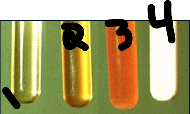
what is 3?
Hemolyzed
red blood cells damaged

what is 4?
lipemic
pet ate a meal before blood taken
Blood samples (serum or plasma) that are collected postprandial or directly after eating are:
lipemic
All of the following statements are correct concerning EDTA, except:
it is the tube of choice to collect blood for chemistry panels
To maintain proper anticoagulant to blood ratio, sample tubes should be filled to what capacity?
90
All can be sources of error when running a PCV, except:
averaging the results of your two tubes to determine the result
Total protein levels are ______ in a dehydrated animals.
increased due to the plasma becoming more concentrated due to the water loss
What quality control measure is important to a well-functioning refractometer?
specific gravity of distilled water
The buffy coat in a spun hematocrit tube consists of:
white blood cells and platelets.
When collecting blood for a coagulation profile, it is very important to:
Cause minimal vascular damage during venipuncture
What is the difference between a red top tube and a tiger top tube?
one serum separator gel and the other does not
Blood cells continue to utilize glucose at a rate of _____ per hour if allowed to remain in contact with the serum.
10%
Hyperglycemia reading on glucometer can be due to:
diabetes
Which of the following samples should NEVER be frozen?
whole blood
Which tube yields plasma via centrifugation?
lithium heparin and EDTA
Yellow plasma can be associated with a problem in which organ?
liver
How long do we leave the drop of blood on the Azostix?
60 seconds
How long should you allow a serum separator tube clot?
15-30 mins
The refractometer is used to determine which value?
total protein
Hemolysis occurs when red blood cells break or become damaged and release their contents into blood serum or plasma all the following are causes of in vitro hemolysis except:
the separation of plasma
If you are unable to perform a CBC within 1 hour of collection, which of the following is the best preservation?
refrigerate the sample
The spun hematocrit tube can allow you to determine the following:
PCV, total protein, plasma color and buffy coat analysis
While working in the lab, a vet tech notices a gray vacutainer tube, on the counter the vacutainer the tech is looking at contains the anticoagulant:
Oxalate fluoride
The most common cause of an elevated packed cell volume (PCV) is:
Dehydration
Plasma that appears white or milky is said to be:
lipemic
Which tube does not need to be inverted after collection of the blood sample?
Red top tube
What is a normal PCV value for a dog?
47%
Azostix results that are are coming up dark green at 80 mg/dL, may indicate
kidney disease
what does a complete hematology profile or complete blood count include
Red blood cell count
white blood cell count
hemoglobin concentration ( Hb)
hematocrit or packed cell volume ( manually done)
total protein/total solids
platelet estimates
white blood cell film examination
calculated absolute and erythrocytes indices