Psychopharmacology of drug addiction
 Call Kai
 Learn
 Practice Test
 Spaced Repetition
 Match
 Flashcards
 Knowt Play
1/99

Earn XP
Description and Tags
Flashcards made from what was covered in lecture, modified w/ textbook information: Pharmacology Exam 3
100 Terms
What is SUD?
Substance use disorder
SUD in the US statistics
About 48.7million people aged 12+ or around 17% off the US population
Individual consequences of SUD
Psychological: Depression, anxiety, psychosis, insomnia
Physical: Liver damage, cardiovascular problems, cancer, eating disorders, etc
Social:
With the advancement of chemistry, we were able to ….
… isolate compounds/increase their potency, which allowed us to use them for multiple different things. For example, “cocaine toothache drops”, before we realize how addictive the substances were.
Describe the schedule of controlled substances

Features of drug abuse/addiction
Complex phenomenon/hard to precisely define but can include the following:
Physical dependence: Abstinence leads to highly unpleaseant withdrawal symptoms that motivate to return to drug use
Craving: Strong urge to take the drug often drives the behaviour of an addict
Remission/relapse: Remissions are drug-free periods but are often followed by relapses in which drug use recurs despite the negative consequences.
Define drug addiction
A chronic, relapsing brain disease characterized by compulsive drug seeking and use despite negative consequences
What does the DSM-5 say about SUD?
It is a diagnosis in the DSM 5. Patient should have a maladaptive pattern of substance use for 12+ months that has lead to significant impairment/distress by clinical standards. Also includes behavioural addictions such as gambling, sex, internet, and shopping addiction. A lot of the diagnostic criteria are to show if you meet a threshold for the disease.
Cycles of drug use that can lead to the development of addiction (from George Koob)
Downward spiral: Addiction is a downward spiral starting from preoccupation/anticipation of drugs → constant desire → tolerance withdrawal → social, occupational, recreational activities are compromised → spiral distress → addiction (chronically relapsing disorder that aligns with the following two phases:)
Abstinence: Intermittent periods of reduced use or abstinence (attempts to stop drug use w/o help, participation in a treatment program, or incarceration)
Relapse: Stress, drug cues, or small doses of the drug itself can trigger relapse to drug-taking

Factors that influence the development/maintenance of drug abuse and addiction
Route of administration: Oral/transdermal is slow whilst IV/inhalation is fast, and rapid drug delivery to the brain (faster onset, shorter duration) has the strongest euphoric effects but also a quicker onset of withdrawal symptoms, which is more likely to produce addiction.
Abused drugs are …: Rewarding (positive/euphoric experience associated w/ the drug) and positive reinforcers (consuming the drug strengthens whatever preceding behaviour was performed)
Conditioned cues/craving: Measured physiologically (heart rate, blood pressure, sweating), questionnaire to rate cravings, and brain scans

Animal models that study development/maintenance of drug abuse/addiction
Drug self-administration: In rodents and primates, we can test to see how likely they are to learn a behaviour (after training) so that they can voluntarily use the drugs. Acquisition → maintenance → extinction → reinstatement (relapse). There are environmental cues associated w/ drug-use and discreet cues associated w/ drug delivery. Can use fixed or progressive ratio procedures.
Relapse: Forces abstinence then reintroducing stimuli known to provoke renewed responding (drug priming - delivering a small dose of drug, subjecting them to stress, or environmental cues previously paired w/ drug delivery)

Fixed vs progressive ratio procedures for DSA
Fixed: An experimental schedule where a subject has to perform a predetermined operant response to receive a single dose of a drug
Progressive: Train on fixed ratio, then increase the FR schedule progressively until the animal stops responding which is known as the breaking point (at some point it will become aversive, like inducing anxiety or psychosis or seizures), a ceiling of how much effort animals are willing to put in to get a certain amount of reward back
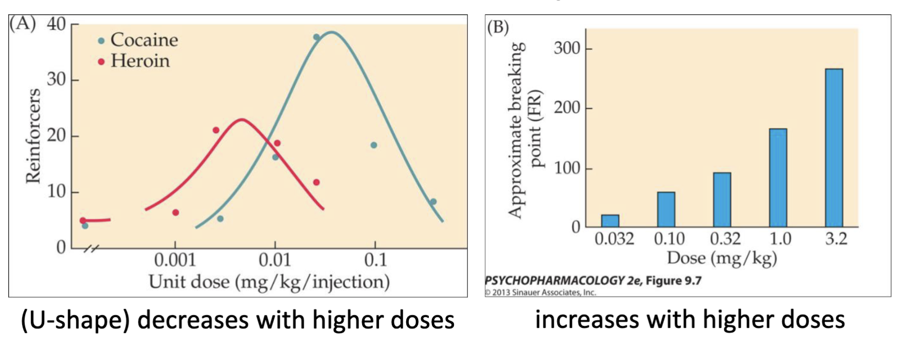
Drug reinforcement in humans
Self-administration tests can also be done in humans, who are usually experienced users. Medications can be tested like methadone for heroin addiction.

Progressive ratio cocaine self-administration & breakpoints and what it tells you
Willing to work more as their experience with a drug increases → addiction

How can we study drug reward? (CPP vs ICSS)
Place conditioning: Animal associates one compartment w/ the rewarding effect of a drug (expectancy of reward). Each compartment is pretty different for easy differentiation. They should show a preference for a specific side after being trained with the drug and once you do it repeatedly (for a drug that rewards them) the animal should choose the compartment that they associate w/ the drug.
Electrical self-stimulation: Of the brain’s reward circuit on performing an operant response. The threshold is reduced when animals have been treated acutely w/ drugs of abuse. There is intracranial self-stimulation (ICSS) where animals stimulate themselves w/ an electrode that descends onto dopamine fibers and stimulates those axons (mesolimbic pathways) which trains them. W/ ICSS, if a drug is rewarding, animals will be willing to push the lever at lower milliamps which shows that the drug is giving them dopamine.

What are aversive effects (in general)?
Drugs of abuse can be highly unpleasant withdrawal symptoms (abstinence syndrome) and can motivate to take the drug again. These are aversive effects, which differ by drug.

Theory for transition to compulsive drug-taking
Drug-taking behavior progresses from an “impulsive” stage (primary motivation is the substance’s positive reinforcing effects) → to a “compulsive” stage (primary motivation is the negative reinforcement obtained by relief from drug withdrawal). So basically it goes from positively reinforcing (attain the feeling of being high) to negatively reinforcing (avoidance of withdrawal).

Conditioned cues and craving
Withdrawal symptoms can be triggered by exposure to the conditional stimuli, such as cocaine-dependent subjects watching a video of someone smoking crack cocaine, which increases craving/desire for a drug.
Risk factors
Drug use pattern: Many individuals who take drugs once or a few times stop before they develop a compulsive pattern and become addicted. The more frequent the schedule → more dependent → more likely to be addicted
Genetic variation: Genes can influence susceptibility to addiction since some people have clusters of alleles that carry increased risk of developing substance use disorders like SNPs (single nucleotide polymorphism) in alcoholism/smoking, and differences in D2/D3 receptors in the nucleus accumbens that can predict impulsivity differences
Comorbidity: Anxiety, mood, and personality disorders (self-medication, shared etiology, causal hypothesis)
Personality variables: Behavioural disinhibition (linked to traits like impulsivity, antisociality, unconventionality, aggressiveness, low constraint/harm avoidance), stress reactivity (high scores on traits like stress reactivity, anxiety, and neuroticism are indicative of heightened vulnerability to stress), and reward sensitivity (sensation/reward seeking, extraversion, gregariousness etc.)
Familial/sociocultural influences: Familial factors have been most studied with alcoholism. Adult children of alcoholics are at increased risk. When consumed in a group setting, drugs may enhance social bonds. The user escapes from normal roles and responsibilities
Neurobiology of addiction
Reward circuit: Responsible for acute rewarding of reinforcing effects of abused drugs.
Dopamine: Reward is a complex phenomenon involving emotion and affect (feelings of pleasure), as well as the fact that dopamine impacts motivation and learning and cognition.
Neuroadaptations: Repeated exposure to a drug results in long-term changes in the brain that lead to a motivational transition. There’s numerous types of neuroadaptive changes that have been proposed.

Expand on the reward circuit and studies that have been done
Activation of it via monetary rewards has been showing positive correlations with the expected value of the reward. Neuroimaging confirms that human brains have a reward circuit similar to that identified in animals, and it is activated by both drug and non-drug rewards.
Expand on dopamine’s involvement w/ rewards + learning
It is involved in more than one aspect of the reward circuit; the role may vary depending on the context. may encode pleasure during drug use, mediate incentive salience when the animal is evaluating incentives, and act as a reward-prediction error signal when the animal is expecting a reward. You LEARN to get the REWARD, though DA isn’t inherently always rewarding.
Expand on the brain changes in context of neurobiology of addiction
Change in striatum areas: Other changes in neural circuitry that contribute to the development of addiction include the transition from the NAcc (ventral striatum) to the dorsal striatum (caudate-putamen) as a key control area for drug-taking behaviour. Impulsive drug taking/seeking the reward → compulsive/habitual drug-taking. Goes from the goal-directed, rewarding mesolimbic pathway (honeymoon phase of reinforcement) to a nigrostriatal pathway (habitual, but more so in terms of motor behaviours, showing that it has become a habit)
Glutaminergic pathways: There is also a functional disruption of the glutamatergic pathway from the PFC to the NAcc. By interfering with normal transmission in this pathway, the changes impair the ability of the PFC to mediate response inhibition and impulse control.

Neuroadaptations shown by brain imaging studies
Structural and functional abnormalities in the PFC and STR in drug-addicted subjects. Structural MRI - reduced gray matter volume in many drug addicts, sometimes even after long-term abstinence. PET imaging with [18F]FDG, - decrease in regional glucose metabolism. Decreased DA D2 receptor binding in the striatum.
Neuroadaptiations – genes and proteins
Different changes in gene expression can explain some of the behaviours we see.


Volkow reading — Figure 1 shows PET measurements of cocaine, methylphenidate, and methamphetamine pharmacokinetics in the human brain and their relationship to subjective "high." How do differences in the rate of brain uptake and clearance of cocaine, methylphenidate, and methamphetamine influence their reinforcing effects, and what do these findings suggest about the role of rapid dopamine increases in drug reward?
Cocaine vs Methylphenidate: The activity of methylphenidate lasts longer but does not get as characteristically “high” as cocaine does, which suggests that the stronger the dopamine rush is then the higher the reinforcing addictive effect is going to be (predictive of addictive potential). Methylphenidate has slower kinetics than does cocaine, which relates to the experience (again, of being “high”). Methylphenidate results in a gradual dopamine rise and milder/delayed subjective response unless given rapidly via IV. The intensity of the subjective high does not strictly match the total amount of drug in the brain or the peak number of occupied receptors; instead, it correlates closely with the rate at which dopamine increases in the synapse.
Cocaine vs Methamphetamine:

Volkow reading — Figure 2 demonstrates PET imaging with [¹¹C]raclopride to compare methylphenidate-induced dopamine release in healthy controls and active cocaine users. Why might active cocaine-addicted individuals show markedly blunted dopamine responses to intravenous methylphenidate despite continuing compulsive drug use, and how does PET imaging help reveal this neuroadaptation?
Raclopride: Radiotracer that binds to receptors and competes with dopamine. Itself is a molecule that is a selective D2-receptor ligand that is a competitive antagonist. Old-school anti-psychotic drug, but not really used for that anymore. Competes directly w/ endogenous DA at the same receptor binding site. An increase in extracellular DA displaces the radioligand. Can be used to measure DA release (researchers infer changes in the synaptic dopamine levels by measuring the decrease in [¹¹C]raclopride binding potential (BPND) before and after a pharmacological/behavioural change).
Study: Methylphenidate was used as a dopamine kick because it is unethical to give naive subjects cocaine.
Cocaine-addicts: Didn’t respond as much to the methylphenidate because [FINISH HERE]. There was less binding potential as a baseline in cocaine-addicted subjects (less binding sites) because [FINISH HERE]. In their systems, the dopamine receptors have been down-regulated and the response to the stimulus that drives the response is blunted. Perhaps they developed a tolerance to such systems, which caused the downregulation of the D2 receptors (and maybe others but we aren’t sure since the radiotracer used only bindind to D2).
PET imaging:

Volkow reading — Figure 3 shows that cocaine-related cues increased striatal dopamine and that the magnitude of dopamine release correlated with self-reported craving. What does the stronger dopamine response to cocaine-conditioned cues compared with pharmacological drug stimulation suggest about the neural mechanisms of craving, habit formation, and relapse risk in addiction?
Positive correlation between the change in cravings and % change in BPND putamen (non-displaceable binding potential).
In cocaine-addicted subjects, the drug that increases dopamine is not driving cravings — it is the CUES that are driving the craving, which is related to psychology since even though the cocaine has lost pharmacological power, the psychological impact of a cue is so strong that it brings them back to the drug.
[FINISH HERE]

Volkow reading — Figure 4 illustrates correlations between reduced striatal D2 receptor availability and decreased metabolism in prefrontal regions such as the orbitofrontal cortex (OFC). How might reductions in striatal D2 receptor availability contribute to impaired inhibitory control and decision-making in addiction, and what advantages does PET imaging provide for investigating these brain-behavior relationships?
Reductions in Striatal D2: [FINISH HERE]
Advantages of PET: [FINISH HERE]
Volkow reading — Other takeaways
Figure 1 shows acute drug effects; we don’t know if the patients are addicts or not.
Craving can be tested in a human lab by using cues that are related to the drug-taking habits/environment (watching someone use the drug), etc. (it has to be a conditioned/drug-associated cue).
What other scan shows you hyperfrontality and what is it: [FINISH HERE]
[FINISH HERE]
Brief history of alcohol (in class)
Alcohol is the second most commonly used psychoactive drug in America (after caffeine).
Merits of beer drinking vs evils of gin drinking image — high proof can’t come from just fermentation (max ~ 15%), distillation, etc. were accessible for a long time. With the creation of Gin used distillation to increase the content of alcohol in the 17th century (and credited with the start of serious alcohol abuse in Europe). Recognized as starting societal issues.
Absorption of Alchohol
Characteristics: Easily absorbed, passive diffusion across membranes from higher concentration in the GI tract to lower concentration in the blood, tissues, and brain. Ethanol diffuses across the biological membrane by moving through the lipid bilayer itself and by moving through water pores and spaces created by proteins.
Affected by: Dose, concentration, food in the stomach, body size/composition, gender differences, ethnicity, drinks w/ alcohol concentration 10-30% are absorbed the fastest, medications, and diseases or stress

What is BAC?
Blood alcohol concentration
0.04% BAC or 0.4 mgs per mL shows measurable behavioural effects
States impose penalties for driving with greater than 0.08% in personal vehicles, 0.04% commercial vehicles, and 0.01% for buses.
Elaborate on gender differences in alcohol absorption
Alcohol dehydrogenase in gastric fluid is 60% more active in men than women so alcohol is absorbed more rapidly in women. And: the same amount of alcohol is more concentrated in the average woman than the average man since her fluid volume is much smaller
Overview of Metabolism of Alcohol + draw the pathway
95% is metabolized by the liver’s ADH, 15% by gastric ADH which depends on if the stomach is full or empty: on a full stomach the gastric emptying slows down so gastric ADH has more time to break it down which lowers BAC.
There’s another metabolic route with Cytochrome P450 in the liver, but that is secondary. The remaining 5% of excreted unchanged by the lungs and can be measured with a breathalyzer (estimates BAC).
Regulated by acetaldehyde; increased levels when acetaldehyde dehydrogenase is less active, causing: flushing, nausea, headache, and increased heart rate.
Genetic variations: 50% of people of northeast asian descent and 5-10% of blond-haired blue-eyed people of northern European descent might have a dominant mutation in their acetaldehyde dehydrogenase gene, making this enzyme less effective.

Ethanol vs methanol
Can metabolize ethanol OR methanol (ethyl alcohol vs methyl alcohol) but methanol results in formic acid which is toxic to our bodies (from formaldehyde) with many adverse effects (10 mL= permanent blindness, 30+ mL = potentially fatal, 100 mL = median lethal dose) or ocular toxicity (damage to the optic nerve which causes the blindness).
Genetic diversity in the ADH genes
Genetic variations: 50% of people of northeast asian descent and 5-10% of blond-haired blue-eyed people of northern European descent might have a dominant mutation in their acetaldehyde dehydrogenase gene, making this enzyme less effective.
Combination of enzymes super active ADH + less active ALDH = more acetaldehyde = protection from alcohol dependence BUT ADH1B*1 (normal) ALDH2*1 (fast) = increased risk of alcohol dependence

Tolerance of Alcohol (acquired tolerance after chronic alcohol use)
Metabolic tolerance: Enzyme induction; number and activity of P450 increases = increases rate of metabolism of alcohol (and other drugs!) …but, prolonged heavy use of alcohol causes liver damage that significantly impairs metabolism of alcohol and many other drugs. Prolonged, heavy use of alcohol causes liver damage that impairs metabolism of many drugs not just alcohol.
Pharmacodynamic tolerance: Neurons adapt to the continued presence of alcohol by making compensatory changes in cell function. It is a GABAa receptor.
Behavioral tolerance: Practicing behaviors while under the influence of alcohol allows adjustment and compensation (someone who is used to driving while drunk will do field sobriety tests better than a first-time drunk driver due to learning to compensate for the impairment via practice)
Behavioural effects of alcohol (anxiety, memory, motor, OD)
Anxiety: At low doses, relaxation and less anxiety (reduce social inhibition, reduced judgment and overconfidence may increase risk-taking behaviors, sexual encounters more likely…). Short-term it increases GABA activity → temporary relaxation, but as BAC drops (or raises too high) → emotional instability. Long-term/during the sober state it has a rebound “hangxiety” effect where the brain relies on alcohol to feel calm so you are stressed when sober.
Memory: Acute effects on memory vary with dose and task difficulty (high doses, blackout – total amnesia for events that occur during intoxication). High doses impair the prefrontal cortex, leading to severe clouding of judgment, inability to concentrate, delayed information processing, and a total loss of impulse control.
Motor: Reduced coordination and impaired motor skills, delayed reaction time, increased drowsiness. Cerebellar impairment → less control of balance, voluntary movement, or spatial orientation: slurred speech, staggering, slow reaction times, poor fine motor skills.
OD: High doses result in unconsciousness and death/depression of respiratory system in the brain - death. Sypmtoms of alcohol poisoning include severe confusion, inability to wake up, seizures, hypothermia, suppressed gag reflex, and respiratory depression.
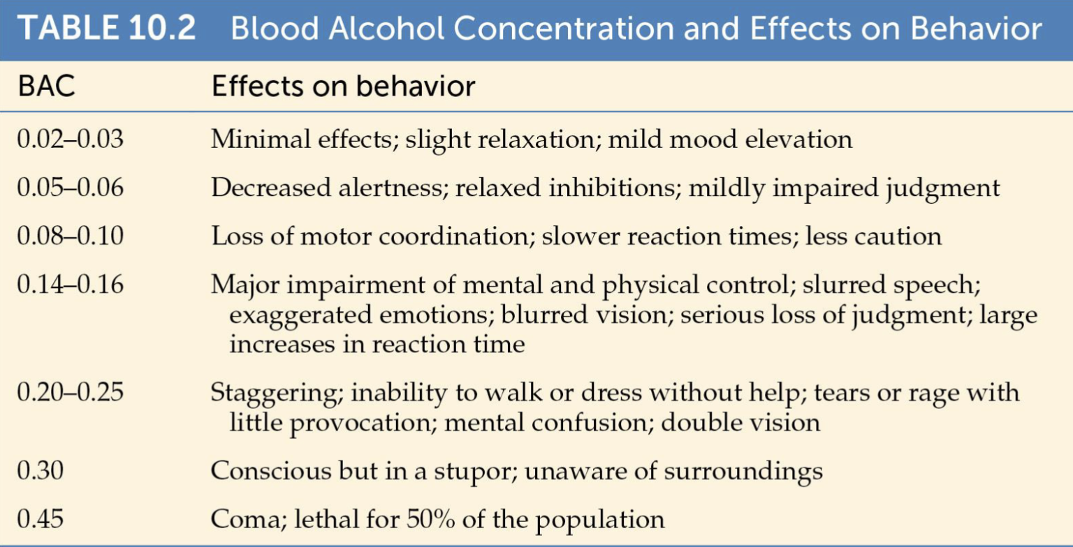
Subjective alcohol effects
Effects of alcohol are influenced by an individual’s environment and expectations.
In the US it is expected that alcohol will increase sociability, reduce anxiety/tension, increase aggression, and enhance sexual responses.
Pronounced behavioural effects occur under placebo conditions when the individual believes that they ingested alcohol, shown by multiple experiments of giving participants alcohol-free drinks and them ACTING as though they were drunk despite not having any.
Physical dependence of alcohol
What it is: Prolonged use of alcohol produces physical dependence where subjects develop tolerance (which by itself does not always mean dependence) and withdrawal symptoms (severity of which depends on the amount taken and duration of drug taking).
Symptoms: Hangovers, aches, gastric irritation due to residual acetaldehyde, tremors (“the shakes”), anxiety, high BP, tachycardia, sweating, hyperventilation, nausea, and delirium tremens in extreme cases.
Cross-dependence: Alcohol has cross-dependence with other drugs in the same sedative-hypnotic class like barbituates and BZDs (both also work on the GABAa receptor).
Consequences of chronic alcohol use
Brain damage: Takes many years to develop brain damage from alcohol consumption (extreme). Can also be exacerbated by elevated acetaldehyde, liver deficiency, and inadequate nutrition. Deficiency of vitamin B1 (thiamine — vital to metabolism of brain glucose) leads to cell death.
Wernicke-Korsakoff syndrome: Confusion, disorientation, amnesia, tremors, dementia, poor coordination, ataxia. Treated with thiamine.
Brain ventricles: Enlarged ventricles are indicative of brain tissue shrinkage in alcoholics.
Peripheral effects: See other card.

Peripheral effects of chronic alcohol use
Low doses: MAY improve cognitive function in older adults through vasodilation in the brain or reduce risk of heart disease because it increases the amount of “good” cholesterol in the blood while reducing the “bad” which reduces the incidence of blood clots and stroke.
High doses: Fatty liver (TAG), Alcoholic hepatitis (high levels of acetaldehyde), alcoholic cirrhosis—death of liver cells stimulates scar formation and blood vessels carrying oxygen are cut off.
What is FAS? Describe it.
What it is: Fetal alcohol syndrome
Causes: Prenatal alcohol exposure aka placental transfer from mother’s bloodstream → umbilical cord → fetus (slow metabolism, keeps it in fetus’s system longer → traps in cycle in amniotic fluid) → interferes w/ division/growth of cells like brain/cns development and other physical structures
Symptoms: Small eye openings, smooth philtrum, thin upper lip, small head, low birth weight, short, structural defects in the heart/kidneys/bones/ears, vision/hearing problems, short attention span, learning disabilities, poor coordination/balanace, bad fine-motor skills, trouble with memory and judgement and reasoning, mental health struggles

Neurochemical effects of alcohol (specific vs nonspecific)
Nonspecific: Disturbs the relationship of protein in the membrane, interacts w/ polar heads of phospholipids, alters lipid composition
Specific: Acts at NT binding sites, modifies gating mechanisms inside the channel, direct interaction with channel proteins, stimulates Gs which is linked to adenylyl cyclase

Neurochemical effects of alcohol (GABA vs glutamate)
GABA: Inhibitory. Alcohol is a GABA positive allosteric modulator (PAM). Ethanol potentiates GABAa responses, but drecreases the excitability of neurons through increased chloride (Cl⁻) influx, which hyperpolarizes the membrane and pushes the neuron further from its firing threshold. This underlying cellular inhibition is a CNS depressant! Acutely: alcohol binds (most but not all) GABAa receptors; increases GABA-mediated Cl- flux to hyperpolarize the membrane, and stimulates GABA release. Chronically: repeated exposure to ethanol GABAa-mediated Cl- flux (higher sensitivity to seizure-inducing doses of the GABA antagonist bicuculline may contribute to tolerance and to some of the signs of withdrawal.).
Glutamate: Acutely, alcohol acts as a glutamate antagonist that inhibits NMDA receptor function, reduces glutamate release in some brain areas (like LTP in the hippocampus). Chronically, there will be up-regulation of NMDA receptor function and density as the brain attempts to compensate for chronic suppression. During withdrawal, this excess of NMDA receptors causes massive glutamate hyperexcitability, leading to excitotoxicity (cell damage) and severe withdrawal seizures.
Neurochemical effects of Alcohol Withdrawal
During withdrawal, glutamate release that is normally reduced by alcohol, increases after about 10 hrs—correlated with CNS hyperexcitability and seizures typical of alcohol abstinence syndrome. This is a REBOUND EFFECT. Also - monoaminergic dysregulation, hypothalamic-pituiary-adrenal axis is hyperactive → increased emotional/physical stress, and increased in voltage-gated Ca2+ channels → downregulations of protective K+ channels → toxic amounts of Ca2+ rushes into neurons.

Dopaminergic neurochemical effects of alcohol
Studies: Mesolimbic DA system plays a significant role in the reinforcement and motivational mechanisms. Responsible for transition of voluntary drinking to compulsive use by changing from the ventral striatum (voluntary, impulsive, goal-directed, PFC regulates it) to dorsal striatum (habitual, “autopilot”).
Acute: Induces DA release in the NAcc and activates the mesolimbic DA pathway.
Chronic: Tolerance - maybe, mostly changes how it manifests (shift to dorsal striatum makes tolerance go from acute to behavioural/habitual).
Withdrawal: Reduces the firing rate of mesolimbic DA neurons and decreased DA release in the Nacc which contributes to behavioural signs of withdrawal.

Neurochemical effects of alcohol on opiod peptides (endorphins)
Overview: Endorphins contribute to the reinforcing effects of alcohol because alcohol releases them, which drives rewarding/reinforcing properties. Blocking opioid receptors reduces alcohol drinking. Drug: naloxone, naltrexone in animals. Some success in Humans (see later). Depends on genes (like having an “addictive personality”).
Acute/low: Increases endogenous opioid production and release (endorphin and enkephalin). In low/moderate drinking, the peptides bind to mu and delta receptors → inhibits GABAergic interneurons → disinhibits DA neurons → more DA in the nucleus accumbens → euphoria.
Chronic/Withdrawal: Reduces production, which may contribute to the dysphoria that accompanies withdrawal. Receptor densitization/neuroadaptation and central opioid deficiency from chronic drinking lead to negative reinforcement w/o alcohol.

Know Table 10.3 from the textbook
View graph

Alcoholism
Definition: A complex phenomenon neurobiological and psychological, genetic, and sociocultural factors, making it difficult to define and treat. Defined by the frequency/pattern and total alcohol consumed. Similarly related to their compulsive alcohol seeking and drinking despite damaging health and social consequences
Binge drinking: 5+ drinks in a row in a given time
Cause: Nothing specific — a variety of factors contribute to the vulnerability of an individual
Linkage studies: Inheritance pattern of genes using DNA analysis and occurrence of alcoholism in a large number of families. One study of a Native American population identified two potential genes for alcoholism, located near genes for DA (D4) and GABAA receptors and TH.
Case-control method: Compare genes of unrelated individuals to see whether members of the affected population have more of a particular allele (ALDH).
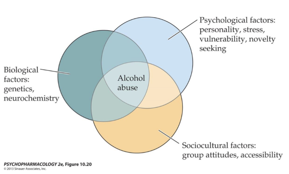
Treatment of alcoholism
Detoxification
: Detoxification. BZDs like chlordiazepoxide (librium) or diazepam (valium) are given to prevent alcohol withdrawal symptoms.
Psychosocial rehabilitation: Helps prevent relapse by interacting with individual/group therapy, residential alcohol-free treatment settings, and self-help groups like alcoholics anonymous
Pharmacotherapeutic treatment: See other flashcard
Pharmacotherapeutic treatment for alcoholism
Making drinking unpleasant: Disulfiram (antabuse) inhibits ALDH which increases acetaldehyde, which means drinking even an ounce of alcohol results in flushing, nausea, tachycardia, and vomiting.
Reduce alcohol’s reward: Naltrexone is an opioid receptor antagonist that reduces alcohol consumption/craving and improving abstinence rates by blocking the effects of alcohol-/cue-induced endorphin release.
Restore excitatory-inhibitory balance: This is disrupted by alcohol withdrawal. The mechanism isn’t entirely clear, but might have to do with NMDA receptor partial antagonist and GABAa receptor partial agonist. Acamprosate (campral) blocks the glutamate increase that occurs during alcohol withdrawal in rats.

Which substance is the primary active ingredient in opium and was the first active compound isolated from a medicinal plant in the early 1800s?
A. Heroin
B. Morphine
C. Methadone
D. Codeine
B. Morphine
It makes up 10%-12% of the raw substance and was first isolated from opium poppy.
Which characteristic makes heroin more potent than morphine?
A. It has a higher affinity for μ-opioid receptors than morphine
B. It is more lipid soluble and reaches the brain more rapidly
C. It cannot be metabolized in the brain
D. It selectively activates δ-opioid receptors
B. It is more lipid soluble and reaches the brain more rapidly
Heroin is chemically altered from morphine w/ +2 acetyl groups attached to it, making it more lipid soluble. The BBB allows fat-soluble substances to pass more easily.
Which statement best distinguishes narcotic analgesics from anesthetics?
A. Narcotic analgesics produce unconsciousness, whereas anesthetics only reduce pain.
B. Narcotic analgesics reduce pain without producing unconsciousness at therapeutic doses, whereas anesthetics block pain and temporarily produce a reversible loss of awareness.
C. Narcotic analgesics and anesthetics have identical effects on consciousness.
D. Anesthetics are used to treat chronic pain, whereas narcotic analgesics are not.
B. Narcotic analgesics reduce pain without producing unconsciousness at therapeutic doses, whereas anesthetics block pain and temporarily produce a reversible loss of awareness.
Narcotic analgesics (like morphine, codeine, or fentanyl) target specific opioid receptors in the nervous system to blunt the perception of pain, allowing the patient to remain awake and conscious at standard medical doses. In contrast, general anesthetics act more broadly on the central nervous system to induce a controlled, reversible state of unconsciousness, muscle relaxation, and total loss of sensation so a patient feels nothing during major surgery.
Which opioid receptor subtype has a high affinity for morphine and is strongly associated with analgesia and positive reinforcement?
A. δ (delta) receptor
B. κ (kappa) receptor
C. μ (mu) receptor
D. NOP receptor
C. μ (mu) receptor
This is the primary binding site for morphine and other classical opioid drugs. Activation is responsible for the strongest pain-relieving effects and euphoria (acts as PR → addiction).
Which brain region gives rise to the descending pathways that contribute to opioid-mediated pain relief?
A. Nucleus accumbens
B. Periaqueductal gray (PAG)
C. Amygdala
D. Hippocampus
B. Periaqueductal gray (PAG)
When opioids bind to the PAG they trigger a descending analgesic pathway to the spinal cord which blocks pain signals from traveling up the brain.
How can opioid drugs produce reward and reinforcement in the brain despite acting through inhibitory opioid receptors?
A. They directly increase dopamine release by exciting dopamine neurons.
B. They inhibit GABA neurons in the ventral tegmental area (VTA), which disinhibits dopamine neurons and increases dopamine release in the nucleus accumbens.
C. They block dopamine receptors in the nucleus accumbens.
D. They increase serotonin release from the raphe nuclei, producing reward.
B. They inhibit GABA neurons in the ventral tegmental area (VTA), which disinhibits dopamine neurons and increases dopamine release in the nucleus accumbens.
Works via disinhibition. With the GABA brake mechanism removed (opioid drugs usually enter the VTA → bind to inhibitory receptors on GABA neurons → shut them down), the DA neurons rapidly fire → floods nucleus accumbens w/ DA → strong sense of reward.
Tolerance to opioids is best defined as:
A. Increased effects of a drug following repeated administration
B. Drug craving that intensifies over time
C. Diminishing effects of a drug with repeated use
D. The appearance of withdrawal symptoms after stopping drug use
C. Diminishing effects of a drug with repeated use
The body will adapt to the opioid’s constant presence so to achieve the same result as before, higher doses are needed. A is sensitization, B defines drug craving, D is specifically physical dependence.
What is the primary advantage of buprenorphine compared with full opioid agonists such as heroin?
A. It completely blocks all opioid receptors
B. It produces stronger euphoria than heroin
C. It is a partial opioid agonist that suppresses withdrawal and cravings with lower abuse potential
D. It reverses overdose within minutes
C. It is a partial opioid agonist that suppresses withdrawal and cravings with lower abuse potential
Buprenorphine is a partial agonist of the μ-opioid receptor (has a ceiling effect on respiratory depression/euphoria/sedation). Satisfies opioid requirement to stop withdrawal symptoms but also satisfy cravings w/o the addictive high.
Acute tolerance of alcohol (psychopharmacology)
Occurs with a single exposure to alcohol since the drug effects are greater while the blood level of alcohol is increasing and smaller while the blood level is falling. The drinker’s perception of intoxication also undergoes acute tolerance (SEE GRAPH).

Draw the pathway of alcohol metabolism
Pathway: alcohol → ADH turns it into acetaldehyde → ALDH turns it into acetic acid → oxidation reaction turns it into carbon dioxide and energy
In the liver, cytochrome P450 2E1 produces a harmful reactive oxygen species that causes oxidative stress and liver injury.

Opioids vs Opiates
Opioids: Umbrella term that include opiates, semisynthetic substances, and endogenous peptides — ALL of which bind to opioid receptors
Opiates: A substance directly derived from the opium poppy
History of opioids
Has always been recognized as a calming agent with pain-relieving properties.
Bayer made Heroin by adding two acetyl groups to morphine which made it more lipid soluble w/ the goal of making a non-addictive pain reliever/cough suppressant. It’s actually more potent.
Harrison narcotics tax act: United States federal law that regulated and taxed the production, importation, manufacture, and distribution of opium, coca leaves, and their derivatives (such as morphine, heroin, and cocaine). Marked a shift to prohibition and black market growth even though there were medical exemptions (people try to garner an exemption by faking a disorder).
Heroin vs Morphine
Heroin is more potent due to higher lipid solubility and crosses the BBB much easier. In the brain, heroin is converted into morphine. Heroin can be thought of as a prodrug; itself has a low affinity for the mu-opioid receptor, 6-MAM is an active metabolite that binds to receptors, and Morphine is a full agonist.

Natural vs Synthetic Opioids
Natural:
Semisynthetic:
Fully synthetic:
Endogenous opioids:
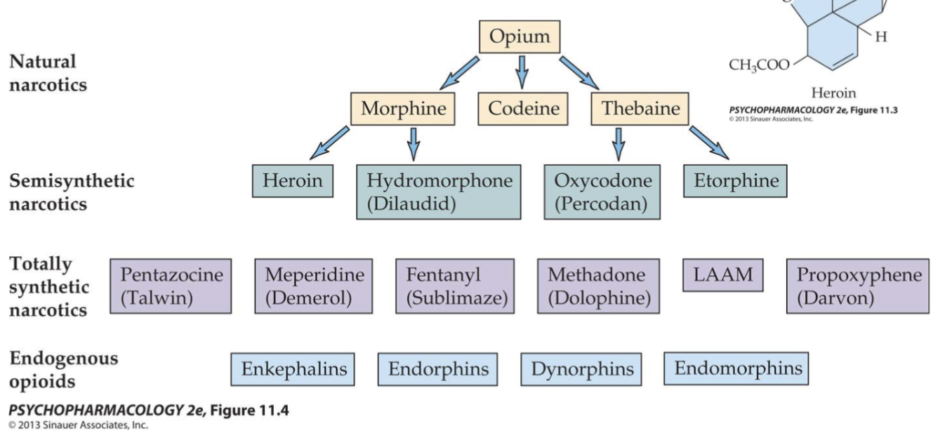
Importance of Endogenous Opioids
Something in the brain that is able to bind endogenous opioids (in the periphery), which the other drugs just hijack. Activate descending inhibitory pathways (like PAQ to raphe nuclei) to blunt the affective (emotional) and physical impact of pain. Regulate HPA axis to adapt to stress and chronic use can blunt/deplete body's natural opioid tone which leads to pain vulnerability and being dependent on it.
Main features of opioids
Narcotic analgesics that, at normal doses, reduce pain w/o producing unconsciousness (except in high doses due to depressing centers that regulate breathing). Compared to anesthetics, which block pain and produce temporary unconsciousness/reversible loss of awareness. At high doses or when mixed with other sedatives like alcohol or BZDs, risk of severe unconsciousness increases.
How can opioids be taken? How fast do they work?
Morphine: IM or orally
Opium: Smoked by recreational users
Heroin: Snorting or subcutaneous administration

Neonatal Abstinence Syndrome
Caused by the use of heroin or other opioid drugs during pregnancy where the newborn experiences withdrawal up to 1 week. Treated with IV fluids, tapering down morphine/methadone doses, phenobarbital (CNS sedation to calm tremors, control seizures, etc., or diazepam (relaxes muscles, sedation, not commonly used today).
Dose-dependent effects of opioids
Low-to-moderate: Pain relief, constricted pupils, drowsiness, inability to concentrate, dreamy sleep, potential relief of anxiety or aggressiveness or feelings of inadequacy which could lead to more drug use.
Higher: Abnormal elation/euphoria. This “rush” acts like a reinforcer to encourage repeated drug use but there could be dysphoria, restlessness and anxiety, nausea and vomiting (area postrema)
Highest: Sedative effects may lead to unconsciousness. Body temperature and blood pressure fall. Pupils become very constricted respiratory failure is the ultimate cause of death in overdose (brainstem respiratory center).
OD and respiratory depression w/ Opioids
This is the direct result from stimulation of μ-opioid receptors in neurons involved in the control of breathing, primarily located in the brainstem. Specifically/particularly in the Nucleus Tractus Solitarius (NTS), Retrotrapezoid Nucleus (RTN), and Median Raphe Nuclei (MRN)

Opioid OD and Narcan
Naloxone is a competitive opioid antagonist that displaces opioids from the receptors, rapidly reversing respiratory depression. Dose-dependent effects of intramuscular (IM) naloxone on fentanyl mu-opioid receptor occupancy (RO) Naloxone works within minutes! Naloxone lasts for about 30 to 90 minutes, whereas many opioids or synthetic variants last much longer, creating a risk of re-overdose once the naloxone wears off (die during “second high” like Dr. Schwendt mentioned in class).

Opioid and constipation
Morphine also affects the GI tract. Opium and morphine have been used to treat diarrhea and can be life-saving to stop fluid loss in severe bacterial and parasitic diseases. Unfortunately, when opioids are used for pain management, constipation is a common side effect.
(McLaughlin lab: counting mouse poop to see constipation as a result from morphine).
How were opioid receptors discovered?
By radioligand binding methods like autoradiography, allowing scientists to see where they were in the brain in 1973. Mid-1970s came the discovery of natural bodily analgesics like enkephalins, endorphins, and dynorphins.
Opioid Receptor Subtypes
μ (MOPR): Targeted primarily for analgesia and reward. MAIN GIRL MAIN GIRL MAIN GIRL. High affinity for morphine. Analgesic effects expressed in the medial thalamus, periaqueductal gray, median raphe, and spinal cord. Feeding/positive reinforcement in the nucleus accumbens. CV and respiratory depression, cough control, nausea/vomitting control in the area postrema of the brain stem. Sensorimotor integration in the thalamus and striatum.
κ (KOPR): Influences stress responses and mood regulation. Found in the striatum and amygdala, hypothalamus and pituitary. Could participate in regulation of pain perception, gut motility, and dysphoria.
δ (DOPR): Involved in pain modulation and emotional processing. Many of these sites are consistent with a possible role for modulation of olfaction, motor integration, reinforcement, and cognitive function. Areas of overlap μ-receptors suggest modulation of both spinal and supraspinal analgesia
NOPR (Nociceptin/Orphanin FQ): Regulates various physiological and sensory functions. Not as important for the exam
Other notes: Subtypes have distinct distributions in the brain and spinal cord which suggests that they mediate a wide variety of effects.
Endogenous Neuropeptides
1970s: Discovery of peptides that could bind to opioid receptors, called endorphins.
Precursor peptides: These propeptides are prodynophin, pro-opiomelanocotin (POMC), proenkephalin, pronociceptin/orphanin FQ. These are then processed into smaller active opioids.
Characteristics of opioid receptors
Metabotropic
Coupled w/ Gi/o proteins (inhibition of nerve activity: opens hyperpolarizing K+ channels, closes Ca2+ channels, inhibition of adenylyl cyclase).
Opioid receptor inhbition
Postsynaptic inhibition: OR activates Gi/o → opens K+ to hyperpolarize the postsynaptic cells → reduces firing rate (depresses firing rate)
Presynaptic inhibition: Autoreceptors activate Gi/o proteins to inhibit voltage-gated Ca2+ channels and reduce release of a co-localized neurotransmitter.
Axoaxonic inhibition: Presynaptic inhibition reduces the release of NTs from a different neuron.
2 components of pain
First/early pain: Immediate sensory component. Signals carried by myelinated Aδ neurons, which conduct action potentials rapidly
Second/late pain: The emotional component which has a slower transmission because the signal carried by thin and unmyelinated C fibers.
3 ways opioids regulate pain
1: Within the spinal cord by small inhibitory interneurons (release of endorphins) to inhibit transmission of pain to the brain.
2: By two descending pathways originating in the periaqueductal gray (PAG)
3: At many higher brain sites, which explains opioid effects on emotional and hormonal aspects of the pain response
Opioid drugs bind to ___ receptors and ___ the ___ action of the ___ at ___ stages of pain transmission
opioid; mimic; inhibitory; endogenous opioids; many
Study about Opioids and Emotional aspects of pain
PET imaging: Carfentanyl (displaceable mu-opioud receptor radiotracer). Sustained pain-stimulus by injection of hypertonic saline into the muscle.
Findings: Significant negative correlation between mu-opioid activity in the nucleus accumbens, amygdala, and thalamus and reported sensory pain scores. Anterior cingulate, NAcc and anterior thalamus had negative correlation w/ affect scores. Thus the endogenous opioids modulate both sensory and emotional compoents of pain, and opioid drugs also act on these sites.

Opioid reward/reinforcement
Impact: SIGNIFICANT(!!!) rewarding/reinforcing properties.
Animal studies: [FINISH HERE]
Mechanism: DA mesolimbic pathway contributes to reinforcement. Beta-endorphin and opioid drugs increase VTA cell firing by inhibiting the inhibitory GABA cells = increased firing and greater DA release in the NAcc which produces the reward/euphoria.
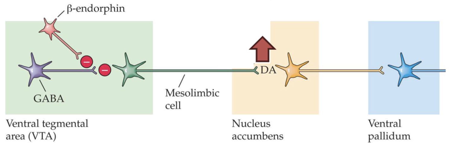
Opioid tolerance/dependence
Chronic use: Neuroadaptive changes which are responsible for tolerance, dependence, and sensitization.
Tolerance: The dimishing effects of a drug w/ repeated use plus cross-tolerance between opioid drugs makes people need to go back for more.
Sensitization: [FINISH HERE]
Physical dependence: When the drug isn’t there anymore, cellular functions return to “normal” but overshoot basal levels. The effects of this are seen in withdrawal symptoms like abstinence syndrome. [FINISH HERE]
[FINISH HERE]
Opioid Withdrawal
Because opioids in general depress CNS function, withdrawal of them result in rebound hyperactivity.
Abstinence signs reflect a loss of inhibitory opioid action at all receptors in the CNS and elsewhere.
Abstinence symptoms depend on the drug (its PK, speed of onset): For ___, the peak is ___ hours after last use. For ___, there is a gradual ___ over several ___ and a gradual ___ over several ___
morphine; 36-48; methadone; increase; days; decrease; weeks
Mechanisms of opioid withdrawal symptoms
1: Acute morphine/heroin — mu-opiod receptors = hyperpolarization, reduced rate of firing LC
2: Chronic morphine/heroin, gradual increases in the firing rates as tolerance developed.
3: Withdrawal excessive firing so withdrawal symptoms are treated by clonidine (alpha2 adrenoreceptor agonist)
Dysphoria of Opioid Withdrawal + Mechanism
Dynorphin acts on kappa-receptors on DA neuron terminals which leads to reduced DA release → dysphoria.
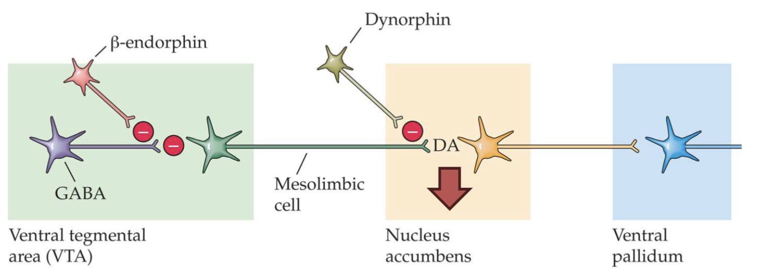
Tolerance/dependence vs addiction
[FINISH HERE]
Commonly abused illicit and prescription opioids
Fentanyl
Heroin
Oxycodone variants: percocet (+acetaminophen), percodan (+aspirin), oxycontin
Morphine
Dilaudid (hydromorphone)
Hydrocodone varients: Vicodin, lorcet, lortab
Demeral (pethidine)
Methadone
Cychlorphine (10x-40x stronger than fentanyl)
Treatments for opioid addiction
[FINISH HERE]
Opioid withdrawal management
Focuses on the specific neurochemical changes that occur when mu-opioid receptors are abruptly cleared. Chronic opioid binding suppresses norepinephrine release in the locus coeruleus; stopping opioids causes a rebound surge in cyclic AMP (cAMP) and norepinephrine, driving autonomic and physical withdrawal symptoms.
Detoxification is the first step which can be assisted by long-acting opioids, such as methadone, that reduce symptoms to a comfortable level (The dose is gradually reduced over 5-7 days). Can also use clonidine, an α2-adrenergic agonist, acts on noradrenergic autoreceptors to reduce NE activity in the locus coeruleus (NE neurons are inhibited by opioids, and the cells increase firing during withdrawal). But clonidine does not relieve all symptoms of withdrawal, and it has other side effects.
___ opioids hijack the ___ pain system by binding directly to ___-opioid receptors with far ___ affinity and potency than natural peptides, flooding the nervous system to produce profound ___, ___, and rapid ___.
Exogenous (from outside of the system); endogenous (inside a system); mu; greater; analgesia; euphoria; tolerance
What is a go circuit vs a no-go circuit?
Go circuit (Direct pathway): Promotes and initiates voluntary actions. It is activated when dopamine binds to D1 receptors in the striatum, which disinhibits the thalamus and allows the motor cortex to execute a movement.
No-go circuit (Indirect pathway): Inhibits, stops, or restrains actions. It is triggered when dopamine binds to D2 receptors, increasing the inhibition of the thalamus and suppressing unwanted or competing movements.
Balance: These two pathways work as a push-and-pull system to ensure smooth, context-appropriate behavior. Imbalance can lead to disorders.

How to use fMRI vs PET to measure baseline brain activity and craving-induced brain activity in addiction?
[FINISH HERE]
Which pathways regulate drug taking vs drug seeking?
[FINISH HERE]