transfusion theory exam (weeks 7-12)
 Call Kai
 Learn
 Practice Test
 Spaced Repetition
 Match
 Flashcards
 Knowt Play
1/123

There's no tags or description
Looks like no tags are added yet.
124 Terms
Antibodies can be found in blood, extravascular fluid and secretions → TRUE or FALSE?
TRUE
Why?
Antibodies are produced by plasma cells & distributed throughout the body to provide immune protection
Blood: Antibodies circulate to recognise foreign antigens
Extravascular fluid: Mainly IgG protects surrounding tissues
Secretions: Mainly IgA protects mucosal surfaces
In newborn blood group testing, only the forward group technique is used → TRUE or FALSE
TRUE
Why?
Anti-A & Anti-B antibodies are absent at birth and they only appear 10-15 days after birth
Blood group AB is known as the universal recipient → TRUE or FALSE
FALSE
Why?
Blood group AB-RhD (pos) is known as a universal recipient
ABO blood group phenotype frequencies;
O and AB have the lowest frequency
O is the most frequent (46%)
A1 is the most frequent (46%)
A2 has the lowest frequency (2.3%)
A & B have a similar frequency
O is most frequent (46%)
Why?
Blood group O is the most frequent (46%) and the A1B blood group has the lowest frequency (2.3%)
From the two subgroups of the A blood group, only A1 react strongly with the anti-A reagent → TRUE or FALSE
FALSE
Why?
Both A1 and A2 subgroups react strongly with the anti-A blood grouping reagent
Secretor status;
The secretor gene consists of only one allele responsible for the expression of the H antigen
The secretor gene consists of 2 alleles responsible for the expression of A & B antigens on RBCs
The secretor gene consists of 2 alleles responsible for the expression of Glucosamine on RBCs
The secretor gene consists of 2 alleles responsible for the expression of the H antigen
The secretor gene consists of 2 alleles responsible for the expression of the H antigen
Why?
The secretor gene (FUT2) has two alleles: Se (dominant) & se (recessive)
SeSe or Sese: Secretor - produces H substance in secretions, allowing A/B substances to be expressed depending on ABO type
sese: Non-secretor - does not express ABH substances in secretions
*Secretor status determines ABH antigen expression in secretions (e.g., saliva), not on RBCs. H antigen expression on RBCs is primarily controlled by FUT1
Blood group antigens are generally found in serum, and antibodies (directed towards blood group antigens) found on cells → TRUE or FALSE
FALSE
Why?
Blood group antigens are generally found on cells, and antibodies found in serum
Forward group/ing refers to the identification of:
Antibodies in an individual’s plasma with commercially available reagents
Antibodies on an individual’s red cells with commercially available reagents
Antibodies in an individual’s plasma using the patient’s own red cells
Antigens on an individual’s red cells with commercially available reagents
Antigens on an individual’s red cells with commercially available reagents
Why?
The forward group identifies A & B antigens on RBCs using known antisera (anti-A & anti-B)
Agglutination (+): Antigen is present
No agglutination (-): Antigen is absent
ABO genes encode for
Transferases that add a sugar moiety to pre-formed H substance
A, B or O antigens (direct gene products)
Transferases that add a sugar moiety to pre-formed A, B or O substance
Transferases which add a protein moiety to pre-formed H substance
Transferases that add a sugar moiety to a pre-formed H substance
Why?
ABO genes encode glycosyltransferases (enzymes) that add specific sugars to the H antigen on RBCs, determining blood group
A gene: Adds N-acetylgalactosamine → A antigen
B gene: Adds galactose → B antigen
O gene: Produces a non-functional enzyme → H antigen remains unchanged
*ABO genes encode enzymes, NOT the A & B antigens directly
If no H substance = Bombay blood group, but the individual may have A and B genes that produce a transferase → TRUE or FALSE?
TRUE
Why?
The Bombay blood group (Oh) occurs when an individual cannot produce the H antigen on their RBCs due to the hh genotype
H antigen: Required as the precursor for A & B antigens
A/B genes: May still be present & produce functional transferases
Without H antigen: Transferases cannot form A or B antigens, even if the genes are present
Therefore, Bombay individuals appear as group O on routine forward ABO grouping despite potentially carrying A or B genes
A person with blood type A can receive blood from which of the following donors?
Only type A
Only type B
Types A and AB
Types A and O
Types A and O
Why?
Individuals with blood group A have A antigens on their RBCs & anti-B antibodies in their plasma
Type A: Compatible because its RBCs have A antigens
Type O: Compatible because its RBCs have neither A nor B antigens
Types B & AB: Incompatible because their B antigens react with the recipient’s anti-B antibodies, causing haemolysis
Which blood type is considered the universal donor?
A
B
AB
O
O
Why?
Blood group O is considered the universal red blood cell donor because its RBCs lack both A & B antigens
Type O: No A or B antigens → minimises ABO incompatibility
Type A, B & AB: Have A and/or B antigens that may react with recipient antibodies
*O RhD negative is the universal RBC donor type when considering both ABO & RhD
What is the genotype of an individual with blood type AB?
AA
BB
AB
OO
AB
Why?
Blood group AB has the genotype IAIB, where both A & B alleles are codominant
A allele: Produces A antigens
B allele: Produces B antigens
Codominance: Both alleles are expressed equally, resulting in both A & B antigens on RBCs
What type of antibodies does a person with blood type B have in their plasma?
Anti-A antibodies
Anti-B antibodies
Both anti-A and anti-B antibodies
No antibodies
Anti-A antibodies
Why?
Individuals with blood group B have B antigens on their RBCs & anti-A antibodies in their plasma
B antigens: Recognised as self, so anti-B antibodies are not normally produced
A antigens: Recognised as foreign, so anti-A antibodies are naturally present
If a father is blood type A and mother is blood type B, what blood types could their offspring have?
A and O
A, B, and AB
A, B, AB, and O
B and O
A, B, AB and O
Why?
A father with blood type A can have genotype AA or AO, while a mother with blood type B can have BB or BO
If the father is AO & the mother is BO, their offspring can inherit any of the four ABO blood groups
According the table in the image, each blood group has a 25% probability if both parents heterozygous (AO x BO)

Which blood type is known as the “universal recipient”?
A
B
AB
O
AB
Why?
Blood group AB is the universal red blood cell recipient because individuals have both A & B antigens on their RBCs but neither anti-A nor anti-B antibodies in their plasma
Type A: Can receive A & O RBCs
Type B: Can receive B & O RBCs
Type AB: Can receive A, B, AB & O RBCs
Type O: Can receive only O RBCs
What happens if a recipient receives blood with an incompatible ABO type?
No adverse effects
Haemolysis (destruction of red blood cells)
Mild fever
Enhanced oxygen transport
Haemolysis (destruction of red blood cells)
Why?
An ABO-incompatible RBC transfusion causes the recipient’s anti-A or anti-B antibodies (mainly IgM) to bind to the donor RBC antigens
Antigen-antibody binding: Recipient antibodies attack incompatible donor RBCs
Complement activation: IgM strongly activates the complement system
Intravascular haemolysis: Donor RBCs are destroyed within blood vessels, potentially causing a life-threatening acute haemolytic transfusion reaction
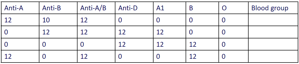
Interpret the following blood group reactions
Interpretations?
Row 1: Anti-A, Anti-B & Anti-A/B pos; Anti-D neg. No anti-A or anti-B in plasma
Row 2: Anti-B & Anti-A/B pos; Anti-D pos. Anti-A present in plasma
Row 3: No A or B antigens; Anti-D pos. Both anti-A & anti-B present in plasma
Row 4: Anti-A & Anti-A/B pos; Anti-D neg. Anti-B present in plasma

What defines the ABO blood group system?
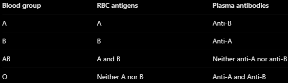
The ABO blood group system is defined by the presence or absence of A & B antigens on the surface of RBCs (see table for layout)
What are the enzymes that confers carbohydrate specificity?
Glycosyltransferases
Why?
Glycosyltransferases are enzymes that add specific sugars to the H antigen, determining ABO blood group specificity
A transferase: Adds N-acetylgalactosamine → A antigen
B transferase: Adds galactose → B antigen
O allele: Produces no functional transferase → H antigen remains unchanged
What carbohydrates define ABO blood group antigens?
ABO blood group antigens are determined by specific terminal sugars attached to the H antigen on RBCs
A antigen = N-acetylgalactosamine (GalNAc)
B antigen = D-galactose
H antigen = L-fucose
O group = H antigen remains unmodified
What ABO antigens are expressed on red blood cells?
ABO antigens are carbohydrate structures on the surface of RBCs, determined by the individual’s ABO blood group (see table for layout)
*H antigen is also present in varying amounts on normal A, B & AB red cells

What ABO antibodies are present in serum?
ABO antibodies are naturally occurring antibodies (mainly IgM) directed against the A or B antigens an individual lacks (see table for layout)

How do we undertake ABO blood grouping in the laboratory?
ABO blood grouping involves two complementary tests using haemagglutination:
Forward grouping: Patient RBCs are tested with anti-A & anti-B reagents to detect A & B antigens
Reverse grouping: Patient plasma is tested with known A1 and B reagent RBCs to detect anti-A & anti-B antibodies
Agglutination (+) indicates an antigen-antibody reaction. Forward & reverse grouping results must agree to confirm the ABO blood group
How do we determine what ABO blood groups can be used for donors & recipients?
ABO compatibility is determined by ensuring the recipient’s plasma antibodies do not react with antigens on the donor’s RBCs, preventing haemolysis (check table for RBC transfusion compatibility)
Why?
Group O: Universal ABO RBC donor because RBCs lack A & B antigens
Group AB: Universal ABO RBC recipient because plasma lacks anti-A & anti-B antibodies
Group A: Cannot receive B antigens due to anti-B
Group B: Cannot receive A antigens due to anti-A

RhD negative phenotype is most common in:
Asians
Africans
None of the above
Caucasians
Caucasians
The haplotype DCe (R1) is most frequent in:
English
None of the above
Nigerians
Chinese
Chinese
The 2 Rh genes, RHD and RHCE, are located on chromosome 1 and are very different in their sequence homology → TRUE or FALSE
FALSE
Why?
The 2 Rh genes, RHD and RHCE, are located on chromosome 1 and are very similar in their sequence homology (94% similarity)
D antigen expression varies in number with different genotypes; Co-expression of C decreases the expression of D → TRUE or FALSE
TRUE
Why?
The no. of D antigens expressed on RBCs varies depending on the individual’s Rh genotype
D antigen density: Different Rh genotypes express different no. of D antigens
C antigen co-expression: Can reduce D antigen expression, particularly when C is inherited in trans to D (on the opposite chromosome)
RhD status: Reduced D antigen density does not necessarily mean the individual is RhD neg
A patient that has developed Anti-D and Anti-C antibodies would need to be transfused with blood with the following haplotype:
dce
DCE
dCe
DcE
dce
Why?
A patient with anti-D & anti-C antibodies must receive donor RBCs that lack both D & C antigens to prevent haemolytic transfusion reactions
dce: Lacks D & C antigens → Compatible
DCE: Contains D & C → Incompatible
dCe: Contains C → Incompatible
DcE: Contains D → Incompatible
*Lowercase d represents the absence of the D antigen, not an actual d antigen
An individual with the following haplotype ‘DCe’ may develop antibodies towards:
E antigen only
E and C antigens
E and C antibodies
c antigen only
D antigens
E and c antigens
Why?
The DCe haplotype (R1) expresses three Rh antigens: D, C & e
D = Present → Normally no anti-D
C = Present → Normally no anti-C
e = Present → Normally no anti-e
E = Absent from this haplotype → May develop anti-E
c = Absent from this haplotype → May develop anti-c
Therefore, an individual lacking E & c antigens may develop anti-E & anti-c antibodies following exposure through transfusion or pregnancy
*Both inherited haplotypes determine the complete Rh phenotype, in this case, assuming the other haplotype also lacks E & c
Antibodies towards the Rh antigens are high immunogenic. The order of immunogenicity includes (from highest to lowest):
e > c > E > C > D
D > E > c > C > e
D > c > E > C > e
D > E > e > C > c
D > c > E > C > e
*Immunogenicity refers to an antigen’s ability to stimulate an immune response & antibody production
k is highly immunogenic with low frequency of the antigen in the general population and therefore many individuals can make Anti-k antibodies → TRUE or FALSE?
FALSE
Why?
k is highly immunogenic with high frequency of the antigen in the general population and therefore only approx. 0.2% or less of individuals can make Anti-k antibodies
About the Duffy blood group system:
Mostly fixes complement
Elute well in the acid glycine technique
The antigens are poorly developed at birth
React best in the indirect antiglobulin test
React best in the indirect antiglobulin test
Why?
Duffy antibodies (anti-Fya & anti-Fyb) are predominantly IgG antibodies that react best at 37°C & are detected during the IAT (AHG) phase
IgG antibodies: Usually produced following transfusion or pregnancy
IAT: IgG antibodies reacting with RBC antigens
Complement: Duffy antibodies usually do not fix complement
Enzymes: Ficin, papain & bromelain destroy Duffy antigens
Clinical significance: Can cause haemolytic transfusion reactions (HTRs) & occasionally HDFN
Kidd antigens;
Poorly developed at birth
Significance long life-time antibodies are produced against Ags
Are only present on the red cells
Antibodies show dosage
Antibodies show dosage
Why?
Kidd antibodies (anti-Jka & anti-Jkb) demonstrate the dosage effect, meaning they react more strongly with RBCs expressing a homozygous antigen than with a heterozygous antigen
Homozygous (JkaJka) → Stronger reaction with anti-Jka
Heterozygous (JkaJka) → Weaker reaction with anti-Jka
Clinical significance → Kidd antibodies can become undetectable over time & cause delayed haemolytic transfusion reactions (DHTRs)
The Rh system genes are:
RHD and RHCE
RHD and LW
RHD and RHAG
RHCE and RHAG
RHD and RHCE
Why?
The Rh blood group is controlled by two closely linked genes on chromosome 1:
RHD gene: Encodes the RhD protein, responsible for D antigen expression
RHCE gene: Encodes the RhCE protein, responsible for C, c, R & e antigen expression
Both genes determine the five major Rh antigens involved in transfusion compatibility
What type of molecules are Rh antigens?
Glycophorins
Simple sugars
Proteins
Lipids
Proteins
Why?
Rh antigens are non-glycosylated transmembrane proteins found on the surface of RBCs
RHD gene → Encodes the D antigen
RHCE gene → Encodes the C, c, E & e antigens
Function → Help maintain RBC membrane integrity & may contribute to ammonium transport
Unlike ABO antigens, which are carbohydrates, Rh antigens are proteins
Rh antibodies are primarily of which immunoglobulin class?
IgA
IgD
IgG
IgM
IgG
Why?
Rh antibodies are primarily IgG immunoglobulins, produced following exposure to foreign Rh antigens through transfusion or pregnancy
IgG → Reacts best at 37°C & is detected using the Indirect Antiglobulin Test (IAT)
Crosses the placenta → Can cause haemolytic disease of the fetus & newborn (HDFN)
Extravascular haemolysis → IgG-coated RBCs are predominantly destroyed by macrophages in the spleen
Clinical significance → Can cause haemolytic transfusion reactions (HTRs)
An individual has the following serological reactions: D+C+E+c+e+. What is the most probable genotype?
R0R1
R1r
R1R2
rr
R1R2
Why?
The patient’s phenotype is D+C+E+c+e+, meaning all five major Rh antigens are present on their RBCs
The R1R2 genotype consists of two haplotypes:
R1 = DCe → Expresses D, C & e
R2 = DcE → Expresses D, c & E
Together, they express D, C, E, c & e, matching the patient’s serological results
Outline the difference between ABO & Rh antibodies
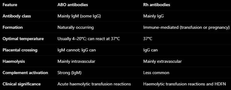

Indicate the expected reactions for the following blood groups
Key: + = agglutination; 0 = no agglutination
\*Bombay phenotype does not determine RhD status, so Anti-D may be pos or neg
\**A2 individuals may produce anti-A1, resulting in a pos reaction with A1 cells, although most do not
Why?
O pos → No A/B antigens; anti-A & anti-B in plasma; D antigen present
AB neg → Both A/B antigens; neither anti-A nor anti-B; D antigen absent
B pos → B antigen; anti-A in plasma; D antigen present
Bombay → Lacks H antigen on RBCs, preventing A/B expression. Plasma typically contains anti-A, anti-B & anti-H, which reacts strongly with O cells
A2 RhD neg → Expresses A antigen with reduced A antigen density compared with A1; has anti-B in plasma; D antigen absent


Based on the forward & reverse grouping results (see image), the patient has the following blood group:
O RhD negative
B RhD positive
A RhD positive
O RhD positive
AB RhD positive
O RhD positive
Why?
No A or B antigens on the red blood cells and anti-A and anti-B antibodies in plasma; D antigens present on red cells → O RhD positive
Anti-M and anti-N antibodies are usually of clinical significance because they are usually IgM antibodies that can cross the placenta → TRUE or FALSE
FALSE
Why?
Anti-M and anti-N antibodies are not usually of clinical significance because they are usually IgM antibodies that cannot cross the placenta & work best in the cold
Zeta potential is defined as the:
Repulsive force generated by the negative charges on the erythrocyte surface
Attracting force generated by negative & positive charges on the erythrocyte surface
Repulsive force generated by positive charges on the erythrocyte surface
Repulsive force generated by negative charges on the thrombocyte surface
Repulsive force generated by negative charges on the erythrocyte surface
Why?
RBCs have a negative surface charge, primarily due to sialic acid on their membranes
Negative charges → Cause RBCs to repel each other
Zeta potential → The electrical potential associated with this surface charge, contributing to RBC repulsion
Agglutination → Reducing zeta potential allows RBCs to move closer together, making antibody-mediated agglutination easier
Antibodies to the ABO blood group system generally can be best detected:
At very high pH levels (i.e. pH 10-11)
At 37 degrees
At very low pH levels (i.e. pH 2-3)
At room temperature
At room temperature
Why?
ABO antibodies are predominantly IgM antibodies, which generally react strongly at room temp
IgM → Reacts well at room temp
IgG → Generally reacts best at 37 degrees
ABO antibodies → Can also react at 37 degrees & cause severe haemolytic transfusion reactions
IgG antibodies characteristically react optimally at 37 degrees, but usually cannot cause agglutination in NaCl at room temperature → TRUE or FALSE?
TRUE
Why?
IgG antibodies react optimally at 37 degrees, but usually cannot directly cause RBC agglutination in saline (NaCl) at room temp
IgG → Small, monomeric antibodies that generally cannot bridge the gap between RBCs due to zeta potential
Zeta potential → Negative surface charges cause RBCs to repel each other
AHG (Anti-Human Globulin) → Bridges IgG-coated RBCs, allowing visible agglutination in the antiglobulin test
Screening cells are always blood group:
AB
O
A
B
O
Why?
Antibody screening cells are always group O RBCs because they lack A & B antigens, preventing interference from naturally occurring anti-A & anti-B antibodies
Group O cells → Lack A & B antigens
Prevents interference → Anti-A & anti-B will not react with screening cells
Purpose → Allows detection of unexpected RBC antibodies, such as anti-D, anti-K & anti-Jka
Sample labelling for blood banking test must include:
Full name, UR or DOB, signature of person taking the blood, date of sampling
Full name, UR or DOB, signature of person taking the blood, date and time of sampling
A first name and surname
A first name, surname & date of birth (DOB)
A first name, surname & UR number
Full name, UR or DOB, signature of person taking the blood, date and time of sampling
All negative anti-human globulin (AHG) test results needs to be confirmed/checked with AHG control cells to identify:
None of the above
If the washing steps were performed
All of the above
If the red cells were washed adequately
If the patient’s plasma was added
All of the above
Why?
AHG control cells (Coombs check cells) are IgG-coated RBCs added after a negative AHG test to confirm that the negative result is valid
Adequate washing → Ensures unbound IgG has been removed so it cannot neutralise AHG
Active AHG reagent → Confirms AHG was added & remains functional
Positive agglutination → Check cells must agglutinate to validate the negative AHG result
No agglutination → Indicates an invalid test that must be repeated
*Little wonky cause technically AHG control cells cannot reliably confirm that the patient’s plasma was added. They primarily validate AHG activity & adequate washing
Papain reduces the zeta potential and increases the reactivity of some antigen-antibody complexes, but also DESTROYS some antigens (Ags) → TRUE or FALSE?
TRUE
Why?
Papain is a proteolytic enzyme that modifies RBC surface proteins, reducing zeta potential & allowing RBCs to move closer together, enhancing certain antigen-antibody reactions
Reduces zeta potential → Decreases RBC repulsion, facilitating agglutination
Enhances antigens → Rh, Kidd, Lewis & P1 antigen-antibody reactions
Destroys antigens → Duffy (Fya, Fyb) & certain MNS antigens (M, N, S, s)
Conclusive evidence of antibody identity (from an antibody panel) occurs when reactivity with 2 antigen-positive cells is shown and no reactivity with 2 antigen-negative cells → TRUE or FALSE
FALSE
Why?
Conclusive evidence of antibody identity (from an antibody panel) occurs when reactivity with 3 antigen-positive cells is shown and no reactivity with 3 antigen-negative cells
Which blood group system is associated with resistance to P. vivax?
Rh
Kell
Duffy
Kidd
Duffy
Why?
The Duffy blood group system is associated with resistance to Plasmodium vivax malaria because Duffy antigens act as receptors for parasite invasion of RBCs
Duffy antigens (Fya/Fyb) → Can act as receptors for P. vivax
Duffy-null phenotype [Fy(a-b-)] → Lacks Duffy antigen expression on RBCs, providing strong resistance to P. vivax invasion
Clinical significance → Duffy antibodies (mainly IgG) can cause haemolytic transfusion reactions & HDFN
Which antibody is most commonly associated with delayed haemolytic transfusion reactions?
Anti-s
Anti-k
Anti-D
Anti-Jka
Anti-Jka
Why?
Anti-Jka (Kidd blood group system) is strongly associated with delayed haemolytic transfusion reactions (DHTRs)
Antibody evanescence → Kidd antibodies can decrease to undetectable levels over time
Anamnestic response → Re-exposure to Jka-positive RBCs triggers rapid antibody production
Delayed haemolysis → Newly produced antibodies destroy transfused RBCs, typically days after transfusion
Which of the following best describes MN antigens and antibodies?
Well developed at birth and susceptible to enzymes
Poorly developed at birth and susceptible to enzymes
Well developed at birth and not susceptible to enzymes
Poorly developed at birth and susceptible to enzymes
Well developed at birth and susceptible to enzymes
Why?
The M & N antigens (MNS blood group system) are well developed at birth but are sensitive to proteolytic enzymes
Well developed at birth → M & N antigens are expressed on fetal & neonatal RBCs
Enzyme-sensitive → Papain & ficin destroy M & N antigen activity
Antibodies → Anti-M & anti-N are usually naturally occurring IgM antibodies that react best at room temp
Clinical significance → Usually insignificant, although some IgG anti-M antibodies can cause HDFN or haemolytic transfusion reactions
Which antigen is destroyed by enzymes?
P1
K
Fya
Jka
Fya
Why?
Duffy antigens (Fya & Fyb) are destroyed by proteolytic enzymes such as papain & ficin
P1 → Enzyme-enhanced
K (Kell) → Generally unaffected by enzymes
Fya (Duffy) → Destroyed by enzymes
Jka (Kidd) → Enzyme-enhanced
Which of the following characteristics best describes Lewis antibodies?
IgM, naturally occurring, cause HDFN
IgM, naturally occurring, do not cause HDFN
IgG, cause haemolytic transfusion reactions
IgG, do not cause haemolytic transfusion reactions
IgM, naturally occurring, do not cause HDFN
Why?
Lewis antibodies (anti-Lea & anti-Leb) are predominantly naturally occurring IgM antibodies
IgM → Generally cannot cross the placenta
Naturally occurring → Usually develop without prior transfusion or pregnancy
HDFN → Do not cause HDFN because IgM cannot cross the placenta & Lewis antigens are poorly developed on fetal RBCs
Clinical significance → Usually insignificant, although rare haemolytic transfusion reactions can occur
The MNS blood group system is located on which chromosome?
Chromosome 1
Chromosome 4
Chromosome 7
Chromosome 19
Chromosome 4
Why?
The MNS blood group system is controlled by genes located on chromosome 4
GYPA gene → Encodes glycophorin A, carrying M & N antigens
GYPB gene → Encodes glycophorin B, carrying S & s antigens
GYPE gene → Also contributes to genetic variation within the MNS system
Which of the following antigens is NOT part of the Kidd blood group system?
Jka
Jkb
Jkc
Jk3
Jkc
Why?
The Kidd blood group system consists of three recognised antigens:
Jka → Major Kidd antigen
Jkb → Major Kidd antigen
Jk3 → High-prevalence antigen present on RBCs expressing Jka and/or Jkb
Jkc → Does not exist in the Kidd blood group system
Clinical significance → Kidd antibodies (anti-Jka, anti-Jkb & anti-Jk3) can cause haemolytic transfusion reactions, particularly delayed haemolytic transfusion reactions (DHTRs)

Interpret the antibody screen results. What would you do next?
Interpretation: Positive antibody screen → possible anti-Fyb or anti-P1
Why?
The patient’s plasma reacts with screening cells 2 & 3, indicating the presence of an unexpected RBC antibody (or antibodies)
Looking at the antigen composition sheet:
Fyb → Absent on cell 1, present on cells 2 & 3
P1 → Absent on cell 1, present on cells 2 & 3
Both antigens match the reaction pattern. However, anti-Fyb is a stronger initial suspicion because Duffy antibodies are predominantly IgG & react at the IAT phase
What would you do next?
Perform an 11-cell antibody identification panel to determine antibody specificity
Use additional testing if necessary, such as enzyme-treated cells (papain destroys Duffy antigens but enhances P1 reactions)
Confirm the identified antibody through patient phenotyping, where appropriate
If transfusion is required, select appropriate antigen-negative, crossmatch-compatible RBCs for clinically significant antibodies

For clinically significant antibodies: It is necessary to phenotype a patient’s red cells for an antigen that corresponds to the atypical antibody detected/identified in their plasma → TRUE or FALSE?
TRUE
Why?
Phenotyping that patient’s red cells for the corresponding antigen demonstrates whether the patient is capable of forming the corresponding antibody. Phenotyping for multiple red cell antigens is also useful in complex investigations, in order to exclude the possibility of some antibodies (if the patient is suspected to have a mixture of atypical antibodies)
You can reliably phenotype a patient for red cell antigens if they have received a transfusion within the last 3 months → TRUE or FALSE
FALSE
Why?
A patient who has received a red blood cell transfusion within the last 3 months cannot be reliably phenotyped using conventional serological methods
Donor RBCs → May still be circulating in the patient’s blood
Mixed-field reactions → Donor & patient RBCs may have different antigens, causing misleading results
Unreliable phenotyping → Results may reflect donor RBC antigens rather than the patient’s own antigens
Alternative → Molecular genotyping can determine the patient’s inherited RBC antigen profile

Part 1
You have performed a blood group and antibody screen on a patient.
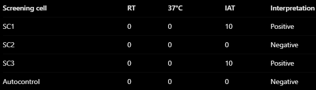
The Patient’s blood group is A Rh(D) positive. The antibody screen results are shown in the image.
What is your interpretation of the antibody screen?
Cold-reacting IgM antibody reacting with screening cells 1 and 3
Warm-reacting IgM antibody reacting with screening cells 1 and 3
Warm-reacting IgG antibody reacting with screening cells 1 and 3
Warm-reacting IgG reacting with all screening cells
Warm-reacting IgG antibody reacting with screening cells 1 and 3
Why?
The antibody screen is positive at the IAT phase, indicating a likely warm-reacting IgG alloantibody
RT & 37°C phases → No visible agglutination
IAT phase → SC1 & SC3 pos, consistent with IgG detected using AHG
Negative autocontrol → Supports an alloantibody rather than an autoantibody
Validation (yes) → Neg IAT reactions were successfully validated

Part 2
Here is the antigram for the screening cells:
What of the following statements is true?
The results of the antibody screen match only with Anti-Jka, however, further testing is required to confirm
The antibody may be Anti-e or Anti-Jka, further testing is required
The antibody is Anti-Jka and we can identify this with matching the pattern of our results. No further testing required
It is likely an antibody to an antigen of the Rh system, as the patient is Rh negative
The antibody is Anti-Jka and we can identify this with matching the pattern of our results. No further testing required
Why?
From Part 1, the patient’s antibody screen showed:
SC1 = Pos (10)
SC2 = Neg (0)
SC3 = Pos (10)
We compare this reaction pattern against the antigens expressed by each screening cell (see table layout)
Both anti-e (Rh) & anti-Jka (Kidd) match the patient’s antibody screen
However, we cannot determine which antibody is present using only these three screening cells


Part 3
You have performed phenotyping on this patient, and the results are as follows (assume controls produced expected results):
Which of the following statements is correct?
The patient is capable of forming Anti-Jka
The phenotyping results show the patient is Rh(D) negative
The patient is capable of forming Anti-Jka because testing shows their red cells have the Jka antigen
The patient likely has Anti-e, as their red cells agglutinated with Anti-e antisera
The patient is capable of forming Anti-Jka
Why?
Jka absent → Patient can develop anti-Jka following exposure to Jka-pos RBCs
e present → Patient would not normally produce anti-e because it is a self-antigen
c & E absent → Patient could also develop anti-c or anti-E following sensitisation
Connection to Part 2
Previously, the antibody screen suggested either anti-e or anti-Jka
Phenotyping now helps distinguish them:
Anti-e → Unlikely because the patient expresses e
Anti-Jka → Possible because the patient lacks Jka
Therefore, anti-Jka is the leading suspected antibody, requiring confirmation with an antibody identification panel

Part 4
You have tested the patient’s plasma against antibody panels, and were able to identify and confirm the atypical antibody detected is Anti-Jka. This patient requires one unit of Packed Red Blood Cells (PRBCs). What further actions will you take?
Grab a random unit of PRBCs from the fridge and assign it. No further testing required
Select and phenotype for Jka negative PRBCs, IAT crossmatch
Select and phenotype for Jka negative PRBCs, Immediate Spin Crossmatch
Perform IAT crossmatch
Select and phenotype for Jka negative PRBCs, IAT crossmatch
Why?
The patient has a confirmed anti-Jka antibody (Kidd system), which is clinically significant & can cause haemolytic transfusion reactions
Select Jka-neg PRBCs → Prevents the patient’s anti-Jka from reacting with donor RBCs
Phenotype donor RBCs → Confirms the selected units lack the Jka antigen
IAT crossmatch → Confirms serological compatibility between the patient’s plasma & donor RBCs at the antiglobulin phase
Immediate spin crossmatch → Insufficient because anti-Jka is predominantly IgG & may only be detectable at the IAT phase
Part 5
What are 2 other things you must consider when selecting compatible PRBCs for this patient?
ABO Rh(D) compatible
Whether another scientist has handled the PRBC recently
PRBCs has not passed expiry date
Whether the PRBCs is fresh (collected <5 days)
ABO Rh(D) compatible & PRBCs has not passed expiry date

You are setting up red cell phenotyping. You wish to use the screening cells for appropriate controls.
Here is the antigram of the screening cells:
Which cell would you select as a positive control for the E antigen?
Cell 3
Cell 1
Cell 2
None of these cells are suitable
None of these cells are suitable
Why?
For selecting a positive control for red cell phenotyping, we must select a cell that has the weakest expression of the antigen we wish to type for. If applicable, we must select a cell that has HETEROZYGOUS expression of that antigen
In this context, the cell we must use as a positive control for the E antigen must be E+e+. None of the screening cells in this antigram have that phenotype

Case study
A 65 yr old male with a history of MVA 5 years ago, for which he received 2 units of RBCs. He is scheduled for a cardiac bypass surgery. His blood bank test results are shown in the image
1) What is the patient’s blood group?
2) What is the interpretation of the antibody screen?
3) What needs to be done next?
1) The patient is AB Rh(D) pos because their RBCs express A, B & D antigens, while their plasma shows no reaction with A1, B or O reagent cells
2) Positive antibody screen, suggesting a warm-reacting IgG alloantibody
Room temp (IS) → No recorded reactions
37°C → No recorded reactions
IAT → Screening cell 2 shows a positive reaction (8), while cell 3 is negative (0)
This suggests an unexpected antibody detectable at the IAT phase, consistent with IgG
Comparing the antigen profiles, anti-E & anti-K are possible candidates, as both E & K are present on screening cell 2 but absent from cells 1 & 3
3) Perform an 11-cell antibody identification panel
The next steps are:
Verify & complete any missing screening results & controls
Perform an 11-cell antibody identification panel to identify the suspected antibody
Phenotype the patient’s RBCs to support antibody identification. His last transfusion was 5 years ago, so recent donor RBC interference is not expected
Select ABO/RhD-compatible, antigen-negative packed RBCs once the clinically significant antibody is identified
Perform an IAT crossmatch to confirm compatibility before surgery

Case study (con’d)
4) What antibody specificity would explain ALL reactions observed in both the antibody screen and antibody identification panel?
5) Are there any additional test that can be done?
6) What blood group should be given to the patient in case of transfusion?
4) Anti-K (Kell blood group system)
Why?
The antibody identification panel shows positive reactions with cells 2, 8 & 9, all of which express the K antigen
The antigen expression matches the reaction pattern as well
Anti-K → Predominantly IgG, detected at the IAT phase
Negative autocontrol → Supports an alloantibody rather than an autoantibody
Clinical significance → Anti-K can cause haemolytic transfusion reactions & HDFN
The previous antibody screen also supports anti-K because only screening cell 2 expressed K & reacted
5) Phenotype the patient’s RBCs for the K antigen
Test the patient’s RBCs using anti-K reagent
Expected result → K-negative, supporting the identification of alloanti-K
Confirm antibody specificity using the rule of three, which is already satisfied by the panel → three K-pos cells react & at least three K-neg cells do not
Because the patient’s last RBC transfusion was five years ago, serological phenotyping should be reliable, assuming no other interference
6) AB Rh(D) pos, K-neg packed RBCs, with a compatible IAT crossmatch
AB RhD pos → Matches the patient’s blood group
K-neg → Prevents anti-K from reacting with donor RBCs
IAT crossmatch → Confirms compatibility before transfusion
Other ABO-compatible RBC groups may also be used if required, provided they are K-neg & crossmatch compatible
At the Australian Red Cross Blood Service (ARCBS) one donation of blood usually includes:
700-850 mls of blood
50-100 mls of blood
200-300 mls of blood
450-500 mls of blood
450-500 mls of blood
At the ARCBS blood is usually anticoagulated into:
Citrate, phosphate, dextrose, adenosine (CPDA)
Lithium heparin
EDTA, phosphate and citrate (EPC)
EDTA
Citrate, phosphate, dextrose, adenosine (CPDA)
Why?
CPDA is an anticoagulant-preservative solution used in blood collection bags to prevent clotting & maintain RBC viability during storage
Citrate → Binds calcium (Ca2+), preventing blood coagulation
Phosphate → Maintains blood pH by acting as a buffer
Dextrose → Provides glucose for RBC metabolism & ATP production
Adenine → Supports ATP synthesis, maintaining RBC viability during storage
*Quiz says adenosine; however, the standard CPDA formulation contains adenine apparently
Viral screening of blood donor units includes testing for:
Hepatitis B and C virus and antibody
HTLV 1&2 antibodies
HIV antibodies
All of the above
All of the above
Why?
Donated blood undergoes viral screening to prevent transmission of blood-borne infections to transfusion recipients
Hepatitis B (HBV) → Screening for viral antigens, antibodies & DNA
Hepatitis C (HCV) → Screening for antibodies & viral DNA
HIV → Screening for antibodies, antigen & viral DNA
HTLV-1 & HLTV-2 → Screening for antibodies
These tests help identify infected donations & prevent potentially infectious blood from being transfused
All platelet donations were screened for bacterial contamination 24 minutes post collection → TRUE or FALSE?
FALSE
Why?
All platelet donations screened for bacterial contamination 24 hours post collection
The ‘window period’ for the detection of the Hepatitis C virus (HCV) at the ARCBS (antibody & RNA testing) is approx.:
30 days
3 days
1 day
10 days
3 days
Why?
The window period is the time between infection & when an infection can first be detected by a lab test
Australian Red Cross Lifeblood uses HCV antibody testing & RNA testing (NAT) to screen donated blood
HCV antibody testing → Detects antibodies produced by the immune system, which take longer to develop
HCV RNA testing (NAT) → Detects viral genetic material much earlier
Combined testing → Reduces the estimated HCV detection window to approximately 3 days
Administration/transfusion of fresh frozen plasma (FFP) units to patients does not require a current group and hold (blood group with 72 hours/3 days validity for transfusion purposes) → TRUE or FALSE?
TRUE
Why?
The transfusion of platelets and frozen products (FFP, cryoprecipitate) only require a historical blood group to be on the patient’s file to file for transfusion. The patient must have a valid group and hold (72 hour/3 day validity) for transfusion of PRBCs
Platelet concentrates:
Are given to recipients to increase their clotting factors
Need to be agitated while in storage
Are usually stored in the fridge
Have a shelf-life of approx. 2 weeks
Needed to be agitated while in storage
Why?
Are stored at room temperature, shelf life of approx 5 days and given to increase platelet counts (not clotting factors)
Blood volume can be estimated as approx 50 mL/kg for adults, 100 mL/kg in children and 150 mL/kg in neonates → TRUE OR FALSE?
FALSE
Why?
Blood volume can be estimated as approx 70 mL/kg for adults, 80 mL/kg in children and 100 mL/kg in neonates
Clinical features (signs & symptoms) of acute blood loss may include:
Paleness
Tachycardia
Hypotension
All of the above
All of the above
Why?
Acute blood loss reduces circulating blood volume, causing hypovolaemia & reduced tissue perfusion
Paleness → Peripheral vasoconstriction reduces blood flow to the skin
Tachycardia → Heart rate increases to compensate for reduced circulating blood volume
Hypotension → Significant blood loss reduces venous return & cardiac output, causing decreased blood pressure
Chronic blood loss that has resulted in the patient developing iron deficiency anaemia (IDA) should be treated with blood transfusions (red cell concentrates) → TRUE or FALSE?
FALSE
Why?
NEVER/RARELY transfuse in IDA; treat the underlying cause (blood loss) & oral iron therapy/intramuscular iron therapy

Case study 2
A 31-year-old female, G2P1 presents for her initial prenatal checkup
1) What is the patient’s blood group?
1) A Rh(D) pos
Why?
Forward grouping: Anti-A pos & Anti-D neg → Group A
Reverse grouping: A1 cells neg & B cells pos → Confirms group A
RhD grouping: Anti-D pos → RhD pos
Control → Neg, supporting valid RhD typing

Case study 2 (cont’d)
2) What is the interpretation of the antibody screen?
3) What needs to be done next?
2) Positive antibody screen - suspected warm-reacting IgG alloantibody
Why?
The patient’s plasma reacts with screening cells 2 & 3, suggesting the presence of an unexpected RBC antibody
Which antibodies are suspected?
According to the table layout, the reaction pattern is consistent with anti-c, anti-Fyb or anti-Jka, but antibody specificity cannot yet be confirmed
3) Perform an 11-cell antibody identification panel

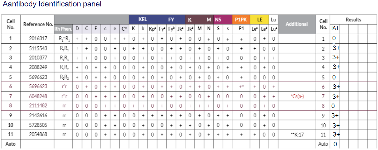
Case study 2 (still cont’d)
4) What antibody specificity would explain ALL reactions observed in both the antibody screen and antibody identification panel?
5) Are there any additional test that can be done?
4) Anti-Fyb (Duffy blood group system)
Why?
The reaction pattern matches anti-Fyb
All Fyb-pos cells react
All Fyb-neg cells are non-reactive
The neg autocontrol supports an alloantibody
Anti-Fyb is predominantly IgG & reacts at the IAT phase
The previous antibody screen also supports anti-Fyb because screening cells 2 & 3 were Fyb-pos & reactive
5) Yes, patient phenotyping & enzyme testing
Patient phenotyping → Test the patient’s RBCs with anti-Fyb. An Fyb-neg result supports the identification of alloanti-Fyb
Enzyme testing (papain) → Duffy antigens are destroyed by papain. Therefore, anti-Fyb reactions should disappear when tested against papain-treated RBCs
Maternal antibody monitoring → Because the patient is pregnant, assess antibody levels & fetal risk according to antenatal protocols. Anti-Fyb can occasionally cause HDFN
A baby was delivered by a mother who is AB Rh(D) negative.
The baby was jaundiced from birth. Upon examination of the baby’s blood film, there were many nucleated red cells and marked polychromasia. The baby’s blood group is A Rh(D) positive.
What is the most likely cause of the baby’s clinical features?
HDFN due to Anti-D
HDFN due to Anti-A
Non-serological cause of jaundice
HDFN due to Anti-Lea
HDFN due to Anti-D
Why?
The mother is AB Rh(D) neg, while the baby is A Rh(D) pos. This RhD incompatibility can cause haemolytic disease of the fetus & newborn (HDFN)
RhD-neg mother → Lacks the D antigen & many produce anti-D following sensitisation
RhD-pos baby → Expresses D antigen inherited from the father
Maternal anti-D (IgG) → Crosses the placenta & binds fetal D-pos RBCs, causing extravascular haemolysis
Jaundice → RBC destruction increases unconjugated bilirubin
Nucleated RBCs & polychromasia → Indicate increased erythropoiesis as the baby compensates for haemolysis
Why not anti-A?
The mother is blood group AB, so she does not normally produce anti-A or anti-B antibodies
A baby was delivered by a mother who is O Rh(D) negative.
The baby experienced increased jaundice after a few days. Upon examination of the baby’s blood film, there were some spherocytes. The baby’s blood group is A Rh(D) positive.
What is the most likely cause of the baby’s clinical features?
HDFN due to Anti-A, but cannot fully exclude possibility of Anti-D also, due to blood group (at this stage)
HDFN due to Anti-A
HDFN due to Anti-D
HDFN due to another Rh antibody
HDFN due to Anti-A, but cannot fully exclude possibility of Anti-D also, due to blood group (at this stage)
Why?
Further testing such as DAT and elution are essential in determining which antibody is responsible for HDFN
Anti-Fya is an IgM antibody and can cause HDFN → TRUE or FALSE?
FALSE
Why?
Anti-Fya is a warm-reacting IgG antibody, therefore is clinically significant (can cross the placenta)
Which test is useful in assessing the degree of foetal-maternal haemorrhage?
Kleihauer test
DCT
Elution
Antibody investigation
Kleihauer test
Why?
The Kleihauer test detects & estimates the amount of fetal RBCs that have entered the maternal circulation, allowing assessment of fetomaternal haemorrhage (FMH)
Principle → Acid elution removes adult haemoglobin (HbA), while fetal haemoglobin (HbF) is resistant
Fetal RBCs → Retain haemoglobin & stain pink/red
Maternal RBCs → Lose haemoglobin & appear pale (ghost cells)
Clinical significance → Helps calculate the amount of anti-D immunoglobulin required for an RhD-neg mother following FMH
Why is Bilirubin, LDH and Haptoglobin part of a transfusion reaction investigation?
To rule out haemolytic transfusion reaction
To specifically identify ABO incompatible transfusion as the cause
To assess TACO
To monitor for infection
To rule out haemolytic transfusion reaction
Why?
Bilirubin, LDH & haptoglobin are biochemical markers of haemolysis (RBC destruction). They help determine whether a transfusion has caused RBC destruction
Bilirubin↑: Increases as haemoglobin is broken down following RBC destruction
LDH↑: Released into the bloodstream when RBCs are damaged or destroyed
Haptoglobin↓: Binds free haemoglobin released from RBCs & is subsequently cleared from circulation
These changes support a diagnosis of haemolytic transfusion reaction, although normal results alone do not completely exclude haemolysis
Anti-E is more commonly associated with what category of transfusion reaction?
Delayed Haemolytic Transfusion Reaction
Acute Transfusion Reaction
Febrile Non-Haemolytic Transfusion Reaction
TRALI
Delayed Haemolytic Transfusion Reaction
Why?
Anti-E → Usually develops following exposure to E-pos RBCs through transfusion or pregnancy
Antibody evanescence → Anti-E levels may decrease below detectable levels over time
Anamnestic response → Re-exposure to E-pos RBCs triggers rapid antibody production
Extravascular haemolysis → IgG-coated donor RBCs are predominantly destroyed by splenic macrophages
Timing → DHTRs typically occur several days to weeks after transfusion
What is not part of a transfusion reaction investigation?
Immediately disposing the suspected donor units in the bin
Crossmatch on pre- and post- transfusion samples
Clerical checks
Visual inspection for haemolysis
Immediately disposing the suspected donor units in the bin
An ABO-incompatible transfusion is related to a
Acute haemolytic transfusion reaction
Delayed haemolytic transfusion reaction
TACO
TRALI
Acute haemolytic transfusion reaction
Why?
An ABO-incompatible RBC transfusion can cause an acute haemolytic transfusion reaction because the recipient’s naturally occurring anti-A or anti-B antibodies (mainly IgM) attack incompatible donor RBCs
IgM binding → Recipient antibodies bind to A or B antigens on donor RBCs
Complement activation → IgM strongly activates the classical complement pathway
Intravascular haemolysis → Donor RBCs are rapidly destroyed within blood vessels
Clinical features → Fevers, chills, hypotension, back pain & haemoglobinuria
Timing → Usually occurs during transfusion or within 24 hrs
A Direct Antiglobulin Test (DAT) tests for:
The presence of antibodies that have bound to red cells in vivo (inside the body)
The presence of antibodies loose in plasma
Antigens on the red blood cells
The degree of foetal-maternal haemorrhage
The presence of antibodies that have bound to red cells in vivo (inside the body)
Why?
The Direct Antiglobulin Test (DAT) detects IgG antibodies and/or complement (C3d) already attached to the patient’s RBCs in vivo
DAT → Detects RBCs sensitised with IgG or complement inside the body
AHG reagent → Binds to IgG and/or complement on RBCs, producing visible agglutination
Clinical significance → Used to investigate haemolytic transfusion reactions, HDFN & autoimmune haemolytic anaemia (AIHA)

Blood tests for a mother and baby were sent to the lab:
Mother: B Rh(D) positive, Anti-E detected
Baby: B Rh(D) positive, DAT positive
Check image for baby’s phenotype results
Which of the following statements is true?
Mother’s Anti-E has crossed the placenta and bound to baby’s red cells, causing a positive DAT
According to the baby’s phenotype results, the mother’s Anti-E is not the cause of the positive DAT
Anti-E is not capable of crossing the placenta because it is an IgM antibody
Mother’s Anti-D has crossed the placenta and bound to the baby’s red cells, causing a positive DAT
Mother’s Anti-E has crossed the placenta and bound to baby’s red cells, causing a positive DAT
Why?
Mother has Anti-E. Anti-E is an IgG antibody and is capable of crossing the placenta
Baby’s red cells do have the E antigen present. The Anti-E antibody is capable of crossing the placenta and binding to the E antigens on the baby’s red cells
The antigen-antibody binding in vivo (inside the body) can be detected by a positive DAT
A patient has a normal PT and a prolonged APTT that corrects after a mixing study. Which finding is most consistent with this result?
Factor VIII deficiency
Factor VIII inhibitor
Disseminated intravascular coagulation
Severe liver disease
Factor VIII deficiency
Why?
A normal PT, prolonged APTT, and correction after a mixing study indicate a deficiency of a factor in the intrinsic coagulation pathway
Normal PT → Extrinsic pathway is functionally normal
Prolonged APTT → Suggests a deficiency or inhibitor affecting the intrinsic pathway (factors VIII, IX, XI or XII)
Correction after mixing → Indicates a factor deficiency, because normal plasma supplies the missing clotting factor
Factor VIII deficiency → Causes prolonged APTT with normal PT, as seen in haemophilia A
Why not Factor VIII inhibitor?
An inhibitor prevents normal clotting factor activity, so the APTT typically fails to correct or becomes prolonged again after incubation
In forward ABO grouping, the patient’s red cells are tested with:
A1 and B reagent cells
Anti-A and anti-B reagents
Patient plasma and group O cells
Anti-human globulin only
Anti-A and anti-B reagents
Why?
Forward ABO grouping detects A & B antigens on the patient’s RBCs using commercially prepared antisera
Anti-A reagent → Detects A antigen on RBCs
Anti-B reagent → Detects B antigen on RBCs
Agglutination (+) → Corresponds antigen is present
No agglutination (0) → Corresponding antigen is absent
Why not A1 and B reagent cells?
A1 and B reagent cells are used for reverse grouping to detect antibodies in the patient’s plasma
A patient’s red cells react with anti-A and anti-B. Their plasma does not react with either A1 or B reagent cells. What is the ABO group?
A
B
AB
O
AB
Why?
The patient has both A & B antigens on their RBCs, but neither anti-A nor anti-B antibodies in their plasma
Anti-A (+) → A antigen present
Anti-B (+) → B antigen present
A1 reagent cells (0) → No anti-A detected
B reagent cells (0) → No anti-B detected
Therefore, forward & reverse grouping confirm blood group AB
Why can group O red cells be incompatible with a patient with the Bombay phenotype A?
Group O cells strongly express H antigen
Group O cells strongly express A antigen
Bombay patients have anti-DD
Bombay patients lack ABO antibodies
Group O cells strongly express H antigen
Why?
Individuals with the Bombay phenotype (Oh) lack H antigen on their RBCs & typically produce anti-H antibodies
Group O RBCs → Have high levels of H antigen because it is not converted into A or B antigens
Bombay phenotype → Lacks H antigen due to the hh genotype
Anti-H phenotype → Reacts with H antigen on normal group O RBCs, potentially causing severe haemolysis
Transfusion → Bombay patients generally require RBCs from another Bombay phenotype donor
Explain the difference between affinity and avidity in antigen-antibody interactions
Affinity → Measures how strongly one antibody binding site attaches to a specific antigen epitope
Avidity → Measures the combined strength of all antigen-antibody interactions, making the overall attachment stronger
For example, IgM has high avidity because its pentameric structure allows multiple antigen-binding interactions

Why should heterozygous cells not always be used to rule out antibodies such as anti-Jka or anti-Fya?
Because anti-Jka & anti-Fya can demonstrate the dosage effect, meaning they react more strongly with homozygous RBCs than heterozygous RBCs
Homozygous cells: Express more of the target antigen → Stronger reactions
Heterozygous cells: Express fewer copies → Weaker or undetectable reactions
Risk → A neg reaction with heterozygous cells may falsely rule out an antibody that is actually present
A patient is group A RhD positive, the current antibody screen is negative. The patient’s transfusion history shows that anti-K was detected 3 years ago.
Why donor red-cell unit is most appropriate, why?
A Rh(D) pos, K-neg donor RBCs with a compatible IAT crossmatch
Why?
Although the patient’s current antibody screen is neg, they have a history of anti-K, a clinically significant IgG alloantibody
Antibody evanescence → Anti-K levels may decrease below detectable levels over time
Immunological memory → The immune system still remembers the K antigen
Anamnestic response → Re-exposure to K-pos RBCs can trigger rapid anti-K production
DHTR → This may cause a delayed haemolytic transfusion reaction
K-neg RBCs → Prevent re-exposure to the K antigen
IAT crossmatch → Confirms serological compatibility before transfusion
*A historically clinically significant antibody must always be considered, even when the current antibody screen is neg

Case study 3
A 65 yr old male with a history of MVA 5 years ago, for which he received 2 units of RBCs. He is scheduled for a cardiac bypass surgery. His blood bank test results are shown in the image.
1) What is the patient’s blood group?
1) A Rh(D) pos
Why?
Forward grouping: Anti-A pos & Anti-B neg → Group A
Reverse grouping: A1 cells neg & B cells pos → Confirms group A
RhD grouping: Anti-D pos → RhD pos