cardiovascular system
 Call Kai
 Learn
Learn Practice Test
 Spaced Repetition
 Match
Match Flashcards
 Knowt Play
1/137

There's no tags or description
Looks like no tags are added yet.
Name | Mastery | Learn | Test | Matching | Spaced | Call with Kai | Chat |
|---|
No analytics yet
Send a link to your students to track their progress
138 Terms
functions of the circulatory system
distribution of dissolved gases and other molecules for nutrition, growth and repair, aste removal
chemical signalling to cells by means of circulating hormones
mediate inflammatory and host defense responses against invading microorganisms, transport clotting factors
conserve or release heat near skin surface
aorta
oxygenated blood coming out of the heart
parallel circuit
blood returns to right atrium via superior and inferior vena cava, pumped out of the right ventricle to the lungs via pulmonary arteries
returns to heart via pulmonary veins, enters the left atrium, pumped out of the left ventricle to the body via aorta

hat affects blood flo?
decreases ith increased resistance
idth, viscoscity, length
increases ith increased difference in pressure
cardiac output
the heart pumps out 5L per minute
blood per beat x beat per minute
flo is the same throughout the entire system if e take a cross section, this flo is divided by e.g. no. of arterioles. if you constrict one, the proportions change, ie more bloodflo through other arterioles
tone
tone in smooth muscles of blood vessels, basal state of muscle contraction
adjust from here
affected by the SNS and local factors, endothelin, myogenic activity, NO

myogenic activity
reflex within the smooth muscle of arterioles
pressure of blood is different in different parts of the body due to eight of blood, changes ith changing posture
in places ith higher pressure (e.g. foot arteriole during standing), the arteriole ill stretch a bit initially, reflex ill cause constriction to protect capillaries
epithelial sodium channel (ENaC) is a pore that allos Na+ to go through, hen the arteriole stretches, the pore opens and Na+ flows into the muscle cells and depolarises them and allos them to constrict
NO
vasodilator
endothelin
vasoconstrictor released from the endothelial cells in response to a variety of chemical and physical signals
highest to loest pressure
aorta
arteries
capillaries
veins
venae cavae
affect of pressure gradient on flow
blood moves from high pressure to lo pressure, flo is proportional to pressure difference

1 and 3, highest pressure difference. overall pressure makes no difference
pressure gradients in blood vessels
pressure is lost due to friction

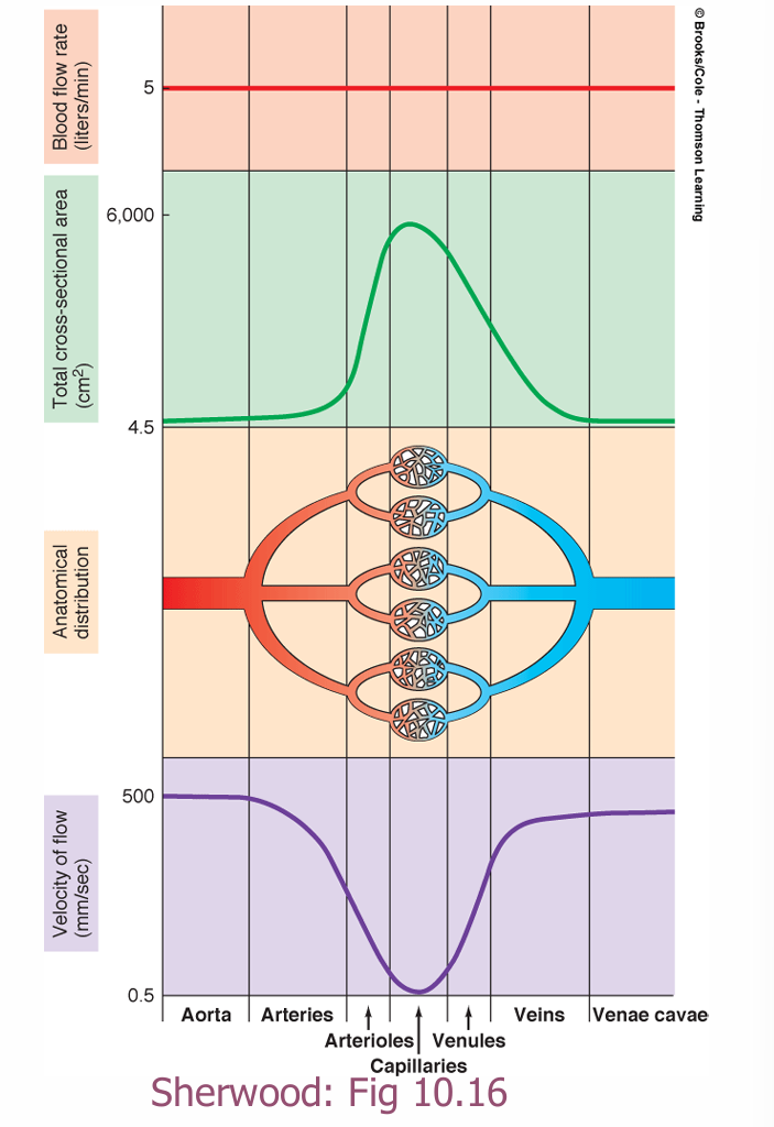
here does blood have its loest velocity?
capillaries
velocity is inversely proportional to cross sectional area, velocity = flo/CSA
capillaries have the highest CSA
if something is thin, to reach 5L/minute, it has to flo faster, if something is thick, to reach this flo, it can flow slower
good for facilitating exchange

velocity vs cross sectional area
blood vessel structure
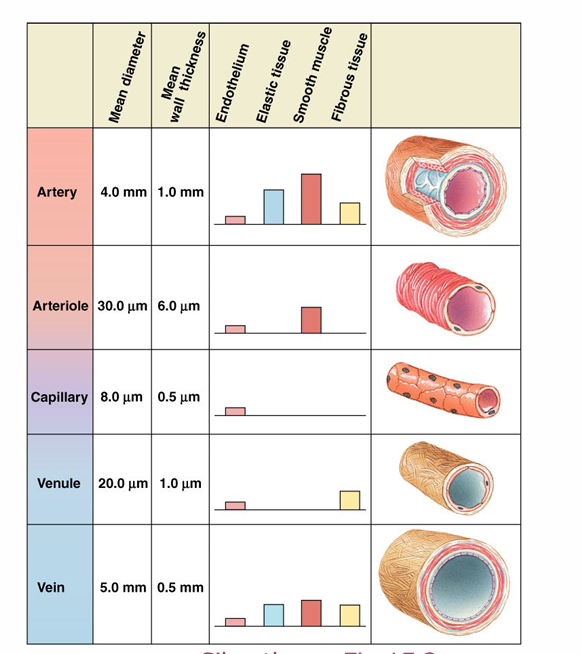
arteries
a lot of elastic tissue, compliant to stretch to allow blood to enter from the heart, then elastically recoils to push the blood forard
arteries hardening means you lose that elastance
a lot of fibrous tissue to strengthen them as they receive blood at a high pressure from the heart

dissection of the aorta
blood gest in beteen the layers of the arterial all, separates the layers and it balloons out
veins
less elastic
ider
expand easily - reserve, most of the blood is in the veins
extra capacity: SNS can constrict these veins and push the blood back out into arterioles during exercise
capillaries
fenestrated
single cell
most cell ithin 0.1 mm of the nearest capillary
vesicular transport
interstitial fluid between cells and capillaries

bulk flow
overall/mass movement of fluid and everything it contains between blood and interstitial fluid
filtration at the arteriole end and absorption at the venous end
direction of flow determined by osmotic pressure (solute conc) and water pressure
absorption
fluid movement from interstitium into capillaries
filtration
movement of fluid out of the capillaries into interstitium
oedema
intersitital fluid accumulation
osmotic/oncotic pressure of blood in capillaries
proteins (albumin exists only in the blood and not in the intestitial fluid). this favours absorption (pulling fluid back into the capillaries)
hat pressures influence direction of flo (bulk flo)?
the balance of osmotic/oncotic pressure and hydrostatic pressure
Starling Forces
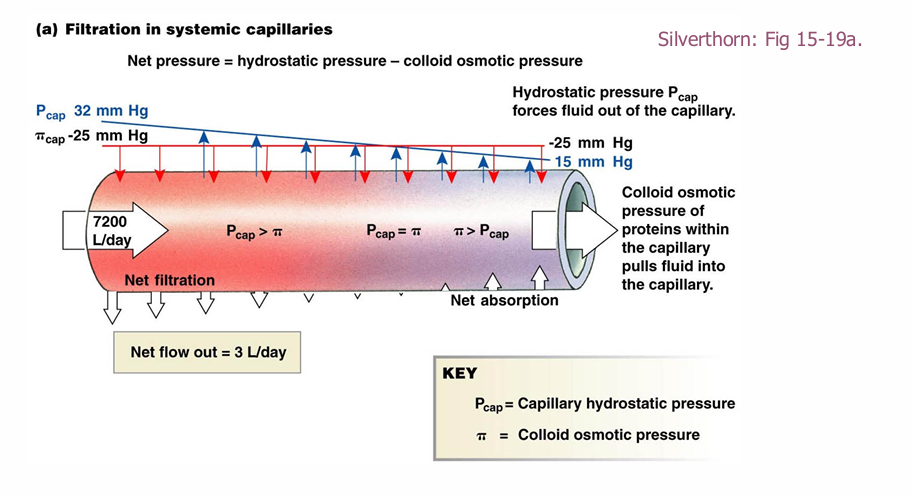
more hydrostatic pressure in the capillary
more oncotic pressure in the interstitial space
at arteriole end, the hydrostatic pressure is higher, as e move through the capillary it decreases due to friction
oncotic pressure stays the same across the capillary (albumin conc stays the same)
hen hydrostatic pressure > oncotic pressure, fluid flos out of capillary
hen hydrostatic pressure < oncotic pressure, fluid flo in

starling forces pressure changes diagram
endothelium
thin, single-cell layer lining the interior of blood vessels hich serves as the interface between the blood and the rest of the vessel wall
metabolic factors hich determine vasomotor tone
arterioles are responsive to local factors such as carbon dioxide, oxygen, lactic acid and heat
chemical mediators hich regulate vasomotor tone
nitric oxide
endothelin
angiotensin II
vasopressin
noradrenaline
adrenaline
histamine
angiotensin II
vasoconstrictor produced by the Renin Angiotensin System
vasopressin
also knon as anti-diuretic hormone
vasoconstrictor involved in fluid balance
adrenaline
released from adrenal glands
vasodilator at lo doses and a vasoconstrictor at high doses
histamine
causes vasodilation
hydrostatic pressure in the capillaries
As plasma travels along the capillary from the arterial to the venous end, capillary hydrostatic pressure decreases. This is due to volume loss resulting from filtration, and energy loss due to friction and resistance inside the capillary.
oncotic pressure
osmotic gradient resulting from albumin, a large protein hich cannot filter from capillaries to ICF, unlike the other solutes in the plasma
imbalance in Starling Forces
there is a natural imbalance in these forces with filtration slightly exceeding absorption.
over the course of a day, this leads to approximately 3L of fluid which is filtered but not absorbed.
Since plasma volume is only 5L, this needs to be reabsorbed somehow
role of the lymphatic system to compensate for Starling Forces
absorbs excess fluid from filtration and drains it back into the bloodstream

oedema
pathological imbalance in the Starling Forces
leads to buildup of fluid in the ISF
leads to an area of the body appearing sollen
factors that cause oedema
increased capillary hydrostatic pressure leading to increased filtration
decreased capillary oncotic pressure reducing absorption
interference ith the lymphatic system, blocking uptake of excess ISF
increased capillary hydrostatic pressure
e.g. heart failure which increases venous pressure
left heart isn’t pumping as ell as it should to clear the pulmonary circulation and thus it gets a buildup of pressure in the capillaries
this causes pulmonary oedema
one of the first signs of heart failure is difficulty breathing due to this fluid buildup in the chest
now the right heart fails as it is pumping against an increased pressure, then results in oedema in the periphery (ankles and legs)
can also result from prolonged inactivity, which leads to a buildup of fluid in the peripheries
decreased capillary oncotic pressure
malnutrition
if a person is severely protein deficient, they will have less albumin in their blood
e.g. Kwashiorkor
disruption to lymphatic system
lymphoedema: oedema caused from blockage of lymphatic system
e.g. obstruction of lymph nodes by parasites or cancerous cells
e.g. mastectomies - removal of a breast generally due to cancer causes oedema in the arm and shoulder. breast tissue contains a lot of lymphatic tissue and the surgery can disrupt that
hat factors favour filtration?
capillary hydrostatic pressure (strong force)
interstitial fluid oncotic pressure (small force)
hat factors favour absorption
interstitial fluid hydrostatic pressure (small force)
plasma oncotic pressure (strong force)
treatment of pulmonary or peripheral oedema
diuretics
hat organ makes albumin?
liver
liver failure can result in oedema (hard belly)
heart anatomy
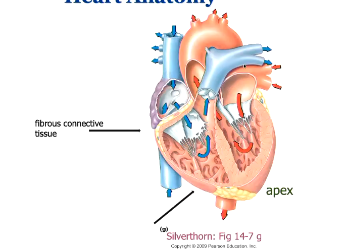
apex at the bottom, base at the top
between the atria and the ventricles is fibrous connective tissue
valves between atria and ventricles
valves between the ventricles and the arteries
pulmonary artery from right heart
aorta from left heart
blood returns to the atria, then enters the ventricles, then pumped out from there
hy does the right ventricle have less muscle mass?
pulmonary resistance is less than systemic resistance
ventricular systole
contraction of the ventricles
ventricular diastole
relaxation of the ventricles, filling ith blood
conduction system of the heart
autorhythmic cells in the sinoatrial node and the atrioventricular node
signal originates in the SA node (pacemaker) and passes through the atria, spread through atrial myocytes to cause a contraction of the atria
insulated connective tissue beteen the atria and the ventricles, the only ay the electrical current can pass into the ventricles is through the AV node
spreads out through the bundles of His and then the Purkinje fibres to the ventricular myocytes
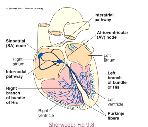
AV node
AV conduction time: a little delay at the AV node before it releases the signal through the bundle of His
compensates for the slo movement of blood, allos for the ventricles to fill ith blood after the atria contract
the AV conduction time decreases ith increased heart rate, stroke volume therefore decreases as e decrease ventricular filling time
damage to SA node
AV node can take over
speed of SA node is 70 bpm, AV node is therefore driven at 70 bpm in normal conditions
however if you remove the SA node, the AV node ill take over but at 50 bpm
bundles of His
carry the signal don the intraventricular septum
delivers signal to the apex to allo the AP to spread back upards from there
Purkinje fibres
stimulate cells in the ventricles (more Purkinje fibres in the left ventricle)
electro-cardio-gram (ECG)
an indicator of hich direction the electricity is travelling (vectors of depolarisation)
body surface recording
usually have 11 leads to give you a 3D picture of electrical conduction in the heart

lead II
rhythm strip
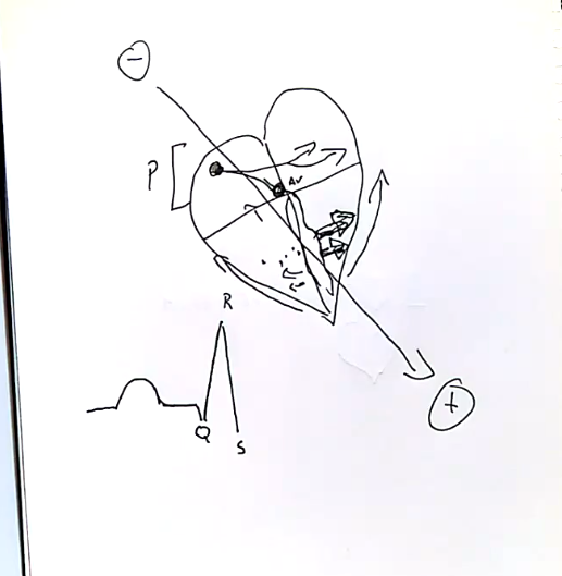
negative electrode on the right rist, positive on left ankle: this diagonal vector from top right to bottom left is in line with the position of the heart. any time a signal is travelling this ay, there will be an upward inflection on the ECG
measurement of direction not amplitude
depending on how well aligned the signal is with this vector, the greater the ECG inflection
how does an ECG work?
3 major waves: P wave, QRS complex and the T wave
P ave
signal starts at SA node and travels to the left (causes an upward inflection)
atrial depolarisation

plateau after P ave
when signal stops at AV node
time = AV conduction time
QRS complex
ventricular depolarisation
signal travels down left and right bundle of His. left bundle fires slightly earlier than right, movement from left to right, so slight donard inflection on ECG
movement across Purkinje fibres, bigger signal moving to left than right as more Purkinje fibres in left heart, causes upard inflection on ECG
then signal moves up walls of ventricles toards base (up and to the right), causes donard inflection
T ave
ventricles repolarisation
travels from apex to base, but since it’s an opposite electrical signal (becoming more negative), the inflection is upards

atrial repolarisation
repolarise, but during the QRS complex so not visible on the ECG

hat can you see on an ECG?
R-R interval = heart rate
doctors ill check that all aves are there, if the rhythm is regular, etc
hat increases heart rate variability?
pathological conditions
alcohol
chronic stress
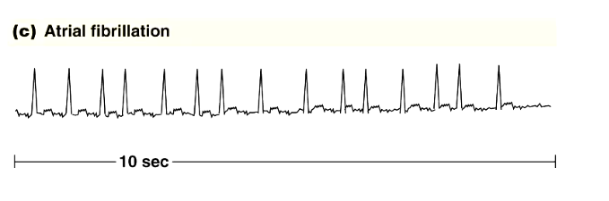
atrial fibrillation
atria do not contract as a unit
usually not a problem as the majority of ventricular filling happens passively, the atrial contraction only squeezes the last bit out
only issue is if blood gets caught in pockets in the atria, it can turn into clots hich can enter the coronary artery and cause a stroke
ventricular fibrillation
ventricle beating on its on from pacemakers in the ventricular muscle, perhaps caused by damaged muscle
need a defibrillator to restore normal rhythm
shocks cells simultaneously to cause them to fire APs simultaneously and come back into rhythm together

treatment for atrial fibrillation
blood thinners to prevent stroke risk
however, in younger people can be caused by rogue cells at base of pulmonary artery which fire spontaneously and throw off the atria, so e can ablate these cells
STEMI
worst kind of myocardial infarction
elevation between S and T
blockage of the coronary vessels leading to the myocardium, causes ischemia of the myocardium
insult to heart muscle is transmural - goes through entire all of the ventricle

hen do clots end up in the brain (Stroke)?
if they originate in the left heart (unless you have a hole in beteen your ventricles, then clot from periphery can reach brain)
clots from periphery
ill end up in the capillaries of the lungs

hat is the abnormality?
the AV node is blocked, not allowing consistent passage of signals to the Purkinje fibres
the electrical signal does not pass through AV node every time, so the QRS complex only happens every so often hen it does pass through
valvues in the heart
beteen atria and ventricles (atrioventricular)
beten ventricles and arteries (arterioventricular)

mitral/bicuspid valve
beteen left atrium and ventricle
tricuspid valve
right atrium and right ventricle
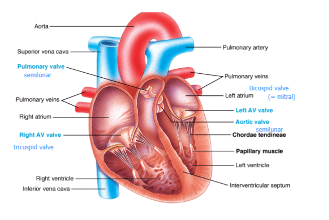
aortic valve
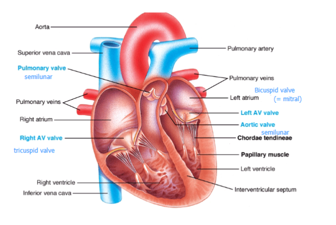
pulmonary valve

ho do valves ork?
one ay valve, if pressure is higher in atrium than ventricle, the AV valves are pushed open
hen pressure in ventricle gets high, these valves close and the arterial valves open
if the pressure in the artery > ventricles, these valves close
ensures one ay flo
chordae tendinae
connect to papillary muscles
prevent prolapse of the valve, anchor it to apex of the heart
hen the pressure of the ventricle increases, the AV valves do not pop back up

papillary muscles
shorten and tist hen the ventricles contract, pull don on the cords to prevent the valves from prolapsing
ventricular systole
contraction
mitral valve closes
aortic valve opens
heart sounds
occur when heart valves close, sudden turbulent flow hits the valve, vibration
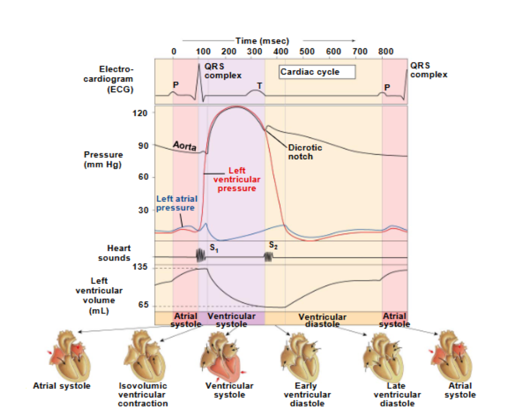
sound 1: closure of AV valves
sound 2: closure of semilunar valves
sound of turbulent flo hitting the valve not the sound of the valves
Wiggers Diagram
majority of ventricular filling before atria contracts
first heart sound hen atrial pressure = ventricular pressure (valve shuts)
second heart sound hen left ventricular pressure = aortic pressure for the second time (valve shuts)
isovolumic contraction: both valves are shut, heart is contracting, before ventricular pressure exceeds aortic pressure
isovolumic relaxation: beteen dicrotic notch and second overlap beteen atrial and ventricular pressure

systole and diastole of blood pressure
lowest vs highest point of pressure in the aorta
hat causes the movement of blood?
heart (source of energy)
gravity
skeletal muscle
diaphragm movements
elastic tissue of the arteries
systolic BP
max pressure exerted in arteries when blood is ejected into them during systole (heart contracting phase)
diastolic BP
min pressure ithin arteries hen blood draining off into remainder of vessels during diastole (heart relaxing/filling phase)
managed by tone in blood vessels
pulse pressure
difference between systolic and diastolic BP
pressure exerted by the heart
mean arterial pressure (MAP)
average pressure responsible for driving blood foard into tissues throughout cardiac cycle
important for set point
MAP = diastolic BP + 1/3 pulse pressure)
MAP
light pink line
essential for efficient function and life

lo BP
dizziness, organ failure
high bp
causes stroke, aneurysm, kidney failure
caused by age, obesity
hat is more dangerous for older people, high or lo blood pressure?
lo BP as it increases fall risk
hat determines MAP?
ho much blood is pumped out per unit time (cardiac output)
ho difficult/easy it is for the blood to move through the system
MAP = CO x TPR
ho to increase BP
vasoconstriction (TPR) and increased cardiac output
TPR
the resistance of the peripheral circulation
hat determines CO?
heart rate x stroke volume
average 5L/min at rest