Patho Exam 2 Full Review
 Call Kai
 Learn
Learn Practice Test
 Spaced Repetition
 Match
Match Flashcards
 Knowt Play
 Podcast
1/99

There's no tags or description
Looks like no tags are added yet.
Name | Mastery | Learn | Test | Matching | Spaced | Call with Kai | Chat |
|---|
No analytics yet
Send a link to your students to track their progress
100 Terms
Intracellular fluid (ICF)
water inside cells; about two-thirds of body water; high in potassium (K+) and some magnesium.
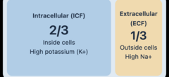
Extracellular fluid (ECF)
Fluid outside cells; about one-third of body water; includes plasma and interstitial fluid; high in sodium (Na+) and chloride (Cl−).
Contains most of the body's sodium.
Transcellular compartment
Specialized fluids such as cerebrospinal, pleural, pericardial, peritoneal and synovial fluids; about 1% of body weight in the outline.
Third spacing
Abnormal accumulation of large amounts of fluid in transcellular spaces; e.g., ascites.
Fluid gets trapped somewhere it cannot effectively help circulation, such as the abdomen (ascites).
Capillary filtration (hydrostatic) pressure
Pushes water OUT of the capillary and into surrounding interstitial tissue.
Capillary colloidal osmotic pressure
Plasma proteins, especially albumin, pull fluid INTO the capillary.
Interstitial hydrostatic pressure
Opposes outward movement of fluid from the capillary.
Tissue colloidal osmotic pressure
Proteins in tissue pull fluid OUT of the capillary.
Lymphatic system
Returns excess interstitial fluid to blood circulation; blockage can cause lymphedema.
Psychogenic polydipsia
Compulsive water drinking; type of inappropriate/false thirst, seen with some psychiatric disorders.
Symptomatic (true) thirst
Thirst from actual body-water loss (e.g., diarrhea, vomiting, diabetes mellitus or diabetes insipidus); relieved by replacing water.
Neurogenic diabetes insipidus (DI)
Impaired synthesis or release of antidiuretic hormone (ADH); severe production of dilute urine and thirst.
Nephrogenic DI
Kidneys fail to respond normally to ADH; large urine losses despite ADH being present.
SIADH
Inappropriately high ADH secretion despite normal osmolality; retained water, reduced urine output and dilutional hyponatremia.
Hypertonic (translocational) hyponatremia
High concentrations of other solutes, such as glucose, draw water from cells into ECF, diluting sodium; example: hyperglycemia.
Hypotonic (dilutional) hyponatremia
Too much water relative to sodium, lowering blood sodium concentration; may occur with water intoxication.
Hypovolemic hypotonic hyponatremia
Loss of sodium and water with net sodium deficit; lecture example: profuse sweating in hot weather.
Euvolemic hypotonic hyponatremia
Water retention dilutes sodium while overall ECF volume is approximately maintained; example: SIADH.
Hypervolemic hypotonic hyponatremia
Excess total fluid with dilution of sodium; lecture examples include heart failure, liver disease and renal disease.
Where is most body fluid?
In the intracellular compartment (ICF), approximately 2/3 of total body water.
Primary route of water loss
Kidneys (urine); other routes include GI tract, skin, and exhaled air.
Hormones regulating Na+
Renin-angiotensin-aldosterone system (especially aldosterone for renal Na+ retention); ADH primarily affects water and thus Na+ concentration.
Hormones regulating K+
Aldosterone promotes kidney elimination of K+; kidneys and redistribution between ICF/ECF maintain levels.
Hormones regulating Ca2+
Parathyroid hormone (PTH) is emphasized by the outline, which links decreased PTH to hypocalcemia; the uploaded outline does not systematically discuss other Ca-regulating hormones.
Primary intracellular buffer
Body proteins (can act as acids or bases).
Primary extracellular buffer
Bicarbonate / carbonic acid (HCO3− / H2CO3); albumin and globulins also buffer plasma.
Respiratory acidosis
CO2 retention: increased pCO2 and carbonic acid, decreased pH; commonly from hypoventilation, lung disease, airway obstruction or respiratory muscle weakness.
Respiratory alkalosis
Excess CO2 loss: decreased pCO2 and carbonic acid, increased pH; the lecture states these changes without detailing causes.
Primary importance of ions
Ions are charged particles; electrolytes support membrane excitability, nerve/muscle function, fluid distribution and acid-base balance (especially K+ in membrane excitability).
Causes of edema
Increased capillary filtration pressure; decreased plasma colloidal osmotic pressure (e.g., low albumin); increased capillary permeability; obstructed lymph drainage.
Useful numerical facts
Normal serum sodium 135-145 mEq/L; potassium 3.5-5.0 mEq/L; arterial pH 7.35-7.45; serum osmolality 280-295 mOsm/L (values stated in lecture). CHAPTERS 26 & 27 • Matching / Compare & Contrast
Stable angina
Chest discomfort from transient myocardial ischemia provoked by activity; relieved by rest and nitroglycerin.
Unstable angina
New, worsening, more frequent or prolonged chest pain, often at rest or with little activity; may last over 20 minutes; higher risk for MI.
Prinzmetal (variant) angina
Coronary-artery vasospasm causes transient ischemic chest pain.
Silent myocardial ischemia
Ischemia without chest pain; can occur in diabetes with autonomic neuropathy.
T-wave inversion
ECG sign of altered myocardial repolarization.
ST-segment elevation
ECG indicator of transmural myocardial injury (full-thickness injury pattern).
ST-segment depression
ECG indicator of subendocardial myocardial injury.
Abnormal Q wave
Related to loss of depolarizing current conduction after myocardial injury.
Dilated cardiomyopathy
Enlarged/dilated heart chambers, weakened pumping ability, possible heart failure; can be related to infections, toxins/alcohol, genetics.
Hypertrophic cardiomyopathy
Abnormally thick ventricular myocardium; may cause dyspnea, chest pain, syncope and sudden death, sometimes in young adults.
Restrictive cardiomyopathy
Rigid ventricular walls prevent normal diastolic filling.
Peripartum cardiomyopathy
Cardiomyopathy associated with pregnancy/postpartum period; symptoms resemble dilated cardiomyopathy; risk factors include hypertensive pregnancy and multiple gestation.
Chylomicrons
Made in small intestine; carry absorbed dietary fats to muscles and adipose tissues.
VLDL
Made mainly in liver; carries large quantities of triglycerides toward peripheral tissues.
IDL
Formed as VLDL loses triglycerides; intermediate particles can become LDL or return to liver.
LDL
Primary carrier of cholesterol to tissues; "bad cholesterol"; higher levels promote atherosclerosis.
HDL
"Good cholesterol"; reverse cholesterol transport from tissues and plaques back to liver.
Mitral valve prolapse
Floppy valve leaflets bulge back into left atrium during systole.
Mitral stenosis
Mitral valve does not open fully; causes left atrial dilation and pulmonary congestion; commonly associated with rheumatic fever.
Mitral regurgitation
Mitral valve fails to close properly; blood flows backward into left atrium during systole; possible pansystolic murmur.
Aortic stenosis
Aortic valve does not open sufficiently; LV must work harder to pump out blood, reducing ejection.
Aortic regurgitation
Leaky aortic valve returns blood to LV; LV must pump both new blood and regurgitant blood; severe cases can widen pulse pressure.
Kawasaki disease
Acute febrile inflammatory vasculitis in children with fever, conjunctivitis, rash, lymph nodes and later skin peeling; can produce coronary aneurysms.
Coarctation of the aorta
Narrowing of aorta; arm pulses/BP stronger than leg pulses/BP.
Transposition of great vessels
Aorta comes off RV and pulmonary artery comes off LV; survival requires an opening between circuits (such as PDA or septal defect).
Patent ductus arteriosus (PDA)
Fetal ductus arteriosus remains open after birth, creating abnormal communication; treated with indomethacin or surgery in lecture.
Endocardial cushion defect
Abnormal formation of AV canals and parts of atrial/ventricular septa; associated with Down syndrome.
Cardiogenic shock
Pump failure: heart cannot eject enough blood, causing hypotension and low tissue perfusion.
Hypovolemic shock
Loss of intravascular volume: blood, plasma, or extracellular fluid.
Obstructive shock
Physical blockage to heart filling or blood outflow; examples: tamponade, pulmonary embolus, tension pneumothorax.
Distributive shock
Widespread vasodilation/increased vascular space despite circulating fluid; includes neurogenic, anaphylactic and septic shock.
Anaphylactic shock
A distributive shock triggered by released vasodilating substances during severe allergic response.
High-output heart failure
Heart output may be elevated but still inadequate for unusually high tissue metabolic demand.
Low-output heart failure
Inadequate output because heart cannot pump sufficiently.
Systolic failure
Impaired ventricular ejection (pumping).
Diastolic failure
Impaired ventricular filling (relaxation).
Right-sided failure
Blood backs up into systemic circulation: peripheral edema, enlarged congested liver, ascites.
Left-sided failure
Blood backs up into pulmonary circulation: shortness of breath, chronic nonproductive cough and lung congestion.
Berry aneurysm
Small round bulge at vessel branch point, e.g., circle of Willis.
Fusiform aneurysm
Progressive expansion of the entire circumference of a vessel.
Saccular aneurysm
Pouch-like dilation involving only part of vessel wall circumference.
Aortic dissection
Tear in aortic intima allows blood to split layers of vessel wall; classically sudden severe tearing/ripping pain.
Thrombophlebitis
Vein thrombus with inflammatory response, often in leg veins; risk factors include blood stasis, vessel injury and hypercoagulability.
Vasculitides
Inflammatory injury and necrosis of vessel walls; may be due to immune processes, infection or direct injury.
Intermittent claudication
Leg/calf pain during walking due to peripheral arterial disease; often with cool foot and weak pulses.
Buerger disease
Thromboangiitis obliterans; disorder of medium-sized arteries associated with tobacco use and pain/cold sensitivity.
Raynaud disease / phenomenon
Vasospasm in fingers/toes triggered by cold or stress; color changes pallor → cyanosis → red during reperfusion; primary disease vs secondary phenomenon. CHAPTERS 26 & 27 • Other Question Formats
Coronary artery branches
Left main coronary artery branches into left anterior descending and circumflex arteries; right coronary artery gives rise to posterior descending artery in the lecture model.
Electrical conduction components
NOT DESCRIBED in the supplied outlines. The exam review guide requests this topic, but the notes supplied do not explain the conduction pathway; check additional class resources.
ECG features
Lecture focuses on inverted T waves, ST elevation/depression, abnormal Q waves; 12-lead ECG aids diagnosis of MI. Full P-wave/PR/QRS pathway is not detailed in these outlines.
Capillary pressure dynamics
Capillary filtration pressure pushes water out; capillary colloidal osmotic pressure pulls it in; interstitial hydrostatic pressure opposes exit; tissue colloidal osmotic pressure pulls out; lymph returns excess.
Three vascular tunics
Intima = innermost endothelial lining; media = smooth muscle/elastin for vasoconstriction or vasodilation; externa/adventitia = collagen-rich support with nerves/vasa vasorum.
Interpret BP measurements
Lecture: normal
BP measurement errors
Cuff too small overestimates BP; cuff too large underestimates. Rest before reading and avoid caffeine/smoking for 30 minutes.
Role of renin / angiotensin II
Low renal perfusion → renin release → angiotensin II vasoconstriction and aldosterone secretion → sodium and water retention and increased BP.
MI serum markers
Myoglobin increases rapidly (~1 hour; not heart-specific); CK-MB rises ~4-8 h, normalizes ~2-3 days; troponin I/T rises within ~3 h and stays elevated ~7-10 days, per lecture.
Vasodilating chemicals
Adenosine, K+, lactic acid, CO2 and endothelial nitric oxide increase coronary blood flow; endothelins promote constriction.
Acute pericarditis
Inflammation of pericardium; often viral; chest pain, friction rub, ECG changes. May be treated with NSAIDs/aspirin and cause-specific treatment.
Chronic pericarditis with effusion
Persistent inflammatory fluid in pericardial sac beyond acute phase.
Constrictive pericarditis
Scar tissue between pericardial layers restricts diastolic ventricular filling.
Cardiac tamponade cause
Blood, fluid or pus in pericardial sac compresses heart, reducing venous return, stroke volume and cardiac output; may cause shock.
Cardiac tamponade relief
Pericardiocentesis to drain fluid; echocardiography can evaluate compression.
Causes of valvular disease
Rheumatic fever commonly causes mitral stenosis and other valve injury; congenital valve malformations can cause aortic stenosis; chordae/papillary muscle injury can cause mitral regurgitation.
Fetal circulation shunts
Ductus venosus bypasses fetal liver; foramen ovale shunts right atrium to left atrium; ductus arteriosus diverts blood from pulmonary artery to aorta. Foramen ovale and ductus arteriosus close after birth.
Heart failure compensations
Frank-Starling mechanism; SNS raises HR/contractility/vascular tone; RAAS conserves fluid and constricts vessels; ANP produces sodium/water excretion; endothelins constrict vessels; myocardial hypertrophy/remodeling.
Distributive shock subtypes
Neurogenic = loss of sympathetic vasomotor tone; anaphylactic = vasodilatory mediators; septic = inflammatory mediators/systemic response.
Antihypertensive drug actions
Diuretics reduce blood volume; beta blockers lower HR/CO/renin; ACE inhibitors block angiotensin I→II; ARBs block angiotensin II receptors; alpha-1 blockers dilate vessels; central alpha-2 agonists reduce SNS outflow; calcium-channel blockers lower smooth muscle calcium entry.
Cardiovascular drug actions
Nitroglycerin → nitric oxide-mediated vasodilation for angina; aspirin → antiplatelet; thrombolytics → break down fibrin-containing clots; statins → decrease hepatic cholesterol synthesis; digoxin, ACE inhibitors, beta blockers and diuretics are identified as heart-failure treatments.
Shock complications
Acute respiratory distress syndrome (ARDS), acute renal failure, GI injury, disseminated intravascular coagulation (DIC) and multiple-organ dysfunction syndrome (MODS).