W6 Clinical Assessment of Pupil Function
 Call Kai
 Learn
 Practice Test
 Spaced Repetition
 Match
 Flashcards
 Knowt Play
1/62

There's no tags or description
Looks like no tags are added yet.
63 Terms
What do we expect normal pupils to be like?
Round
React briskly
Equal in size
Dilate in dark (mydriasis)
Constrict in response to light (miosis)
Constrict in response to accommodation and convergence
Mydriasis occuring more slowly than miosis
RED CM
What happens to pupil diameter as people get older?
Decreases with increasing age (senile miosis)
NOT influenced by gender/iris colour/rx error (only when rx error is corrected as hyperopia can cause miosis when accommodating)
What does the pupil do?
Protects from excess light
Improves image quality
What is the sphincter papillae responsible for?
Responsible for constricting the eye- innervated by parasympathetic system

What is the dilator papillae responsible for?
Widening the eye- innervated by sympathetic system
What does parasympathetic innervation consist of?
4 neurones- including afferent and efferent
What is the afferent pathway in parasympathetic innervation?
Connects the retina with the pre-tectal nucleus at the level of the superior colliculus in the midbrain
Connects the pre-tectal nucleus to both Edinger-Westphal nuclei

What happens to fibres in afferent innervation of the parasympathetic nervous system?
When fibres reach the chiasm, nasal fibres cross over to the contralateral side of the brain
Temporal fibres will remain on the ipsilateral side of the brain

Why do we get a consensual response even if light is only shone in one eye?
Equal innervation in Edinger-Westphal due to crossing over at the chiasm
What does the efferent pathway in parasympathetic innervation look like?
Connects Edinger-Westphal nucleus to the ciliary ganglion
PARASYMPATHETIC FIBRES PASS IN THE INFERIOR DIVISION OF THE IIIRD NERVE
Sphincter papillae is innervated when it leaves the ciliary ganglion and passes with the short ciliary nerves to innervate

What does sympathetic innervation consist of?
3 neurones
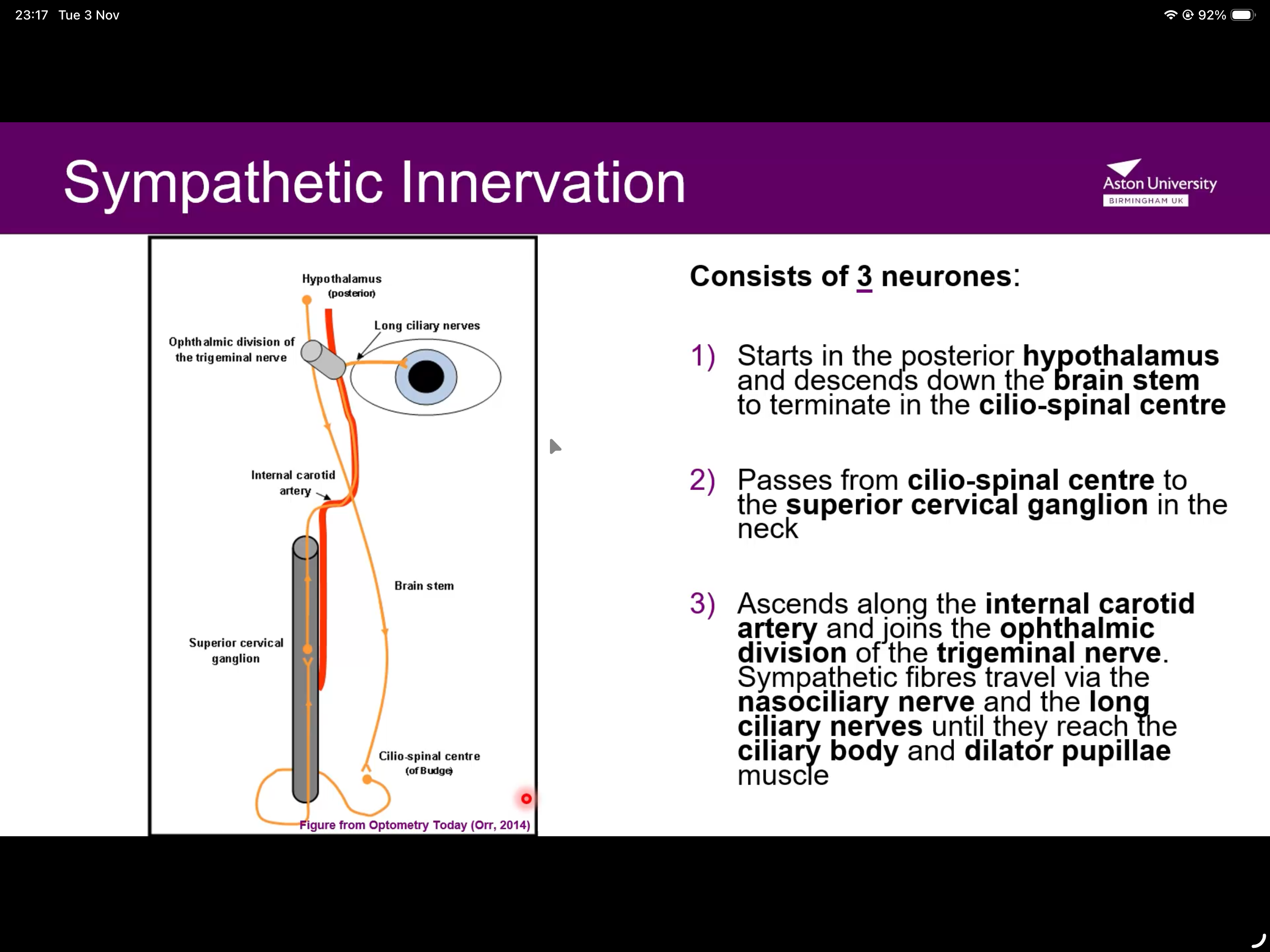
What happens first in sympathetic innervation?
Starts in the posterior hypothalamus and descends down into the brain stem to terminate in the cilio-spinal centre

What happens next in sympathetic innervation?
Passes from cilio-spinal centre to superior cervical ganglion in the neck
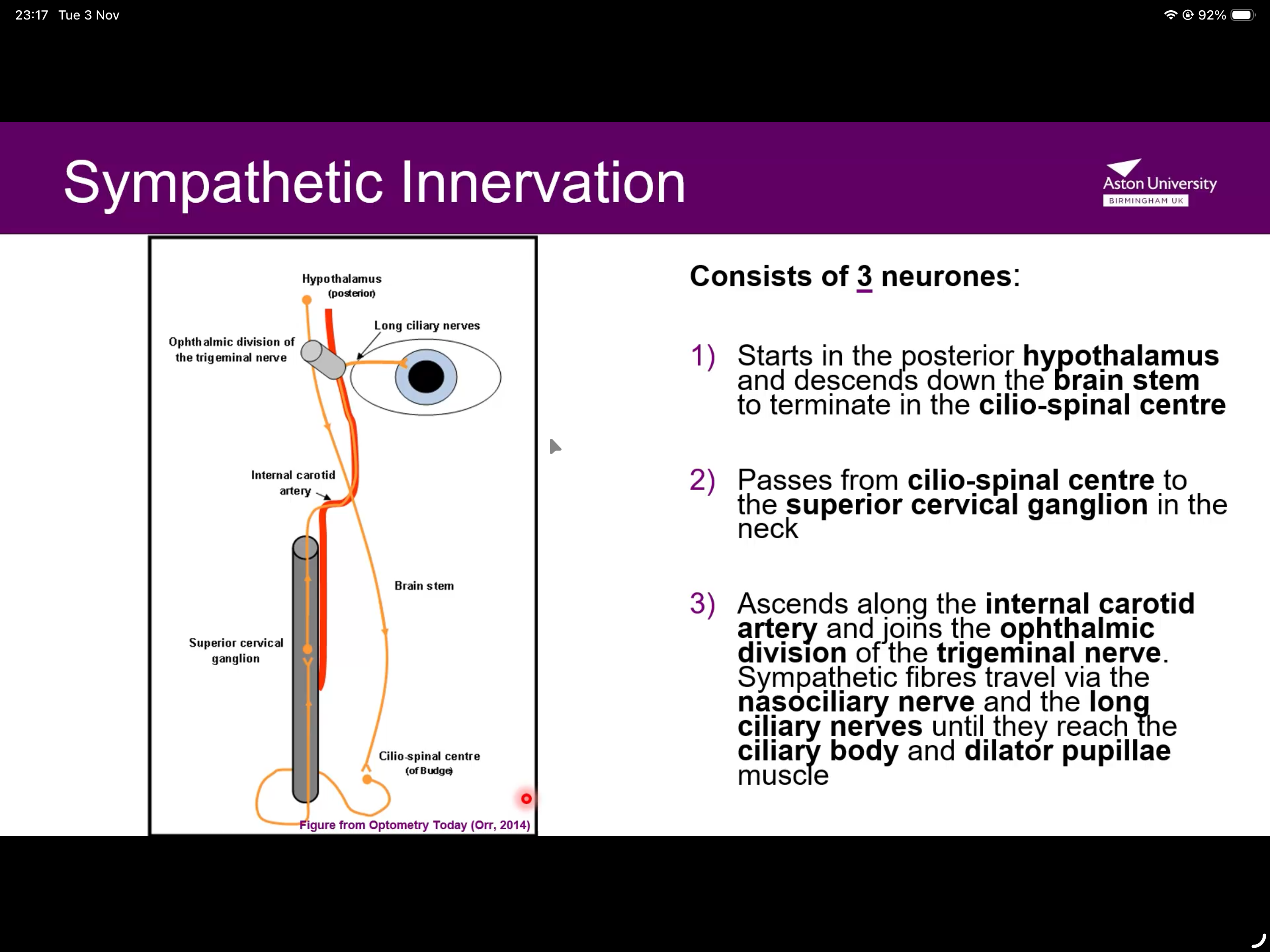
What happens last in sympathetic innervation?
Ascends along internal carotid artery from SCG and joins ophthalmic division from CN V
Sympathetic fibres travel along nasociliary nerve and long ciliary nerves until they reach the ciliary body and dilator papillae muscle
What do we consider when conducting a pupil assessment?
Shape, position and size of pupil
Reaction to light and accommodation
Presence of RAPD
What does RAPD stand for?
Relative afferent pupillary defect
How do we do pupil observation with a direct opthalmoscope?
Large aperture, medium brightness, low dioptric power
Focus on size, shape and centration (off-centre)
Why do you not stand in front of the px when doing direct and consensual?
Px will fixate on you and accommodate
How do we test the near response?
Instruct px to shift gaze from distant target to near target and back to the distant target again
We expect constriction of pupil when looking at near target
What is near light dissociation?
Constriction of pupil to near fixation but not direct light
What are causes of near-light dissociation?
Viral infection (Adie’s tonic pupil)
Damaged pre-tectal area
Damage in the rostral mid brain
What does presence of RAPD indicate?
Significant area of vision is affected (Includes retinal layers, optic nerve, chiasm, mid brain pre-tectal area)
Never seen in px with cataract/high refractive error/anisocoria
How do you do the swinging flash light test?
Perform in DIM room-light
Shine pen torch on right eye for 2/3 secs and observe RE dilate
Quickly move light to LE and hold for 2/3 secs- pupil should remain constricted/dilate and constrict again
An eye with RAPD will dilate when light is shone on it
Repeat several times
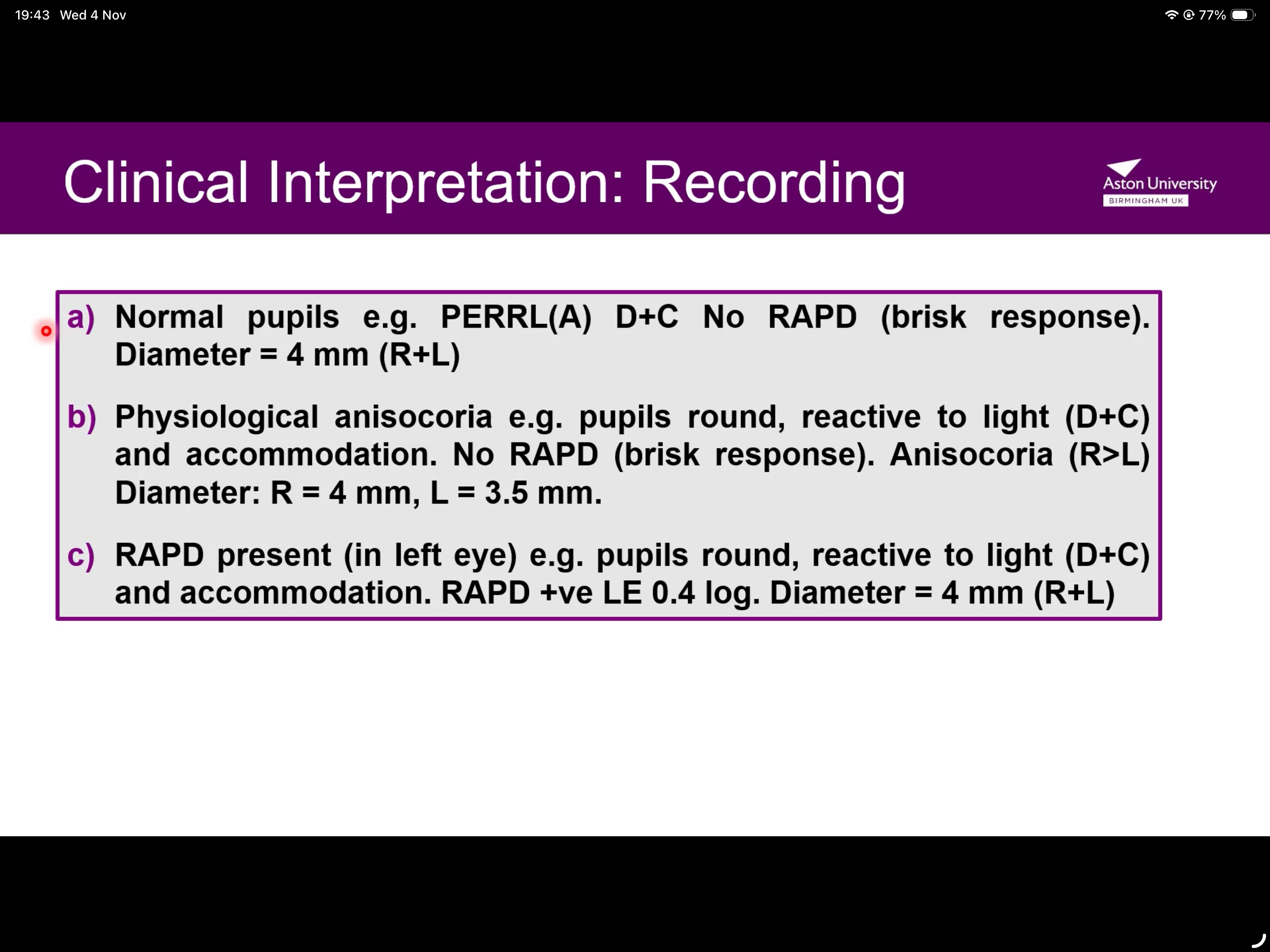
What does a normal record entail?
Normal pupils e.g. PERRL(A) D+C No RAPD (brisk response).
Diameter = 4 mm (R+L)
What defects do we get within the parasympathetic nervous system?
Afferent defects- do not cause anisocoria- results in abnormal light reflex, near reflex is normal
Where do afferent defects occur?
Between photoreceptors and Edinger-Westphal nucleus
How are afferent anomalies caused?
Detects interference with the input of light to the pupils motor system and is caused by defects in
Chiasm
Optic nerve
Optic tract *
Retinal layers
*(Mid brain pre-tectal area)
COOR M.
Can we see a post-chiasmic lesion in RAPD?
PCL rarely produce a clinically detectable RAPD
What does a clinically presentable RAPD imply?
Optic neuritis
AION
Compressive optic neuropathy
Retinal detachment
Advanced glaucoma
CRVO
What is APD?
Absolute pupillary defect- occurs when there has been severe damage to the optic nerve such that the eye is completely blind
When light is shone in the abnormal eye the direct and consensual response is absent
When light is shone in the normal eye both eyes constrict
Where do efferent pupil anomalies occur?
Arises from the efferent parasympathetic pathway or anywhere in the sympathetic pathway
What are efferent pupillary defects associated with?
Damage to the parasympathetic (sphincter) nerves
Damage to sympathetic (dilator) nerves
Mechanical hyper-excitation of sympathetic supply
Direct damage to sphincter/dilator muscles that may cause immobility of the iris
Where do parasympathetic efferent defects occur?
From Edinger-Westphalia nucleus to the sphincter pupillae
Do efferent defects cause anisocoria?
Yes- does not cause RAPD
Where does damage occur in efferent abnormalities?
Interference with contraction or dilation of the pupil can occur due to damage in the
Midbrain
Iris muscles
Peripheral nerve that supplies the iris muscles
What does the extent of anisocoria depend on?
Condition of lighting/near effort
What is physiological anisocoria?
20% of the population has it
Difference is same in dim and bright light (simply more pronounced in dim)
Normal light and near response
If there is a large difference in pupil diff (>1) but reflexes and responses are normal, it remains to be physiological
What is pathological anisocoria?
More marked in dim or bright lighting conditions
What happens if anisocoria increases in bright light?
Parasympathetic problem (third nerve palsy)- larger pupil affected
What happens if anisocoria increases in the dark?
Sympathetic problem (Horner’s syndrome)- smaller pupil affected
Who are miotic (small) pupils commonly found in?
Infants
Elderly px
Long standing diabetics
Brain stem lesions
Anterior iritis
Who are large (mydraitic) pupils found in?
Anxious px
Px who are medicated (anticholinergics)
Afferent pathway legions
Optic neuritis/ optic nerve tumours
How is Adie’s tonic pupil caused?
Disruption of the post-ganglionic supply to sphincter pupillae, linked to viral illnesses
What are symptoms of Adie’s tonic pupil?
Dilation and constriction problems
Affects healthy young adults (women>men)
Reaction to light and accommodation reduced/absent
Redilation of the affected pupil is slow
Segmental damage
What do patients present with when suffering from Adie’s tonic pupil?
Problems at near
Blurred distance VA
Photophobia
Eye pain
Headache
However some px may be asymptomatic
How do we identify Adie’s tonic pupil and distinguish it from 3rd nerve palsy?
Put pilocarpine in affected eye- in Adie’s it will cause pupil reduction
What happens in long standing Adie’s tonic pupil?
Reduction in pupil size can occur
Reaction to light remains sluggish/absent
Goes from unilateral to bilateral (20% of the time)
Light-near dissociation develops after 2 months
What is third cranial nerve palsy?
A lesion that compresses the third cranial nerve also compresses the parasympathetic efferent pupillary nerve fibres which travel along its surface
How does third cranial nerve palsy present itself?
Complete Ptosis
Exotropia and vertical deviation
Diplopia (pupils no longer aligned)
Inhibits normal constriction of pupil and results in dilation
Extent of anisocoria increases in bright light
CEDIE
What are causes of third cranial nerve palsy?
Enlarging aneurysm of the posterior communicating artery
Tumours/space occupying lesions
Vascular disease such as hypertension and diabetes mellitus (only without pupil involvement)
Upper eyelid elevates as px looks down (recovering nerve palsy)
What is Argyll-Robertson pupil?
Occurs in neurosyphillis
Arises from an interruption of fibres from Edinger-Westphal nucleus that connect pretectal nuclei
Both pupils are involved but anisocoria is present
What do pupils look like in argyll-Robertson pupils?
irregular in shape
Very miotic (small)
Express light near dissociation/normal near reflex
How does Horner’s syndrome present itself?
Unilateral
Mild ipsilateral ptosis
Lower lid elevation
Miosis
Dilation lag in affected eye
Normal light and near pupil response
Educed ipsilateral sweating

How do you diagnose Horner’s syndrome?
Instill topical cocaine 4%, causing dilation of the mitotic pupil in physiological anisocoria but makes minimal difference to the mitotic eye in Horners
When do we do urgent referral for Horner’s?
Disrupted sympathetic pathway
Pancoast tumour
Carotid aneurysm
Lesions in the neck
Idiopathic
Features of an ectopic pupil.
Creates an abnormally shaped pupil
Either congenital or acquired
If acquired, it can result from ocular trauma, surgery or inflammation
Features of iris coloboma.
Congenital defect caused by incomplete closure of foetal fissure during embryonic stage of development
Characterised by unilateral/bilateral partial inferior iris defect
What is polycoria?
Multiple pupils- congenital or acquired (results from iris trauma, atrophy or inflammation)
What’s ischaemic damage?
Direct damage to the iris;
acute angle closure glaucoma
Herpes zoster infections
Surgical procedures (cataract surgery)
When do we immediately refer to eye casualty?
Acute onset abnormalities in pupil shape
What is iritis/anterior uveitis?
Inflammation of the iris
Painful red eye
Small pupil due to iris/ciliary spasm
Mechanical limitation
Inflammation may lead to posterior synechiae- iris sticks to front part of crystalline lens

If iritis is left untreated what can happen?
Causes:
angle closure
Macula oedema
Cataract
IMPORTANT RULES OF THUMB
…
