Anatomy- Heart/Thorax
 Call Kai
 Learn
Learn Practice Test
 Spaced Repetition
 Match
Match Flashcards
 Knowt Play
1/78

There's no tags or description
Looks like no tags are added yet.
Name | Mastery | Learn | Test | Matching | Spaced | Call with Kai | Chat |
|---|
No analytics yet
Send a link to your students to track their progress
79 Terms
Artery
a thick-walled blood vessel that carries OXYGENATED blood away from the heart to the rest of the body

Vein
a blood vessel that carries DEoxygenated blood back to the heart from the rest of the body

Tunica intima
the innermost layer of a blood vessel that is in direct contact with flowing blood, (intimate), smooth and provides low friction for blood to flow easily
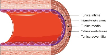
Tunica media
the middle layer of tissue in the wall of a blood vessel, regulates blood pressure

Tunica adventitia
the outermost layer of a blood vessel wall, acts as an anchor and protective sheild

Lumen
the inside hollow space or channel of a blood vessel where blood flows
Atherosclerosis
the gradual buildup of sticky plaque made of fat, cholesterol, and calcium inside the walls of your arteries, which narrows the blood vessels and restricts blood flow

Pulse point
a specific spot on the body where a major artery runs close enough to the skin to produce a palpable pulse

Papillary Muscle
small, cone-shaped muscles located inside the lower chambers (ventricles) of the heart, they anchor the chordae tendinae to stop backflow

Trabeculae carnae
irregular, muscular ridges and columns that project from the inner walls of the heart's ventricles

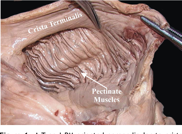
Pectinate muscle
parallel muscular ridges on the inner walls of the heart's atria that look like the teeth of a comb
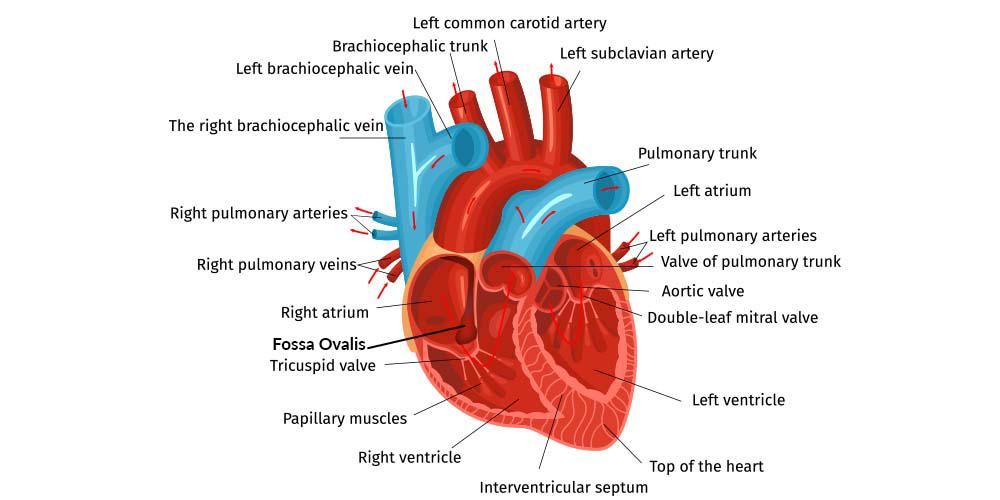
Foramen ovale
a normal, temporary opening in the wall (septum) between the left and right upper chambers (atria) of a fetus's heart

Fossa ovalis
an oval-shaped depression formed after the baby is born and takes its first breath, lung pressure drops and pressure change slams a tiny tissue flap shut, sealing the opening permanently to seperate the L and R atrium
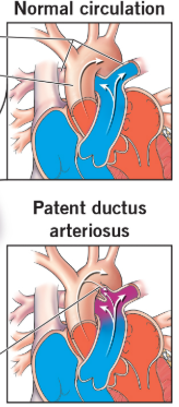
Ductus arteriosus
a temporary blood vessel in a developing fetus that connects the main pulmonary artery to the aorta, allowing blood to bypass the unfunctioning lungs
Ductus venosus
a temporary blood vessel in a fetus that lets oxygen-rich blood from the umbilical vein bypass the liver and flow straight to the heart
Ligamentum arteriosum
the fetal remenant found between the pulmonary trunk and aorta

Vitelline system
the early system of blood vessels that circulates blood between a developing embryo and its yolk sac
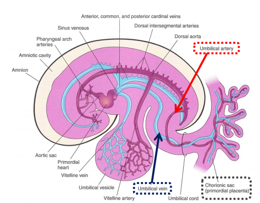
Placental (Umbillical) system
2 arteries and 1 single vein, two arteries deliver DEOXYGENATED blood and waste to the placenta.
Singular vein transports oxygenated and nutrient-rich blood from the placenta back into the baby.

Intraembryonic system
a horseshoe-shaped, fluid-filled space inside the developing embryo that forms the future body cavities

Erythrocyte
Red blood cells: These cells make up a large part of your blood cells and contain hemoglobin which transport oxygen

Leukocyte
White blood cells: These are a key part of your immune system. They find and destroy germs like bacteria and viruses to protect you from illness

Platelet
tiny cell fragments. They stick together to form clots when you get a cut or scrape, which helps stop the bleeding.

Plasma
pale-yellow liquid that makes up about 55% of your blood volume. It carries water, salts, enzymes, nutrients, and hormones through your body.

Oblique sinus
a blind-ending pocket in the serous pericardium located on the back of the heart, gives the left atrium room to move and expand as it fills with blood from the lungs

Transverse sinus
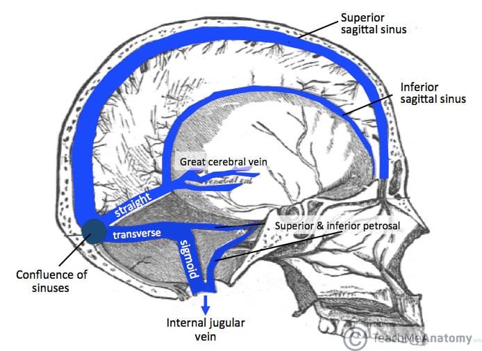
two large dural venous sinuses—channels located inside the head between layers of the dura mater—that drain venous blood from the back of the brain
Pulmonary circuit
The flow of blood between the heart and lungs
Superior and inferior vena cava → right atrium → right ventricle → pulmonary trunk → lungs

Systemic Circuit
The flow of blood between the heart and the rest of the body
lungs → pulmonary veins → left atrium → left ventricle → aorta → body

AV (Atrioventricular) valves
The two one-way valves that connect the atrium to the ventricle
Left side: Bicuspid valve (mitral) Right side: Tricuspid valve
Semilunar valves
The two one-way valves that connect the ventricles to the great vessels
Aortic valve: Sits between the left ventricle and the aorta. It sends oxygen-rich blood to the body
Pulmonary valve: Sits between the right ventricle and the pulmonary artery. It sends oxygen-poor blood to the lungs
Sinoatrial node
the heart's natural pacemaker (located in the upper wall of the right atrium) generates electrical impulses to start every heartbeat

How many embryonic chambers? What is there function?
4, perform sequential pumping and directional blood flow before splitting into the different chambers of the heart
Sinus venosus
Acts as the inflow tract, collecting all venous blood from the body and embryo, and eventually forms the posterior part of the right atrium, the sinoatrial (SA) node, and the coronary sinus
Primitive Atrium
Serves as the early collecting chamber that receives blood from the sinus venosus and distributes it forward; it develops into the anterior portions of the definitive left and right atria and auricles.
Primitive Ventricle
Functions as the primary early pumping chamber and expands to form the shape of the left ventricle
Bulbus cordis
Works in tandem with the primitive ventricle to propel blood forward, specifically giving rise to the right ventricle and parts of the outflow tract.
Cardiac looping
the essential embryonic process where the straight, linear heart tube bends and twists asymmetrically into an S-shaped loop

Chordae tendinae (heartstrings)
inelastic cords of fibrous connective tissue that connect the papillary muscles to the tricuspid valve and the mitral valve in the heart.

What are the great vessels? How many?
There are 8 great vessels, and they are the major arteries and veins that connect the heart to the rest of the body
Aorta
Carries oxygen-rich blood away from the left ventricle of the heart to the rest of the body.

Superior Vena Cava
Brings oxygen-poor blood from the head, neck, arms, and upper chest back to the right atrium of the heart.

Inferior Vena Cava
Brings oxygen-poor blood from the lower chest, abdomen, and legs back to the right atrium of the heart.

Pulmonary Trunk (arteries)
Carries oxygen-poor blood away from the right ventricle toward the lungs.

Right Pulmonary Artery & Left Pulmonary Artery
Directs oxygen-poor blood separately into the right and left lungs for gas exchange.

Right Pulmonary Veins (Two)
Carries oxygen-rich blood from the right lung back to the left atrium of the heart.

Left Pulmonary Veins (Two)
Carries oxygen-rich blood from the left lung back to the left atrium of the heart.

Pericardium (pericardial sac)
The 3-layered protective covering of the heart
Fibrous pericardium
the tough, outer layer of connective tissue that surrounds and protects the heart

Serous pericardium
a thin, double-layered membrane inside the fibrous sac that surrounds the heart
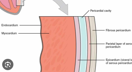
Pericardial cavity
the fluid-filled potential space between the parietal and visceral layers of the serous pericardium that surrounds the heart

Brachiocephalic artery
The first and largest major branch coming off the aortic arch, carrying oxygen-rich blood to the right side of the head, neck, and right arm
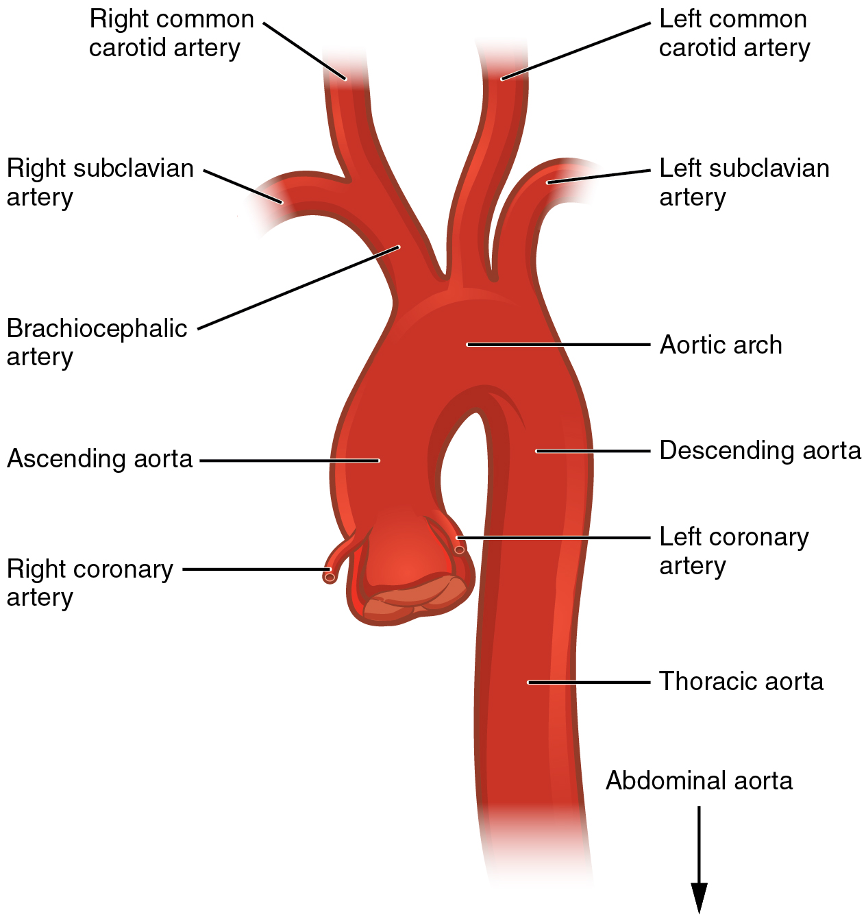
Left common carotid artery
A branch of the aorta that supplies oxygen-rich blood to the left side of the head, neck, and brain

Left subclavian artery
A branch of the aorta that supplies blood to the left arm, chest, and brain

Right coronary artery
originates from the aorta and supplies oxygen-rich blood to the right side of the heart
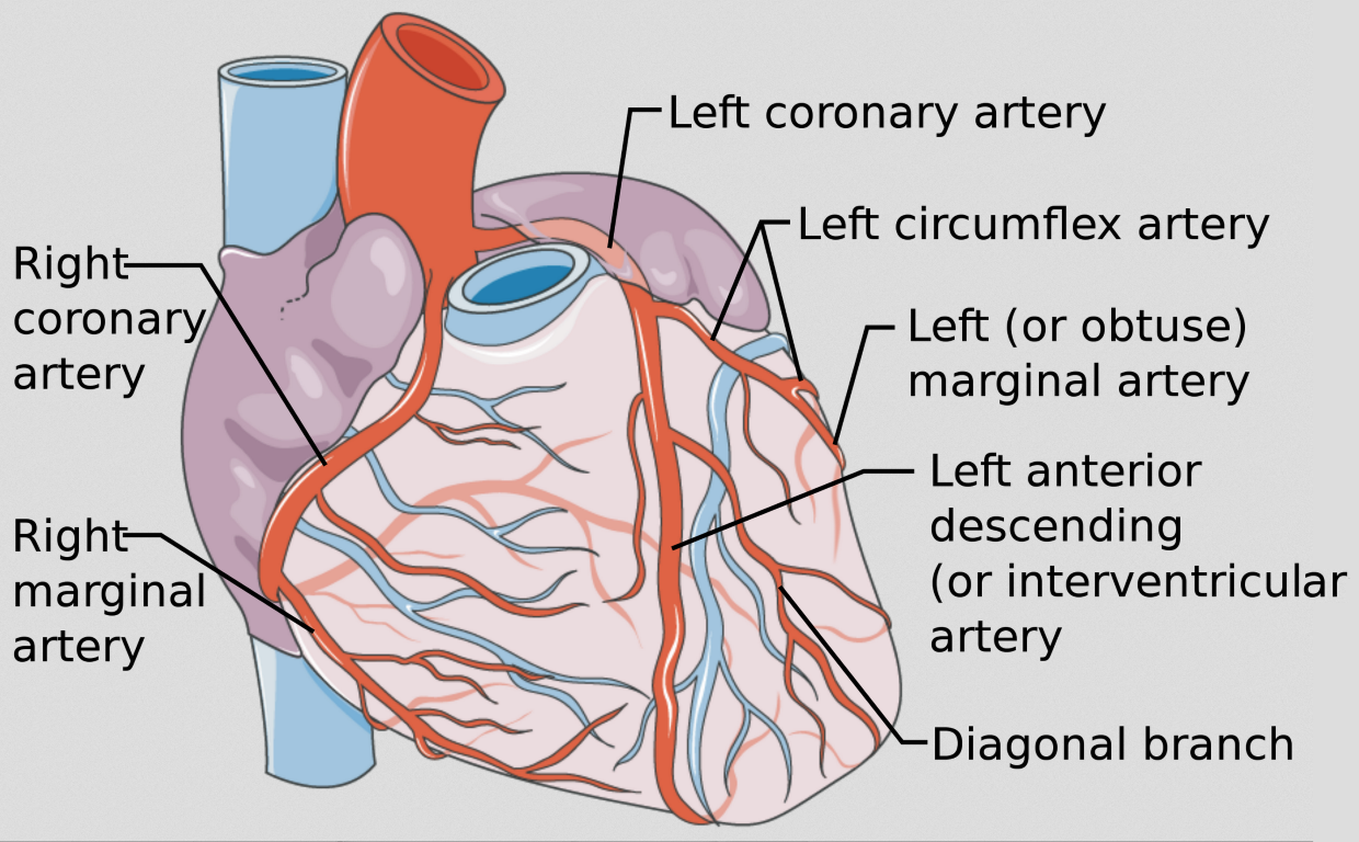
Left coronary artery
originates from the aorta and supplies oxygen-rich blood to the left side of the heart

Left circumflex artery
a major branch of the left main coronary artery that supplies oxygen-rich blood to the side and back of the heart

Left anterior descending LAD (anterior interventricular artery)
originates from the aorta and supplies about half of the blood to your heart's main pumping chamber

Coronary sinus
largest vein of the heart and drains DEoxygenated blood from the wall of the heart

The Azygous System
drains blood from the thoracic wall and some thoracic viscera into the superior vena cava (SVC)
azygous vein 2. hemiazygous vein 3. accessory hemiazygous vein
Hepatic Portal Venous System
drains deoxygenated nutrient-rich blood to liver via hepatic portal vein for processing
Why are the heart muscle fibers looped?
optimizes mechanical efficiency, fluid dynamics, and uniform workload distribution
Ribs
24 flat, curved bones arranged in 12 pairs that protect internal organs and assist with breathing

True Ribs
Connect directly to the breastbone (sternum) in the front using individual pieces of flexible costal cartilage (pairs 1-7)
False Ribs
Connect indirectly to the sternum by sharing cartilage that attaches to the rib above them (pairs 8-10)
Floating Ribs
Do not attach to the sternum at all and end in the abdominal muscles (pairs 11-12)
Vertebral Foramen
opening in spinal cord where spinal cord passes through
Head of rib
the wedge-shaped posterior end of a rib that connects the rib cage to the thoracic spine

Tubercle of rib
a small bump on the back part of most ribs where the neck meets the main shaft

Shaft of rib
the long, thin, flat, and curved portion that spans between the neck (near the tubercle) and the anterior costal cartilage

Costal groove
a small shallow depression located along the inner, lower border of most human ribs, contains the intercostal vein, intercostal artery, and intercostal nerve

anastomoses
connections between blood vessels, intestines, or other tubular structures in the body (provides an alternate route in case the vessel backs up)

Mediastinum
the central compartment of the thoracic cavity located in the middle of the chest between the two lungs

External Intercostal muscles
down and forwards toward the midline/front connect rib above to rib below, help with inhalation

Internal Intercostal muscles
down and backwards toward the posterior, help with expiration

Innermost Intercostal muscles
fibers run slightly down and backward but are almost vertical

Auricle
a small, ear-shaped muscular pouch attached to the outer front surface of each atrium (left and right)
function: expands to increase the blood-collecting and pumping capacity of the atrium

Cristia terminalis
smooth, C-shaped muscular ridge located on the inner wall of the heart's right atrium
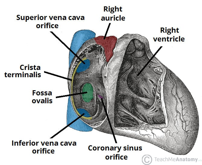
Trebeculae Carnae
irregular muscular ridges and columns that project from the inner walls of both ventricles of the heart

Marginal branch
key blood vessels that supply oxygen-rich blood to the walls of the heart

Posterior interventricular branch
coronary artery branch that runs along the bottom surface of the heart to supply oxygen-rich blood to the posterior third of the septum and the lower walls of both ventricles
