AP Biology Unit 4 MCQ (168 questions, formatted)
 Call Kai
 Learn
 Practice Test
 Spaced Repetition
 Match
 Flashcards
1/167

There's no tags or description
Looks like no tags are added yet.
168 Terms
In mammals, an increase in the concentration of sodium in the blood triggers the release of antidiuretic hormone (ADH) from the pituitary gland. As the concentration of sodium in the blood returns to previous levels, the release of ADH from the pituitary gland is reduced.
Q1. Based on the information presented, which of the following describes the most likely role of ADH in maintaining blood osmolarity?
a) ADH promotes an increase in the movement of sodium into the bloodstream. b) ADH promotes an increase in the movement of water into the bloodstream. c) ADH promotes an increase in the excretion of water from the body. d) ADH promotes an increase in the secretion of additional ADH from the pituitary gland.
b) ADH promotes an increase in the movement of water into the bloodstream.
Researchers have discovered details about apoptosis (programmed cell death) by studying embryologic development of a nematode worm, Caenorhabditis elegans. Apoptosis is a normal developmental process in C. elegans. They found several genes involved in apoptosis, including ced-9 and ced-3. The ced-3 gene was found to promote cell death, and ced-9 to inhibit it. The ced-9 gene serves as a regulator that prevents apoptosis in the absence of a signal promoting apoptosis.
Q2. Which of the following statements best justifies the claim that changes in the expression of ced-9 in C. elegans can affect regulation of apoptosis in the cell?
a) An experiment showed that a mutation in the ced-9 gene led to excessive cell death in C. elegans. b) An experiment showed that the ced-9 gene normally produces a protein that promotes excessive cell death in C. elegans. c) A mutation in ced-3 will cause ced-9 to be incorrectly transcribed. d) Apoptosis is dependent on a signal from the ced-9 gene in C. elegans.
a) An experiment showed that a mutation in the ced-9 gene led to excessive cell death in C. elegans.

Scientists compared the chemical structure of several molecules that various bacterial species use for quorum sensing. Quorum sensing is an ability some bacteria have to detect the number of related cells nearby. The chemical structure of some of these molecules found in certain species of bacteria are shown in Figure 1.
[Figure 1.
[See figure]
Q3. The chemical structure of several molecules used for quorum sensing in three species of bacteria]
Which of the following research questions would best guide an investigation of the link between the structure of the signaling molecules and the evolution of quorum sensing?
a) Do these molecules require the same receptors in each bacteria species to generate a response? b) Did these species evolve from a common ancestor that used a similar signaling molecule? c) Do these species all perform the same action when the concentration of the signaling molecules is high enough? d) Did these species evolve from the same common ancestor that is still living today and uses the same receptors?
b) Did these species evolve from a common ancestor that used a similar signaling molecule?

In response to elevated blood glucose levels, beta (β) cells in the pancreas release insulin, a regulatory hormone. Insulin signals body cells to take up glucose from the blood, which returns blood glucose levels back to normal. Type 1 diabetes is an autoimmune disorder that destroys β-cells, resulting in elevated blood glucose levels.
Researchers have proposed that diabetes could be treated by implanting human embryonic stem cells (hESCs) that have been induced to develop into β-cells (hESC-β). To test the proposed treatment, the researchers set up two groups of genetically identical mice and implanted the mice from one group with hESC-β cells. Several weeks after the hESC-β implant, both groups of mice were given a drug (STZ) that selectively destroys the naturally occurring mouse β-cells but does not affect the implanted hESC-β cells. Figure 1 shows a comparison of average blood glucose levels in both groups of mice.
[Figure 1. Average blood glucose levels in mice after STZ treatment. Error bars indicate standard deviation.]
In a continuation of the experiment, the researchers removed the hESC-β implant from one of the mice 16 weeks after STZ treatment. Figure 2 shows the blood glucose levels in the mouse over the duration of the experiment.
[Figure 2. Blood glucose levels for an individual mouse over the duration of the experiment.]
[See figure]
Q4. Based on the data in Figure 2, which of the following best justifies the researchers’ claim that hESC-β implants can functionally replace the mouse’s naturally occurring β-cells?
a) The mouse with an hESC-β implant demonstrated a decrease in blood glucose levels for the duration of the experiment. b) The mouse with an hESC-β implant demonstrated an increase in blood glucose levels before STZ treatment. c) The mouse with an hESC-β implant successfully regulated blood glucose levels by regenerating its naturally occurring β-cells. d) The mouse with an hESC-β implant successfully regulated blood glucose levels after STZ treatment but not after the implant was removed.
d) The mouse with an hESC-β implant successfully regulated blood glucose levels after STZ treatment but not after the implant was removed.

In response to elevated blood glucose levels, beta (β) cells in the pancreas release insulin, a regulatory hormone. Insulin signals body cells to take up glucose from the blood, which returns blood glucose levels back to normal. Type 1 diabetes is an autoimmune disorder that destroys β-cells, resulting in elevated blood glucose levels.
Researchers have proposed that diabetes could be treated by implanting human embryonic stem cells (hESCs) that have been induced to develop into β-cells (hESC-β). To test the proposed treatment, the researchers set up two groups of genetically identical mice and implanted the mice from one group with hESC-β cells. Several weeks after the hESC-β implant, both groups of mice were given a drug (STZ) that selectively destroys the naturally occurring mouse β-cells but does not affect the implanted hESC-β cells. Figure 1 shows a comparison of average blood glucose levels in both groups of mice.
[Figure 1. Average blood glucose levels in mice after STZ treatment. Error bars indicate standard deviation.]
In a continuation of the experiment, the researchers removed the hESC-β implant from one of the mice 16 weeks after STZ treatment. Figure 2 shows the blood glucose levels in the mouse over the duration of the experiment.
[Figure 2. Blood glucose levels for an individual mouse over the duration of the experiment.]
[See figure]
Q5. Which of the following modifications to the experimental design would improve the reliability of the results presented in Figure 1?
a) Measuring blood glucose levels in human patients who have type 1 diabetes b) Measuring blood glucose levels in a group of mice that were not given STZ c) Using a drug that destroys both mouse β-cells and hESC-β cells d) Injecting the mice in the experiment with sucrose or fructose
b) Measuring blood glucose levels in a group of mice that were not given STZ

In response to elevated blood glucose levels, beta (β) cells in the pancreas release insulin, a regulatory hormone. Insulin signals body cells to take up glucose from the blood, which returns blood glucose levels back to normal. Type 1 diabetes is an autoimmune disorder that destroys β-cells, resulting in elevated blood glucose levels.
Researchers have proposed that diabetes could be treated by implanting human embryonic stem cells (hESCs) that have been induced to develop into β-cells (hESC-β). To test the proposed treatment, the researchers set up two groups of genetically identical mice and implanted the mice from one group with hESC-β cells. Several weeks after the hESC-β implant, both groups of mice were given a drug (STZ) that selectively destroys the naturally occurring mouse β-cells but does not affect the implanted hESC-β cells. Figure 1 shows a comparison of average blood glucose levels in both groups of mice.
[Figure 1. Average blood glucose levels in mice after STZ treatment. Error bars indicate standard deviation.]
In a continuation of the experiment, the researchers removed the hESC-β implant from one of the mice 16 weeks after STZ treatment. Figure 2 shows the blood glucose levels in the mouse over the duration of the experiment.
[Figure 2. Blood glucose levels for an individual mouse over the duration of the experiment.]
[See figure]
Q6. Which of the following conclusions is best supported by the data presented in Figure 1?
a) Mouse β-cells are more effective than implanted hESC-β cells at regulating blood glucose levels. b) Implanted hESC-β cells are effective at regulating blood glucose levels in the absence of mouse β-cells. c) Mouse b-cells do not regulate blood glucose levels in the presence of implanted hESC-β cells. d) Implanted hESC-β cells cause type 1 diabetes in laboratory mice.
b) Implanted hESC-β cells are effective at regulating blood glucose levels in the absence of mouse β-cells.

In response to elevated blood glucose levels, beta (β) cells in the pancreas release insulin, a regulatory hormone. Insulin signals body cells to take up glucose from the blood, which returns blood glucose levels back to normal. Type 1 diabetes is an autoimmune disorder that destroys β-cells, resulting in elevated blood glucose levels.
Researchers have proposed that diabetes could be treated by implanting human embryonic stem cells (hESCs) that have been induced to develop into β-cells (hESC-β). To test the proposed treatment, the researchers set up two groups of genetically identical mice and implanted the mice from one group with hESC-β cells. Several weeks after the hESC-β implant, both groups of mice were given a drug (STZ) that selectively destroys the naturally occurring mouse β-cells but does not affect the implanted hESC-β cells. Figure 1 shows a comparison of average blood glucose levels in both groups of mice.
[Figure 1. Average blood glucose levels in mice after STZ treatment. Error bars indicate standard deviation.]
In a continuation of the experiment, the researchers removed the hESC-β implant from one of the mice 16 weeks after STZ treatment. Figure 2 shows the blood glucose levels in the mouse over the duration of the experiment.
[Figure 2. Blood glucose levels for an individual mouse over the duration of the experiment.]
In contrast to type 1 diabetes, where there is no insulin production, type 2 diabetes is characterized by a failure of body cells to respond normally to insulin.
[See figure]
Q7. Based on the results of the experiment, which of the following best predicts the effectiveness of using hESC-β implantation to treat type 2 diabetes?
a) The treatment would fail because insulin produced by implanted hESC-β cells will not stimulate glucose uptake by body cells. b) The treatment would fail because the implanted hESC-β cells would require more glucose than the body can produce. c) The treatment would succeed because the implanted hESC-β cells can express the gene that codes for human insulin. d) The treatment would succeed because the body cells would secrete more insulin in the presence of the implanted hESC-β cells.
a) The treatment would fail because insulin produced by implanted hESC-β cells will not stimulate glucose uptake by body cells.

In response to elevated blood glucose levels, beta (β) cells in the pancreas release insulin, a regulatory hormone. Insulin signals body cells to take up glucose from the blood, which returns blood glucose levels back to normal. Type 1 diabetes is an autoimmune disorder that destroys β-cells, resulting in elevated blood glucose levels.
Researchers have proposed that diabetes could be treated by implanting human embryonic stem cells (hESCs) that have been induced to develop into β-cells (hESC-β). To test the proposed treatment, the researchers set up two groups of genetically identical mice and implanted the mice from one group with hESC-β cells. Several weeks after the hESC-β implant, both groups of mice were given a drug (STZ) that selectively destroys the naturally occurring mouse β-cells but does not affect the implanted hESC-β cells. Figure 1 shows a comparison of average blood glucose levels in both groups of mice.
[Figure 1. Average blood glucose levels in mice after STZ treatment. Error bars indicate standard deviation.]
In a continuation of the experiment, the researchers removed the hESC-β implant from one of the mice 16 weeks after STZ treatment. Figure 2 shows the blood glucose levels in the mouse over the duration of the experiment.
[Figure 2. Blood glucose levels for an individual mouse over the duration of the experiment.]
[See figure]
Q8. Based on the data, which of the following best explains how the mice with the implanted hESC-β cells use negative feedback to return blood glucose levels to normal if blood glucose levels increase?
a) Decreased insulin secretion by body cells → increased glucose uptake by mouse β-cells → increased insulin secretion by body cells b) Increased insulin production by mouse β-cells → increased glucose uptake by hESC-β cells → decreased glucose metabolism by body cells c) Increased insulin secretion by hESC-β cells → increased glucose uptake by body cells → decreased insulin secretion by hESC-β cells d) Increased metabolism of glucose by hESC-β cells → differentiation of hESC-β cell into body cells → increased glucose production by body cells
c) Increased insulin secretion by hESC-β cells → increased glucose uptake by body cells → decreased insulin secretion by hESC-β cells

Insulin, a hormone secreted by pancreatic cells, stimulates glucose uptake in skeletal muscle cells by mobilizing glucose transporter proteins (GLUT4) to the plasma membrane. As depicted in Figure 1, binding of insulin to the insulin receptor triggers an intracellular signaling cascade in which certain molecules activate other molecules in a relay of the hormone signal to cell targets. One outcome of the signaling cascade is mobilization of GLUT4 from vesicle storage sites in the cytoplasm to sites at the cell surface, where GLUT4 allows glucose to enter the cell.
In type 2 diabetes, the cellular response to insulin is disrupted, and individuals with type 2 diabetes cannot properly regulate their blood glucose levels. In an investigation of the insulin signaling pathway, samples of skeletal muscle were isolated from individuals who have type 2 diabetes and from individuals who do not. The results of several experiments that were performed on the muscle samples are shown in Figure 2, Figure 3, and Figure 4.
[See figure]
Q9. Based on the experimental results, which of the following explains the most likely defect in muscle cells of patients with type 2 diabetes?
a) Insulin receptor proteins do not reach the cell surface. b) Insulin does not activate its receptor. c) IRS-1 activation is reduced at high insulin concentrations. d) GLUT4 blocks glucose from entering cells.
c) IRS-1 activation is reduced at high insulin concentrations.

Insulin, a hormone secreted by pancreatic cells, stimulates glucose uptake in skeletal muscle cells by mobilizing glucose transporter proteins (GLUT4) to the plasma membrane. As depicted in Figure 1, binding of insulin to the insulin receptor triggers an intracellular signaling cascade in which certain molecules activate other molecules in a relay of the hormone signal to cell targets. One outcome of the signaling cascade is mobilization of GLUT4 from vesicle storage sites in the cytoplasm to sites at the cell surface, where GLUT4 allows glucose to enter the cell.
In type 2 diabetes, the cellular response to insulin is disrupted, and individuals with type 2 diabetes cannot properly regulate their blood glucose levels. In an investigation of the insulin signaling pathway, samples of skeletal muscle were isolated from individuals who have type 2 diabetes and from individuals who do not. The results of several experiments that were performed on the muscle samples are shown in Figure 2, Figure 3, and Figure 4.
[See figure]
Q10. Based on the information presented, which of the following genetic changes in an individual without diabetes is most likely to result in a disrupted cellular response to insulin signaling similar to that of an individual with type 2 diabetes?
a) A deletion in the gene encoding the insulin receptor that removes only the cytoplasmic domain of the protein b) Duplication of the gene encoding a PI-3 kinase that results in synthesis of a muscle-specific variant of the enzyme in skin cells as well as in muscle cells c) A mutation in the gene encoding IRS-1 that causes the protein to be active in muscle cells even in the absence of insulin signaling d) Insertion of a small segment of DNA into the promoter of the Glut4 gene that results in increased synthesis of GLUT4 proteins in muscle cells
a) A deletion in the gene encoding the insulin receptor that removes only the cytoplasmic domain of the protein

Insulin, a hormone secreted by pancreatic cells, stimulates glucose uptake in skeletal muscle cells by mobilizing glucose transporter proteins (GLUT4) to the plasma membrane. As depicted in Figure 1, binding of insulin to the insulin receptor triggers an intracellular signaling cascade in which certain molecules activate other molecules in a relay of the hormone signal to cell targets. One outcome of the signaling cascade is mobilization of GLUT4 from vesicle storage sites in the cytoplasm to sites at the cell surface, where GLUT4 allows glucose to enter the cell.
In type 2 diabetes, the cellular response to insulin is disrupted, and individuals with type 2 diabetes cannot properly regulate their blood glucose levels. In an investigation of the insulin signaling pathway, samples of skeletal muscle were isolated from individuals who have type 2 diabetes and from individuals who do not. The results of several experiments that were performed on the muscle samples are shown in Figure 2, Figure 3, and Figure 4.
[See figure]
Q11. Which of the following is a valid interpretation of the experimental results that explains how individuals with type 2 diabetes differ from individuals without diabetes?
a) The relatively low levels of glucose uptake in individuals with type 2 diabetes indicate that mobilization of GLUT4 to the cell surface is reduced in muscle cells of those individuals. b) The relatively low levels of glucose uptake in individuals with type 2 diabetes indicate that no functional GLUT4 protein is produced in the muscle cells of those individuals. c) The absence of activated insulin receptors in individuals with type 2 diabetes indicates that no insulin is secreted by the pancreatic cells of those individuals. d) The absence of activated IRS-1 in individuals with type 2 diabetes indicates that no functional insulin receptor protein is produced in the muscle cells of those individuals.
a) The relatively low levels of glucose uptake in individuals with type 2 diabetes indicate that mobilization of GLUT4 to the cell surface is reduced in muscle cells of those individuals.

Excess intracellular iron is toxic to cells (iron-induced toxicity). Ferritin is an intracellular iron storage protein that binds excess iron. The presence of ferritin can protect cells from iron-induced toxicity.
In an experiment to investigate the effects of dietary iron intake on ferritin synthesis, rats were given food containing different amounts of iron. Subsequently, the levels of ferritin protein in the liver were measured. The results are shown in Figure 1.
Based on these and other data, researchers have developed the following model demonstrating how ferritin synthesis is regulated by iron. When iron levels are low, a repressor of translation, iron response protein (IRP), binds to an iron response element (IRE), which is a stem-loop structure near the 5′ end of ferritin mRNA. When iron levels are high, intracellular iron binds to the IRP, and the iron-IRP complex dissociates from the IRE, permitting ribosomes to proceed with the translation of ferritin mRNA. Figure 2 represents the model of the regulation of ferritin mRNA translation by iron.
[Figure 2. Model of regulation of ferritin synthesis by iron]
[See figure]
Q12. Based on the model of ferritin synthesis presented in Figure 2, which of the following best describes the mechanism whereby iron most likely regulates ferritin production?
a) Translation occurs under low intracellular iron concentration when the IRP recruits ribosomes to the ferritin mRNA. b) Translation occurs under low intracellular iron concentration when the IRP stabilizes the stem-loop structure in the ferritin mRNA. c) Translation occurs under high intracellular iron concentration when the IRP-iron complex dissociates from ferritin mRNA, permitting ribosomes access to the ferritin coding region. d) Translation occurs under high intracellular iron concentration when the IRP-iron complex brings the 5′ end of the mRNA closer to the ferritin coding region.
c) Translation occurs under high intracellular iron concentration when the IRP-iron complex dissociates from ferritin mRNA, permitting ribosomes access to the ferritin coding region.

Excess intracellular iron is toxic to cells (iron-induced toxicity). Ferritin is an intracellular iron storage protein that binds excess iron. The presence of ferritin can protect cells from iron-induced toxicity.
In an experiment to investigate the effects of dietary iron intake on ferritin synthesis, rats were given food containing different amounts of iron. Subsequently, the levels of ferritin protein in the liver were measured. The results are shown in Figure 1.
Based on these and other data, researchers have developed the following model demonstrating how ferritin synthesis is regulated by iron. When iron levels are low, a repressor of translation, iron response protein (IRP), binds to an iron response element (IRE), which is a stem-loop structure near the 5′ end of ferritin mRNA. When iron levels are high, intracellular iron binds to the IRP, and the iron-IRP complex dissociates from the IRE, permitting ribosomes to proceed with the translation of ferritin mRNA. Figure 2 represents the model of the regulation of ferritin mRNA translation by iron.
[Figure 2. Model of regulation of ferritin synthesis by iron]
[See figure]
Q13. Based on the model of ferritin synthesis presented in Figure 2, which of the following describes the role of feedback on the control of intracellular iron levels?
a) A decrease in iron levels activates the IRP. The IRP in turn activates iron transport proteins in the cell membrane, thereby returning free iron levels to normal. b) A decrease in iron levels activates synthesis of ferritin protein. Ferritin protein in turn releases bound iron, thereby returning free iron levels to normal. c) An increase in iron levels activates the IRP. The IRP in turn binds iron, thereby decreasing both free iron levels and ferritin synthesis. d) An increase in iron levels activates synthesis of ferritin protein. Ferritin protein in turn binds iron, thereby decreasing both free iron levels and ferritin synthesis.
d) An increase in iron levels activates synthesis of ferritin protein. Ferritin protein in turn binds iron, thereby decreasing both free iron levels and ferritin synthesis.

The beta-2 adrenergic receptor is a membrane-bound protein that regulates several cellular processes, including the synthesis and breakdown of glycogen. The receptor binds specifically to the hormone epinephrine. The binding of epinephrine to the beta-2 adrenergic receptor triggers a signal transduction cascade that controls glycogen synthesis and breakdown in the cell. A simplified model of the signal transduction cascade is represented in Figure 1.
[Figure 1.
[See figure]
Q14. A simplified model of the signal transduction cascade triggered by epinephrine binding to the beta-2 adrenergic receptor]
Which of the following outcomes will most likely result from the inactivation of the beta-2 adrenergic receptor?
a) The cellular concentration of cyclic AMP will increase. b) The enzymatic activity of protein kinase A will increase. c) The activation of glycogen phosphorylase will increase. d) The rate of glycogen synthesis in the cell will increase.
d) The rate of glycogen synthesis in the cell will increase.
G proteins are a family of receptor proteins that are involved in transmitting signals from outside a cell to inside a cell. When a signaling molecule binds to a G protein, the G protein is activated. The G protein then activates an enzyme that produces a second messenger called cAMP.
Q15. Which of the following describes a critical role of cAMP during the transduction stage of a G protein signal transduction pathway?
a) cAMP carries the signal to the nucleus of the cell and results in new sequences of nucleotides being added to the cell’s DNA. b) cAMP binds the extracellular signal molecule and carries it to the intracellular target specified by the signal. c) cAMP modifies a specific monomer so that it can be added to an elongating structural macromolecule. d) cAMP results in the activation of an enzyme that amplifies the signal by acting on many substrate molecules.
d) cAMP results in the activation of an enzyme that amplifies the signal by acting on many substrate molecules.
An antigen can induce an immune response in a host organism. Antigens are targeted by antibodies that are produced by the organism’s immune system in response to contact with the antigen. Antibodies are specific to antigens. Many different cell types are involved in immune responses.
Q16. Which of the following best explains the role of cell-to-cell communication during a response to an invasion of antigens?
a) Chemicals that are secreted from antigen-presenting cells then activate helper T cells. b) A macrophage cell engulfs a pathogen in the blood. c) Antigens attaching to receptors on memory B cells stimulate the memory B cells to become plasma cells. d) Antigen-presenting cells engulf antigens at the first exposure.
a) Chemicals that are secreted from antigen-presenting cells then activate helper T cells.
Q17. Which of the following best explains how a group of cells can send chemical signals to cells in another part of the body?
a) The chemical is released into the extracellular space and immediately binds to specific receptors on the surface of the target cells that released it. b) The chemical diffuses through the extracellular space until it reaches target cells with specific receptors for that chemical. c) The chemical is released into blood vessels, where it circulates until it reaches target cells that have specific receptors for that chemical. d) The chemical is sent from the group of cells through neurons to the target cells, where it binds to specific receptors.
c) The chemical is released into blood vessels, where it circulates until it reaches target cells that have specific receptors for that chemical.
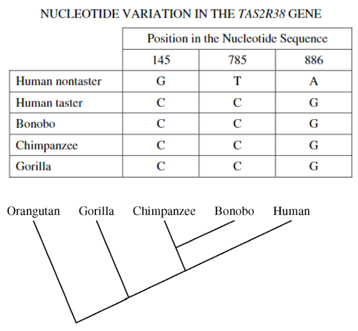
The TAS2R38 gene encodes a receptor protein that influences the ability to taste bitterness. The gene has two alleles: a dominant, wild-type allele that enables an individual (taster) to taste bitterness and a recessive, mutant allele that interferes with the ability of an individual (nontaster) to taste bitterness. Three single nucleotide mutations in the coding region of the TAS2R38 gene are associated with the nontaster allele. The nucleotides present at the three positions are shown in the table below. A cladogram representing the evolutionary relatedness of selected primates is shown below. The TAS2R38 receptor protein has been detected on the surface of cells from individuals who are homozygous for the nontaster allele of the TAS2R38 gene.
[See figure]
Q18. Which of the following is the most likely effect of the mutations associated with the nontaster allele on TAS2R38 gene expression?
a) The mutations change the primary structure of the encoded receptor protein. b) The mutations increase the stability of the TAS2R38 mRNA. c) The mutations prevent transcription of the TAS2R38 gene. d) The mutations prevent translation of the TAS2R38 mRNA.
a) The mutations change the primary structure of the encoded receptor protein.
Cortisol is a hormone produced in response to stress, including starvation, in humans.
Q19. Which of the following is most likely an immediate effect of a starvation-induced increase in cortisol secretion?
a) Increased activation of the immune system b) Increased urine production by the kidneys c) Increased bone and collagen formation d) Increased mobilization of fatty acids from fat cells
d) Increased mobilization of fatty acids from fat cells
Human epidermal growth factor (EGF) is a protein that stimulates cell division and growth by binding to its receptor, human epidermal growth factor receptor 2 (HER2). Certain types of cancer are caused when the overexpression of EGF activates a cell-signaling pathway, leading to uncontrolled cell division.
Q20. Which of the following best explains how a drug can be used to inhibit tumor growth caused by EGF overexpression?
a) The drug binds to HER2, preventing the activation of the signaling pathway. b) The drug acts as a positive regulator of transcription of the gene encoding EGF. c) The drug activates the receptor in the absence of EGF, leading to the activation of the signaling pathway. d) The drug enables the cells to progress through the S/G₂ checkpoint.
a) The drug binds to HER2, preventing the activation of the signaling pathway.
A cell signaling pathway begins when a hormone binds to its receptor. After binding, relay molecules within the cell become activated and trigger cellular responses. When drug K is added to cells, the cellular response is heightened and prolonged.
Q21. Which of the following mechanisms best explains how drug K increases the cellular response?
a) Drug K inhibits the activation of the receptor protein. b) Drug K promotes the activation of relay molecules. c) Drug K inhibits the deactivation of the receptor protein. d) Drug K promotes the circulation of relay molecules.
b) Drug K promotes the activation of relay molecules.
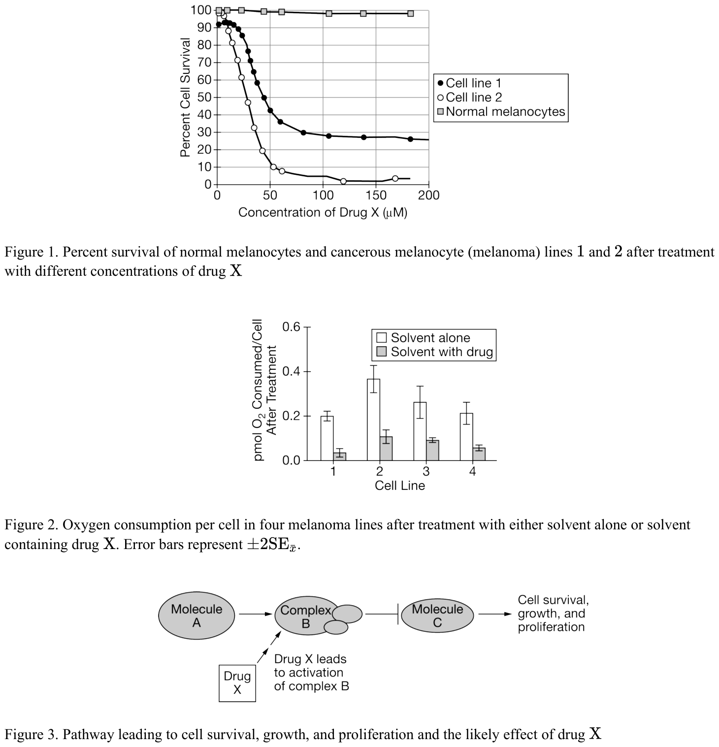
Melanocytes are skin cells that can become cancerous and develop into a cancer known as melanoma. Some cancerous melanocytes have developed resistance to the drugs currently used to treat melanoma. As a result, researchers are investigating the effects of a new compound (drug X) on four different melanoma cell lines. Researchers analyzed cell survival in two cell lines (Figure 1) and oxygen consumption in the presence of drug X in all four cell lines (Figure 2). Figure 3 shows the proposed mechanism by which drug X affects cells.
[Figure 1. Percent survival of normal melanocytes and cancerous melanocyte (melanoma) lines 1 and 2 after treatment with different concentrations of drug X]
[Figure 2. Oxygen consumption per cell in four melanoma lines after treatment with either solvent alone or solvent containing drug X. Error bars represent ±2SEx̄.]
[Figure 3. Pathway leading to cell survival, growth, and proliferation and the likely effect of drug X]
[See figure]
Q22. Which of the following best describes the data in Figure 1?
a) As the concentration of drug X increases, there is an increase in melanoma cell survival. b) At a concentration above 10 μM, drug X reduces melanoma cell survival. c) At a concentration below 25 μM, drug X increases survival in all melanoma cell lines. d) At a concentration of 25 μM, drug X has a greater effect on melanoma line 1 than on melanoma line 2.
b) At a concentration above 10 μM, drug X reduces melanoma cell survival.

Melanocytes are skin cells that can become cancerous and develop into a cancer known as melanoma. Some cancerous melanocytes have developed resistance to the drugs currently used to treat melanoma. As a result, researchers are investigating the effects of a new compound (drug X) on four different melanoma cell lines. Researchers analyzed cell survival in two cell lines (Figure 1) and oxygen consumption in the presence of drug X in all four cell lines (Figure 2). Figure 3 shows the proposed mechanism by which drug X affects cells.
[Figure 1. Percent survival of normal melanocytes and cancerous melanocyte (melanoma) lines 1 and 2 after treatment with different concentrations of drug X]
[Figure 2. Oxygen consumption per cell in four melanoma lines after treatment with either solvent alone or solvent containing drug X. Error bars represent ±2SEx̄.]
[Figure 3. Pathway leading to cell survival, growth, and proliferation and the likely effect of drug X]
A researcher has identified a compound that reverses the effect of drug X.
[See figure]
Q23. Based on Figure 3, which of the following best explains how the compound acts in the pathway to reverse the effects of drug X?
a) [image: see option (A) in figure] b) [image: see option (B) in figure] c) [image: see option (C) in figure] d) [image: see option (D) in figure]
c) [image: see option (C) in figure]

Melanocytes are skin cells that can become cancerous and develop into a cancer known as melanoma. Some cancerous melanocytes have developed resistance to the drugs currently used to treat melanoma. As a result, researchers are investigating the effects of a new compound (drug X) on four different melanoma cell lines. Researchers analyzed cell survival in two cell lines (Figure 1) and oxygen consumption in the presence of drug X in all four cell lines (Figure 2). Figure 3 shows the proposed mechanism by which drug X affects cells.
[Figure 1. Percent survival of normal melanocytes and cancerous melanocyte (melanoma) lines 1 and 2 after treatment with different concentrations of drug X]
[Figure 2. Oxygen consumption per cell in four melanoma lines after treatment with either solvent alone or solvent containing drug X. Error bars represent ±2SEx̄.]
[Figure 3. Pathway leading to cell survival, growth, and proliferation and the likely effect of drug X]
[See figure]
Q24. Based on Figure 2, which of the following best supports the claim that drug X inhibits oxygen consumption?
a) In the absence of drug X, melanoma lines 1 and 4 consume similar amounts of oxygen. b) In the presence of drug X, melanoma line 2 consumes statistically more oxygen per cell than does melanoma line 3. c) Melanoma line 3 consumes statistically less oxygen per cell in the presence of drug X than it does in the presence of the solvent alone. d) Melanoma line 2 in the presence of drug X consumes statistically less oxygen than does melanoma line 4 in the absence of drug X.
c) Melanoma line 3 consumes statistically less oxygen per cell in the presence of drug X than it does in the presence of the solvent alone.

[Figure 1. A model of epinephrine signaling]
Two types of cells, alpha and beta cells, produce signaling molecules that affect blood sugar levels in opposite ways (Figure 1). Epinephrine is a chemical, often released during periods of exercise, that ultimately causes an increase in blood sugar levels in the body.
[See figure]
Q25. Based on Figure 1, which of the following best explains how exercise causes blood glucose levels to rise?
a) Epinephrine inhibits alpha cells, causing the release of glucagon, and activates beta cells, blocking the release of insulin. b) Epinephrine activates alpha cells, blocking the release of glucagon, and inhibits beta cells, causing the release of insulin. c) Epinephrine activates alpha cells, causing the release of glucagon, and inhibits beta cells, blocking the release of insulin. d) Epinephrine inhibits alpha cells, blocking the release of glucagon, and activates beta cells, causing the release of insulin.
c) Epinephrine activates alpha cells, causing the release of glucagon, and inhibits beta cells, blocking the release of insulin.

The diagram above shows a developing worm embryo at the four-cell stage. Experiments have shown that when cell 3 divides, the anterior daughter cell gives rise to muscle and gonads and the posterior daughter cell gives rise to the intestine. However, if the cells of the embryo are separated from one another early during the four-cell stage, no intestine will form. Other experiments have shown that if cell 3 and cell 4 are recombined after the initial separation, the posterior daughter cell of cell 3 will once again give rise to normal intestine.
[See figure]
Q26. Which of the following is the most plausible explanation for these findings?
a) A cell surface protein on cell 4 signals cell 3 to induce formation of the worm’s intestine. b) The plasma membrane of cell 4 interacts with the plasma membrane of the posterior portion of cell 3, causing invaginations that become microvilli. c) Cell 3 passes an electrical signal to cell 4, which induces differentiation in cell 4. d) Cell 4 transfers genetic material to cell 3, which directs the development of intestinal cells.
a) A cell surface protein on cell 4 signals cell 3 to induce formation of the worm’s intestine.
A particular cell signaling pathway requires a hormone that is released into the bloodstream and travels to target cells. Scientists learned that not all cells respond to the hormone.
Q27. Which of the following best explains why some cells do not respond to the hormone?
a) Some cells lack hormone receptors. b) Some cells lack regulators. c) Some cells lack cholesterol. d) Some cells lack embedded proteins.
a) Some cells lack hormone receptors.
During a fight-or-flight response, epinephrine is released into the body’s circulatory system and transported throughout the body. Some cells exhibit a response to the epinephrine while other cells do not.
Q28. Which of the following justifies the claim that differences in components of cell signaling pathways explain the different responses to epinephrine?
a) Cell signaling depends on the ability to detect a signal molecule. Not all cells have receptors for epinephrine. Only cells with such receptors are capable of responding. b) Cell signaling depends on the transduction of a received signal by the nervous system. Not all cells are close enough to a synapse to receive the signal and respond. c) Cell signaling depends on the signal being able to diffuse through the cell membrane. Epinephrine is incapable of diffusing through some plasma membranes because of the membrane’s phospholipid composition. d) Cell signaling requires reception, transduction, and response. All cells can receive epinephrine, all cells respond with a pathway, but only select cells have the proper coding in their DNA to respond.
a) Cell signaling depends on the ability to detect a signal molecule. Not all cells have receptors for epinephrine. Only cells with such receptors are capable of responding.
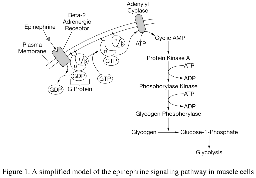
The epinephrine signaling pathway plays a role in regulating glucose homeostasis in muscle cells. The signaling pathway is activated by the binding of epinephrine to the beta-2 adrenergic receptor. A simplified model of the epinephrine signaling pathway is represented in Figure 1.
[Figure 1. A simplified model of the epinephrine signaling pathway in muscle cells]
[See figure]
Q29. Based on Figure 1, which of the following statements best describes the epinephrine signaling pathway?
a) It involves the opening and closing of ion channels. b) In involves enzymes activating other enzymes. c) It involves changes in the expression of target genes. d) It involves protons moving down a concentration gradient.
b) In involves enzymes activating other enzymes.

The epinephrine signaling pathway plays a role in regulating glucose homeostasis in muscle cells. The signaling pathway is activated by the binding of epinephrine to the beta-2 adrenergic receptor. A simplified model of the epinephrine signaling pathway is represented in Figure 1.
[Figure 1. A simplified model of the epinephrine signaling pathway in muscle cells]
[See figure]
Q30. Which of the following statements best describes the role of adenylyl cyclase in the epinephrine signaling pathway?
a) It converts a polymer to its monomer subunits. b) It moves substances across the plasma membrane. c) It accelerates the production of a second messenger. d) It transfers phosphate groups from ATP to protein substrates.
c) It accelerates the production of a second messenger.

The epinephrine signaling pathway plays a role in regulating glucose homeostasis in muscle cells. The signaling pathway is activated by the binding of epinephrine to the beta-2 adrenergic receptor. A simplified model of the epinephrine signaling pathway is represented in Figure 1.
[Figure 1. A simplified model of the epinephrine signaling pathway in muscle cells]
[See figure]
Q31. Which of the following outcomes will most likely result from the irreversible binding of GDP to the G protein?
a) The intracellular concentration of glycogen will increase. b) The intracellular concentration of activated protein kinase A will increase. c) The intracellular concentration of cyclic AMP will increase. d) The intracellular concentration of glucose-1-phosphate will increase.
a) The intracellular concentration of glycogen will increase.

The epinephrine signaling pathway plays a role in regulating glucose homeostasis in muscle cells. The signaling pathway is activated by the binding of epinephrine to the beta-2 adrenergic receptor. A simplified model of the epinephrine signaling pathway is represented in Figure 1.
[Figure 1. A simplified model of the epinephrine signaling pathway in muscle cells]
A researcher claims that the epinephrine signaling pathway controls a catabolic process in muscle cells.
[See figure]
Q32. Which of the following statements best helps justify the researcher’s claim?
a) Epinephrine is a signaling molecule that binds to a transmembrane protein. b) The G protein in the epinephrine signaling pathway consists of three different subunits. c) Phosphorylase kinase catalyzes the hydrolysis of ATP. d) Glycogen phosphorylase catalyzes the conversion of glycogen to glucose-1-phosphate.
d) Glycogen phosphorylase catalyzes the conversion of glycogen to glucose-1-phosphate.
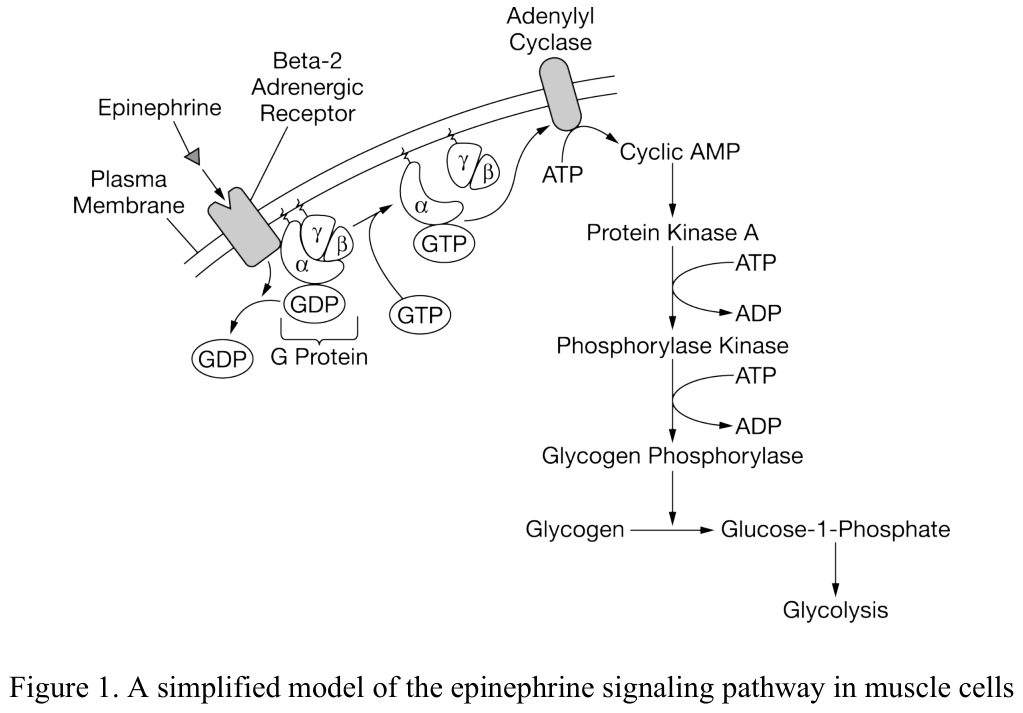
The epinephrine signaling pathway plays a role in regulating glucose homeostasis in muscle cells. The signaling pathway is activated by the binding of epinephrine to the beta-2 adrenergic receptor. A simplified model of the epinephrine signaling pathway is represented in Figure 1.
[Figure 1. A simplified model of the epinephrine signaling pathway in muscle cells]
Cyclic AMP phosphodiesterase is an enzyme that catalyzes the conversion of cyclic AMP to a different molecule.
[See figure]
Q33. Which of the following best predicts the effect of inhibiting cyclic AMP phosphodiesterase in a muscle cell stimulated by epinephrine?
a) The concentration of cyclic AMP will decrease because adenylyl cyclase will no longer be activated. b) The G protein will diffuse out of the cell because it will no longer bind to the plasma membrane. c) Phosphorylase kinase will remain active because protein kinase A will no longer be deactivated. d) Glycolysis will stop because epinephrine signaling will no longer stimulate glycogen breakdown.
c) Phosphorylase kinase will remain active because protein kinase A will no longer be deactivated.
Ethylene causes fruits to ripen. In a signaling pathway, receptors activate transcription factors, which ultimately leads to ripening.
Q34. Which of the following best supports the claim that ethylene initiates the signal transduction pathway that leads to ripening of fruit?
a) Ethylene is a simple gaseous molecule, which makes it easily detected by receptors. b) Fruit will ripen in closed containers without exposure to air. c) Ethylene synthesis is under both positive and negative feedback regulation. d) Loss-of-function mutations in ethylene receptors result in changes to the ripening process.
d) Loss-of-function mutations in ethylene receptors result in changes to the ripening process.

Fibroblast growth factor receptors (FGFRs) are transmembrane proteins that regulate cellular processes such as cell proliferation and differentiation. The extracellular domains of FGFR proteins bind specifically to signaling molecules called fibroblast growth factors. The intracellular domains of FGFR proteins function as protein kinases, enzymes that transfer phosphate groups from ATP to protein substrates. FGFR activation occurs when binding by fibroblast growth factors causes FGFR proteins in the plasma membrane to become closely associated with each other. The association of two FGFR proteins stimulates protein kinase activity, which triggers the activation of intracellular signaling pathways. A simplified model of FGFR activation is represented in Figure 1.
[Figure 1.
[See figure]
Q35. A simplified model of FGFR activation]
Which of the following changes in the FGFR signaling pathway is most likely to result in uncontrolled cell proliferation?
a) The irreversible association of FGFR proteins b) The loss of the FGFR protein kinase function c) A decrease in the intracellular concentration of ATP d) A decrease in the extracellular concentrations of fibroblast growth factors
a) The irreversible association of FGFR proteins
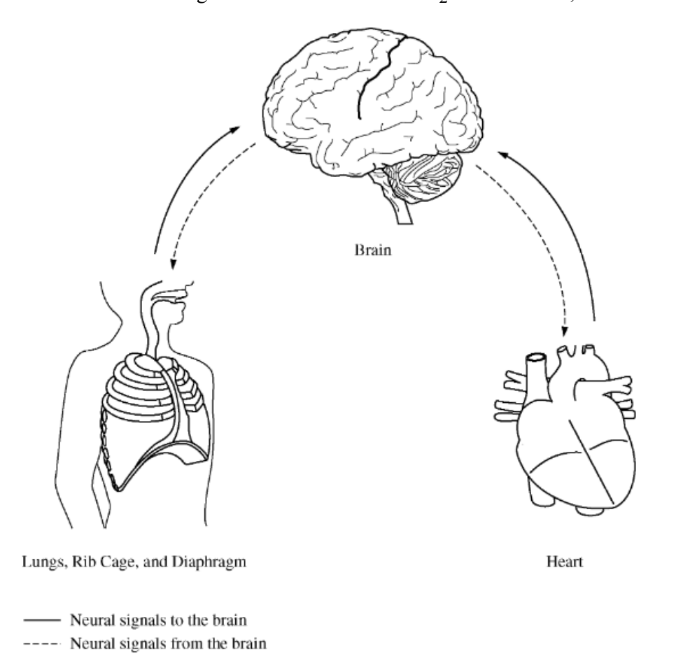
The brain coordinates the circulatory and respiratory systems of the human body. The control of breathing, for example, involves neural pathways among the structures represented in the figure above. One important stimulus in the control of breathing is an increase in blood CO₂ concentration, which is detected as a decrease in blood pH.
[See figure]
Q36. Which of the following best explains the physiological response to an overall increase in cellular respiration in the body?
a) In response to depleted blood CO₂ levels, the pH sensors send signals directly to the rib muscles, resulting in an increase in the rate of CO₂ uptake by the lungs and a decrease in CO₂ utilization by the brain. b) In response to low blood pH, the pH sensors send a signal to the brain, which then sends a signal to the diaphragm, resulting in an increased rate of breathing to help eliminate excess blood CO₂. c) In response to high blood pH, the pH sensors send a signal directly to the lungs, resulting in a slower rate of breathing, and the lungs send a signal back to the heart once CO₂ availability has been restored. d) In response to an increased rate of breathing, the rib muscles send a signal to the brain, which then sends a signal to the heart, resulting in a decrease in heart activity and slower flow of blood through the body.
b) In response to low blood pH, the pH sensors send a signal to the brain, which then sends a signal to the diaphragm, resulting in an increased rate of breathing to help eliminate excess blood CO₂.

Glucocorticoids are steroid hormones that control cellular responses through several different signaling pathways. One of the signaling pathways involves the glucocorticoid receptor, an intracellular protein that is activated by binding to a glucocorticoid molecule. A simplified model of the glucocorticoid receptor signaling pathway is represented in Figure 1.
[Figure 1.
[See figure]
Q37. A simplified model of the glucocorticoid receptor signaling pathway]
Which of the following statements best predicts the effect of a mutation that results in a loss of the glucocorticoid receptor’s ligand binding function?
a) The transduction of the glucocorticoid signal across the plasma membrane will be blocked. b) The glucocorticoid receptor will remain associated with the accessory proteins. c) The rate of diffusion of glucocorticoid molecules into the cell will increase. d) The concentration of glucocorticoid receptors inside the nucleus will increase.
b) The glucocorticoid receptor will remain associated with the accessory proteins.
Over 100 different types of G-protein-linked receptors have been observed in eukaryotes, including scent receptors in animals, mating-signal receptors in yeasts, and light-sensing proteins in vertebrates. The proton pumps of bacteria have protein structures similar to G-protein-linked receptors.
Q38. Based on this information, which of the following scientific questions will best guide research on the evolution of G-protein-linked signal transduction?
a) Did the G-protein-linked signal transduction pathway in eukaryotes arise from a precursor in prokaryotes? b) Are G-protein-linked receptors acquired from the environment as they are needed? c) Do eukaryotes require symbiotic prokaryotes in order to use G-protein-linked receptors? d) Do single-celled organisms have more efficient cell communication than do multicellular organisms?
a) Did the G-protein-linked signal transduction pathway in eukaryotes arise from a precursor in prokaryotes?
Q39. Which of the following statements best describes how a growth factor stimulates cell division from outside a cell?
a) The growth factor binds to other cells in the same area and holds them together to form a large, multicellular structure. b) The growth factor binds to receptors on the cell surface, initiating a signal transduction pathway that activates specific target genes. c) The growth factor binds to sugar molecules in the extracellular fluid and provides them to the cell as a source of energy. d) The growth factor binds to phospholipids in the plasma membrane, creating a channel through which substances enter the cell.
b) The growth factor binds to receptors on the cell surface, initiating a signal transduction pathway that activates specific target genes.

The Hedgehog protein (Hh) plays a critical role during a certain period of embryo development, but it normally has no role in adults except for the maintenance of adult stem cells. However, the Hedgehog protein has been detected in 70 percent of pancreatic cancer cell samples. As illustrated in the figures below, the Hedgehog protein binds to an integral membrane protein receptor known as Patched (Ptc), thus initiating a pathway of gene expression. When Hedgehog is absent, Ptc inhibits another protein known as Smoothened (Smo), which, in turn, blocks the activation of a group of proteins collectively known as the Hedgehog signaling complex (HSC). The inactivation is the result of proteolytic cleavage of one component of the HSC complex, a transcription factor known as Cubitus interruptus (Ci). When Hedgehog is present, it binds to Ptc, which prevents the inhibition of Smo by Ptc. The result is that Ci remains intact and can enter the nucleus, where it binds to and activates certain genes. One approach to treating patients with pancreatic cancer and other cancers in which the Hedgehog protein is detected is to modify the Hedgehog signaling pathway.
[See figure]
Q40. Which of the following is the most useful approach?
a) Treating patients with a molecule that is structurally similar to Hedgehog and that will bind to and interact with Ptc in the same fashion as Hedgehog b) Injecting patients with embryonic cells so that Hedgehog will bind to those cells instead of the cancer cells c) Treating patients with a membrane-soluble compound that can bind to Smo and block its activity d) Injecting patients with a preparation of purified membrane-soluble Ci that will enter the nuclei of the cancer cells and induce gene transcription
c) Treating patients with a membrane-soluble compound that can bind to Smo and block its activity
A hydrophilic peptide hormone is produced in the anterior pituitary gland located at the base of the brain. The hormone targets specific cells in many parts of the body.
Q41. Which of the following best explains a possible mechanism that would enable the hormone to efficiently reach all of the target cells in the body?
a) The hormone interacts with the nerves at the base of the brain and directs signals to the target cells through the nervous system. b) The hormone diffuses into target cells adjacent to the anterior pituitary gland, where the hormone is degraded. c) The hormone is released into the bloodstream where it can be transported to all cells with the correct receptors. d) The hormone moves through cytoplasmic connections between cells until it has reached all cells with the correct intracellular binding sites.
c) The hormone is released into the bloodstream where it can be transported to all cells with the correct receptors.
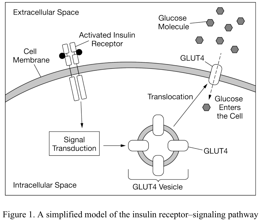
The insulin receptor is a transmembrane protein that plays a role in the regulation of glucose homeostasis. The receptor’s extracellular domain binds specifically to the peptide hormone insulin. The receptor’s intracellular domain interacts with cellular factors. The binding of insulin to the receptor stimulates a signal transduction pathway that results in the subcellular translocation of GLUT4, a glucose transport protein that is stored in vesicles inside the cell. A simplified model of the insulin receptor–signaling pathway is shown in Figure 1.
[Figure 1.
[See figure]
Q42. A simplified model of the insulin receptor–signaling pathway]
Which of the following statements best predicts the effect of a loss of function of the insulin receptor’s intracellular domain?
a) The stimulation of the signal transduction pathway will increase. b) The storage of GLUT4 in vesicles inside the cell will increase. c) The number of GLUT4 molecules in the plasma membrane will increase. d) The concentration of glucose inside the cell will increase.
b) The storage of GLUT4 in vesicles inside the cell will increase.

[Figure 1. Pathway activated by insulin binding to the insulin receptor]
[Figure 2: Blood insulin levels in normal mice and Esp mutant mice after exposure to glucose]
Hormones are chemical signals that are released by cells in one part of the body that travel through the bloodstream to signal cells in another part of the body. Insulin is a hormone that is released by the pancreas that induces the uptake of glucose molecules from the bloodstream into cells. In this way, insulin lowers the overall blood glucose levels of the body. Osteoblasts and osteoclasts are two types of bone cells that play a role in regulating blood glucose levels (Figure 1).
Binding of insulin to the insulin receptor on osteoblasts activates a signaling pathway that results in osteoblasts releasing a molecule, OPG, that binds to neighboring osteoclasts. In response, the osteoclasts release protons (H⁺) and create an area of lower pH outside the cell. This low pH activates osteocalcin, a protein secreted in an inactive form by osteoblasts.
The Esp gene encodes a protein that alters the structure of the insulin receptor on osteoblasts and interferes with the binding of insulin to the receptor. A researcher created a group of osteoblasts with an Esp mutation that prevented the production of a functional Esp product (mutant). The researcher then exposed the mutant strain and a normal strain that expresses Esp to glucose and compared the levels of insulin in the blood near the osteoblasts (Figure 2).
[See figure]
Q43. Based on the information provided, which of the following best justifies the claim that osteocalcin is a hormone?
a) The phosphorylation of the insulin receptor causes a response in osteoblast bone cells. b) The osteoblasts in the bone secrete osteocalcin, which causes cells in the pancreas to change their activity. c) The change in expression of Esp changes the insulin receptor activity of the osteoblast. d) The activation of the osteocalcin by a bone cell is pH dependent.
b) The osteoblasts in the bone secrete osteocalcin, which causes cells in the pancreas to change their activity.

[Figure 1. Pathway activated by insulin binding to the insulin receptor]
[Figure 2: Blood insulin levels in normal mice and Esp mutant mice after exposure to glucose]
Hormones are chemical signals that are released by cells in one part of the body that travel through the bloodstream to signal cells in another part of the body. Insulin is a hormone that is released by the pancreas that induces the uptake of glucose molecules from the bloodstream into cells. In this way, insulin lowers the overall blood glucose levels of the body. Osteoblasts and osteoclasts are two types of bone cells that play a role in regulating blood glucose levels (Figure 1).
Binding of insulin to the insulin receptor on osteoblasts activates a signaling pathway that results in osteoblasts releasing a molecule, OPG, that binds to neighboring osteoclasts. In response, the osteoclasts release protons (H⁺) and create an area of lower pH outside the cell. This low pH activates osteocalcin, a protein secreted in an inactive form by osteoblasts.
The Esp gene encodes a protein that alters the structure of the insulin receptor on osteoblasts and interferes with the binding of insulin to the receptor. A researcher created a group of osteoblasts with an Esp mutation that prevented the production of a functional Esp product (mutant). The researcher then exposed the mutant strain and a normal strain that expresses Esp to glucose and compared the levels of insulin in the blood near the osteoblasts (Figure 2).
[See figure]
Q44. Which of the following best describes the effect of insulin binding to the receptor on the osteoblast cells?
a) Insulin binding ultimately increases pancreatic secretion of additional insulin. b) Insulin binding blocks the release of osteocalcin from the osteoblasts. c) Insulin binding inhibits the expression of Esp. d) Insulin binding increases the pH of the extracellular matrix.
a) Insulin binding ultimately increases pancreatic secretion of additional insulin.

[Figure 1. Pathway activated by insulin binding to the insulin receptor]
[Figure 2: Blood insulin levels in normal mice and Esp mutant mice after exposure to glucose]
Hormones are chemical signals that are released by cells in one part of the body that travel through the bloodstream to signal cells in another part of the body. Insulin is a hormone that is released by the pancreas that induces the uptake of glucose molecules from the bloodstream into cells. In this way, insulin lowers the overall blood glucose levels of the body. Osteoblasts and osteoclasts are two types of bone cells that play a role in regulating blood glucose levels (Figure 1).
Binding of insulin to the insulin receptor on osteoblasts activates a signaling pathway that results in osteoblasts releasing a molecule, OPG, that binds to neighboring osteoclasts. In response, the osteoclasts release protons (H⁺) and create an area of lower pH outside the cell. This low pH activates osteocalcin, a protein secreted in an inactive form by osteoblasts.
The Esp gene encodes a protein that alters the structure of the insulin receptor on osteoblasts and interferes with the binding of insulin to the receptor. A researcher created a group of osteoblasts with an Esp mutation that prevented the production of a functional Esp product (mutant). The researcher then exposed the mutant strain and a normal strain that expresses Esp to glucose and compared the levels of insulin in the blood near the osteoblasts (Figure 2).
[See figure]
Q45. Which of the following claims is most consistent with the data shown in Figure 2?
a) Esp expression is necessary to prevent the overproduction of insulin. b) Esp protein does not regulate blood-sugar levels in normal mice. c) Normal mice require a higher blood concentration of insulin than mutant mice do. d) Mutant mice have a cyclical pattern of insulin secretion.
a) Esp expression is necessary to prevent the overproduction of insulin.

[Figure 1. Pathway activated by insulin binding to the insulin receptor]
[Figure 2: Blood insulin levels in normal mice and Esp mutant mice after exposure to glucose]
Hormones are chemical signals that are released by cells in one part of the body that travel through the bloodstream to signal cells in another part of the body. Insulin is a hormone that is released by the pancreas that induces the uptake of glucose molecules from the bloodstream into cells. In this way, insulin lowers the overall blood glucose levels of the body. Osteoblasts and osteoclasts are two types of bone cells that play a role in regulating blood glucose levels (Figure 1).
Binding of insulin to the insulin receptor on osteoblasts activates a signaling pathway that results in osteoblasts releasing a molecule, OPG, that binds to neighboring osteoclasts. In response, the osteoclasts release protons (H⁺) and create an area of lower pH outside the cell. This low pH activates osteocalcin, a protein secreted in an inactive form by osteoblasts.
The Esp gene encodes a protein that alters the structure of the insulin receptor on osteoblasts and interferes with the binding of insulin to the receptor. A researcher created a group of osteoblasts with an Esp mutation that prevented the production of a functional Esp product (mutant). The researcher then exposed the mutant strain and a normal strain that expresses Esp to glucose and compared the levels of insulin in the blood near the osteoblasts (Figure 2).
[See figure]
Q46. Which of the following was a positive control in the experiment?
a) Minutes after glucose injection b) Blood insulin c) Mutant strain d) Normal strain
d) Normal strain

[Figure 1. Pathway activated by insulin binding to the insulin receptor]
[Figure 2: Blood insulin levels in normal mice and Esp mutant mice after exposure to glucose]
Hormones are chemical signals that are released by cells in one part of the body that travel through the bloodstream to signal cells in another part of the body. Insulin is a hormone that is released by the pancreas that induces the uptake of glucose molecules from the bloodstream into cells. In this way, insulin lowers the overall blood glucose levels of the body. Osteoblasts and osteoclasts are two types of bone cells that play a role in regulating blood glucose levels (Figure 1).
Binding of insulin to the insulin receptor on osteoblasts activates a signaling pathway that results in osteoblasts releasing a molecule, OPG, that binds to neighboring osteoclasts. In response, the osteoclasts release protons (H⁺) and create an area of lower pH outside the cell. This low pH activates osteocalcin, a protein secreted in an inactive form by osteoblasts.
The Esp gene encodes a protein that alters the structure of the insulin receptor on osteoblasts and interferes with the binding of insulin to the receptor. A researcher created a group of osteoblasts with an Esp mutation that prevented the production of a functional Esp product (mutant). The researcher then exposed the mutant strain and a normal strain that expresses Esp to glucose and compared the levels of insulin in the blood near the osteoblasts (Figure 2).
A researcher observes that mice from the mutant strain experience low blood sugar.
[See figure]
Q47. Which of the following best describes the feedback mechanism in the pathway (Figure 1) causing the low blood sugar in the mutant strain?
a) The positive feedback of insulin production b) The negative feedback of inactive osteocalcin production c) The positive feedback of the Esp protein d) The negative feedback of insulin-secreting pancreatic cell proliferation
a) The positive feedback of insulin production
Signal transduction may result in changes in gene expression and cell function, which may alter phenotype in an embryo. An example is the expression of the SRY gene, which triggers the male sexual development pathway in mammals. This gene is found on the Y chromosome.
Q48. Which statement provides the evidence to justify the claim that signal transduction may result in an altered phenotype?
a) If the SRY gene is absent or nonfunctional, the embryo will exhibit male sexual development. b) If the SRY gene is absent or nonfunctional, the embryo will exhibit female sexual development. c) An embryo with a male sex chromosome will always exhibit male sexual development. d) An embryo with two male sex chromosomes will always exhibit male sexual development.
b) If the SRY gene is absent or nonfunctional, the embryo will exhibit female sexual development.
Two types of cholesterol transport proteins, low-density lipoproteins (LDL) and high-density lipoproteins (HDL), bind to cholesterol and carry it through the bloodstream. Familial hypercholesterolemia (FH) is characterized by high cholesterol levels in the blood, which can lead to cardiovascular disease. FH is associated with a loss-of-function mutation of a gene that encodes LDL receptors in liver cells. Individuals who are heterozygous produce lower-than-normal amounts of the LDL receptors, and individuals who are homozygous for the mutant allele have no LDL receptor function. Individuals with FH can be treated with drugs that result in increased production of LDL receptors in liver cells.
Q49. Which of the following best explains the observation that the drugs can effectively control blood cholesterol levels in individuals who are heterozygous but are not effective in individuals homozygous for the mutant allele?
a) The drugs repair the mutant allele by copying the wild-type allele. b) The drugs prevent cholesterol from entering the liver cells in individuals who are heterozygous but not in individuals who are homozygous for the mutant allele. c) Cholesterol molecules primarily bind to HDL receptors in individuals with FH. d) There must be at least one copy of the wild-type LDL receptor allele to produce functional LDL receptors.
d) There must be at least one copy of the wild-type LDL receptor allele to produce functional LDL receptors.
Q50. Which of the following steps in a signaling pathway typically occurs first once a chemical messenger reaches a target cell?
a) Specific genes are activated. b) A second messenger molecule is produced. c) A ligand binds to a receptor. d) Specific proteins are synthesized.
c) A ligand binds to a receptor.
The mechanism of action of many common medications involves interfering with the normal pathways that cells use to respond to hormone signals.
Q51. Which of the following best describes a drug interaction that directly interferes with a signal transduction pathway?
a) A medication causes the cell to absorb more of a particular mineral, eventually poisoning the cell. b) A medication enters the target cell and inhibits an enzyme that normally synthesizes a second messenger. c) A medication enters the target cell’s nucleus and acts as a mutagen. d) A medication interrupts the transcription of ribosomal RNA genes.
b) A medication enters the target cell and inhibits an enzyme that normally synthesizes a second messenger.
Q52. Which of the following best describes a molecule that relays messages from receptors to other molecules only inside the cell?
a) The molecule is a ligand. b) The molecule is a hormone. c) The molecule is a secondary messenger. d) The molecule is a transport protein.
c) The molecule is a secondary messenger.
Metformin is a drug used to treat type 2 diabetes by decreasing glucose production in the liver. AMP-activated protein kinase (AMPK) is a major cellular regulator of glucose metabolism. Metformin activates AMPK in liver cells but cannot cross the plasma membrane. By blocking AMPK with an inhibitor, researchers found that AMPK activation is required for metformin to produce an inhibitory effect on glucose production by liver cells.
Q53. Which of the following best explains the component that metformin represents in a signal transduction pathway that regulates glucose production in the liver?
a) It is a secondary messenger that amplifies a signal through a cascade reaction. b) It is a ligand that activates the signal transduction pathway of the activation of AMPK. c) It is an allosteric regulator that binds to a crucial section of the DNA that makes the enzymes needed for glucose uptake. d) It is a competitive inhibitor that binds to glucose and prevents it from entering the cell.
b) It is a ligand that activates the signal transduction pathway of the activation of AMPK.
Vertebrate immune responses involve communication over short and long distances.
Q54. Which of the following statements best helps explain how cell surface proteins, such as MHC proteins and T cell receptors, mediate cell communication over short distances?
a) The proteins receive electrical signals from nerve cells. b) The proteins leave the cell and travel in the bloodstream to other cells. c) The proteins interact directly with proteins on the surfaces of other cells. d) The proteins bind to molecules secreted by cells located in other parts of the body.
c) The proteins interact directly with proteins on the surfaces of other cells.

[Figure 1. A signaling cascade triggers the relaxation of smooth muscle cells.]
Blood vessels are surrounded by cells called smooth muscle cells. Nitric oxide triggers a signaling cascade in smooth muscle cells that causes the cells to relax (Figure 1).
[See figure]
Q55. Which of the following is represented by the gradual increase in thickness of the arrows from the top to the bottom of Figure 1?
a) The rate at which nitric oxide triggers signaling gradually increases over time. b) The number of signaling molecules that are produced or activated increases with each step in the pathway. c) The size of the proteins in the pathway increases as the signaling cascade moves through the cell. d) The signaling pathway uses an increase in negative feedback to reduce intracellular Ca²⁺ levels and cell sensitivity to Ca²⁺.
b) The number of signaling molecules that are produced or activated increases with each step in the pathway.

Figure 1 shows a model of a signal transduction cascade, initiated by the binding of a ligand to the transmembrane receptor protein A.
[Figure 1. Model of signal transduction cascade incorporating protein A]
A DNA mutation changes the shape of the extracellular domain of transmembrane receptor protein A produced by the cell.
[See figure]
Q56. Which of the following predictions is the most likely consequence of the mutation?
a) Production of activated molecule 1 will stop, but production of activated molecules 2 and 3 will continue. b) The molecule that normally binds to protein A will no longer attach, deactivating the cellular response. c) The molecule that normally binds to protein A will not enter the cell, thus no cellular response will occur. d) Since protein A is embedded in the membrane, the mutation will be silent and not affect the cellular response.
b) The molecule that normally binds to protein A will no longer attach, deactivating the cellular response.
Notch is a receptor protein displayed on the surface of certain cells in developing fruit fly embryos. Notch’s ligand is a membrane-bound protein called Delta that is displayed on the surface of adjacent cells. When Notch is activated by its ligand, the intracellular tail of the Notch protein becomes separated from the rest of the protein. This allows the intracellular tail to move to the cell’s nucleus and alter the expression of specific genes.
Q57. Which of the following statements best explains Delta’s role in regulating cell communication through the Notch signaling pathway?
a) Delta transmits a chemical signal to all the cells of a developing embryo. b) Delta allows the cells of a developing embryo to communicate without making direct contact. c) Delta restricts cell communication to short distances within a developing embryo. d) Delta determines which cells in a developing embryo express the gene that encodes the Notch protein.
c) Delta restricts cell communication to short distances within a developing embryo.
Q58. Which of the following best explains how small molecules move between adjacent cells in a plant shoot?
a) The molecules are actively transported by motor proteins along the cytoskeleton. b) The molecules pass freely through plasmodesmata, which are cytoplasmic strands connecting two cells. c) The molecules are swept along in the extracellular fluid by cilia projecting from cell membranes. d) The molecules bind reversibly to receptors on the cell membranes of xylem.
b) The molecules pass freely through plasmodesmata, which are cytoplasmic strands connecting two cells.
In flowering plants, plasmodesmata are narrow channels through cell walls that connect the cytoplasms of adjacent cells.
Q59. An explanation of how plant cells communicate across cell walls will most likely refer to the diffusion through plasmodesmata of which of the following?
a) Membrane-bound organelles b) Condensed, duplicated chromosomes c) Branched polysaccharides d) Small, water-soluble molecules
d) Small, water-soluble molecules
Adjacent plant cells have narrow channels called plasmodesmata that pass through the cell walls of the connected cells and allow a cytoplasmic connection between the cells.
Q60. Which of the following statements best explains a primary function of plasmodesmata?
a) They allow the movement of molecules from one cell to another, enabling communication between cells. b) They prevent the cell membrane from pulling away from the cell wall during periods of drought. c) They eliminate the need to produce signaling molecules and eliminate the need for cells to have receptors for signaling molecules. d) They increase the surface area available for attachment of ribosomes and thus increase protein synthesis.
a) They allow the movement of molecules from one cell to another, enabling communication between cells.
The binding of erythropoietin to the erythropoietin receptor (EPoR) stimulates intracellular signaling in certain cells, which causes them to differentiate to red blood cells. The EPoR is a transmembrane protein with two cytoplasmic domains, each of which is bound by a kinase protein, JAK2. The binding of erythropoietin to the EPoR causes the cytoplasmic domains of the EPoR to undergo conformational changes that activate the bound JAK2 kinases. The JAK2 kinases then phosphorylate several intracellular proteins, including STAT5, whose resulting activity causes the cells to differentiate to red blood cells.
Q61. Which of the following explains how a mutation that removes a portion of each cytoplasmic domain of EPoR most likely affects differentiation of the cells?
a) The cells will differentiate more rapidly than they typically do because the JAK2 kinases will always be active. b) The cells will mature to another cell type because the JAK2 kinases will phosphorylate inappropriate cellular proteins. c) STAT5 will not be activated because erythropoietin cannot bind to the EPoR. d) STAT5 will not be activated because it will not be phosphorylated by the JAK2 kinases.
d) STAT5 will not be activated because it will not be phosphorylated by the JAK2 kinases.
In a certain signal transduction pathway, the binding of an extracellular molecule to a cell-surface protein results in a rapid increase in the concentration of cyclic AMP inside the cell. The cyclic AMP binds to and activates cytosolic enzymes that then activate other enzymes in the cell.
Q62. Which of the following statements best describes the role of cyclic AMP in the signal transduction pathway?
a) It acts as a signaling molecule that passes the signal from the cell to other cells. b) It acts as a receptor that carries the signal from outside the cell to inside the cell. c) It acts as a second messenger that helps relay and amplify the signal within the cell. d) It acts as a channel protein that transmits the signal across the cell’s nuclear membrane.
c) It acts as a second messenger that helps relay and amplify the signal within the cell.

[See figure]
Q63. Which of the following best represents a signaling pathway that will lead to multiple responses from the same signaling molecule?
a) [image: see option (A) in figure] b) [image: see option (B) in figure] c) [image: see option (C) in figure] d) [image: see option (D) in figure]
c) [image: see option (C) in figure]
A student claims that the Y chromosome contains the sex-determining region gene, known as the SRY gene, which causes male fetuses to develop testes.
Q64. Which of the following provides correct information about cell signaling that supports the claim?
a) The SRY gene produces a protein that binds to specific regions of DNA in certain tissues, which affects the development of these tissues. b) The SRY gene produces a protein that deletes portions of the X chromosome in males so that male characteristics can develop. c) The SRY gene produces an RNA segment that is exported from specific cells and targets the developing gonads. d) The SRY gene is found only in tissues of the developing gonads.
a) The SRY gene produces a protein that binds to specific regions of DNA in certain tissues, which affects the development of these tissues.

The diagram above represents a model of signal transduction pathways (I and II) in a cell that is targeted by two different hormones (H₁ and H₂). The components of the signal transduction pathways are identified in the figure legend. Each cellular molecule in both pathways can exist in an inactive or active form. When the components in pathway I are sequentially activated, the TAP molecules promote cell division. When the components in pathway II are sequentially activated, downstream signaling by the G protein is inhibited.
[See figure]
Q65. Based on the model, which of the following mutations is most likely to result in a cell that will generate a cancerous tumor?
a) A mutation in the gene encoding PP that results in a nonfunctional protein b) A mutation in the gene encoding G-PIP that results in a nonfunctional protein c) A mutation in the gene encoding R₁ so that it is inactive even in the presence of H₁ d) A mutation in the gene encoding R₂ so that it is active even in the absence of H₂
b) A mutation in the gene encoding G-PIP that results in a nonfunctional protein
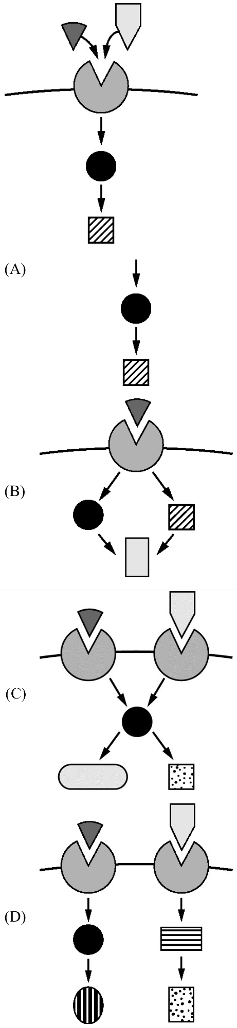
[See figure]
Q66. Which of the following best represents two different signaling pathways that share a second messenger?
a) [image: see option (A) in figure] b) [image: see option (B) in figure] c) [image: see option (C) in figure] d) [image: see option (D) in figure]
c) [image: see option (C) in figure]
Type 1 diabetes results from the destruction of insulin-producing cells in the pancreas. Individuals with type 1 diabetes produce insufficient amounts of insulin, a hormone that regulates the concentration of glucose in the blood.
Q67. Which of the following best explains how treatment with a drug that stimulates the production of insulin receptors on target cells will affect the insulin signaling pathway in an individual with type 1 diabetes?
a) The drug will have little or no effect on the signaling pathway because the receptors will not be activated in the absence of insulin. b) The drug will have little or no effect on the signaling pathway because insulin receptors will not be allowed to enter the cells. c) The drug will restore the function of the signaling pathway because insulin levels will return to normal. d) The drug will restore the function of the signaling pathway because nonpancreatic cells will begin to produce insulin receptors.
a) The drug will have little or no effect on the signaling pathway because the receptors will not be activated in the absence of insulin.

Pyruvate dehydrogenase is an enzyme that converts pyruvate to acetyl-CoA. Acetyl-CoA is further metabolized in the Krebs cycle. A researcher measured the accumulation of acetyl-CoA in a reaction containing pyruvate and pyruvate dehydrogenase under several different conditions (Figure 1).
[Figure 1.
[See figure]
Q68. Accumulation of acetyl-CoA under different conditions]
Which of the following observations provides the best evidence that acetyl-CoA negatively regulates pyruvate dehydrogenase activity?
a) The rate of the pyruvate dehydrogenase–catalyzed reaction is slower in the presence of a higher concentration of acetyl-CoA. b) The gene that encodes pyruvate dehydrogenase is transcribed when excess acetyl-CoA is detected. c) The accumulation of acetyl-CoA stops after 70 seconds, regardless of the reaction mixture. d) Acetyl-CoA is continuously broken down in the Krebs cycle.
a) The rate of the pyruvate dehydrogenase–catalyzed reaction is slower in the presence of a higher concentration of acetyl-CoA.

[Figure 1. Activation of a cyclin-CDK complex]
The regulation of a certain cyclin-CDK complex involves the activity of three enzymes (Figure 1), and the activation of this complex is required for cells to progress through the cell cycle.
[See figure]
Q69. Based on Figure 1, which of the following describes a cell in which cell cycle progression continues?
a) A cell in which enzyme 1 is functional, and enzyme 2 and enzyme 3 are nonfunctional b) A cell in which enzyme 1 is nonfunctional, and enzyme 2 and enzyme 3 are functional c) A cell in which enzyme 1 and enzyme 2 are functional, and enzyme 3 is nonfunctional d) A cell in which enzyme 1 and enzyme 3 are nonfunctional, and enzyme 2 is functional
a) A cell in which enzyme 1 is functional, and enzyme 2 and enzyme 3 are nonfunctional
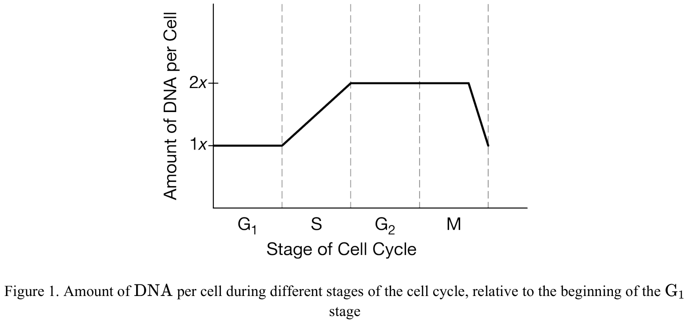
The relative amount of DNA in a cell at various stages of the cell cycle is shown in Figure 1.
[Figure 1.
[See figure]
Q70. Amount of DNA per cell during different stages of the cell cycle, relative to the beginning of the G₁ stage]
Which of the following best describes how the amount of DNA in the cell changes during M phase?
a) The amount of DNA doubles as the DNA is replicated. b) The amount of DNA slightly increases as a result of new organelle synthesis. c) The amount of DNA does not change while the cell grows. d) The amount of DNA is halved as the cell divides into two daughter cells.
d) The amount of DNA is halved as the cell divides into two daughter cells.

Phosphofructokinase (PFK) is a key enzyme in glycolysis. ATP is one of the two substrates for the reaction catalyzed by PFK. ATP is also an allosteric regulator of PFK. Figure 1 shows the enzyme-substrate interactions of PFK.
[Figure 1. The enzyme-substrate interactions of PFK]
A researcher found a mutation that resulted in the PFK enzyme being unable to bind ATP to the allosteric site.
[See figure]
Q71. Which of the following best predicts the effect of the mutation?
a) The activity of the enzyme will not be affected because the active site is not involved in substrate binding at the allosteric site. b) Negative feedback regulation does not occur, so the enzyme will be active when glycolysis is not needed. c) Positive feedback does not occur, and the activity of the enzyme will decrease when glycolysis is needed. d) The activity of the enzyme will fluctuate independent of the ATP concentration.
b) Negative feedback regulation does not occur, so the enzyme will be active when glycolysis is not needed.

Blood clots are formed by a positive feedback loop. Two pathways exist, the extrinsic and intrinsic pathways, which converge during clot formation. There are many clotting factors involved, most of which are proteins. Vitamin K is required for the formation of the active form of several of the clotting factors, including Factor X. A simplified model of the blood clotting process is shown in Figure 1.
[Figure 1. Simplified model of clotting cascade]
Warfarin is a drug used to treat certain blood clots. Warfarin blocks the formation of the active form of vitamin K-dependent clotting factors.
[See figure]
Q72. Based on the model, which of the following best predicts the effects of warfarin on a patient?
a) Fibrinogen will form fibrin, but the clot will not form because Factor XIII will not be synthesized. b) The intrinsic pathway will take over because the clotting factors are part of that pathway. c) Thrombin will be converted to prothrombin because Factor X will reverse the reaction. d) Factor X will not be activated, which will prevent thrombin from forming.
d) Factor X will not be activated, which will prevent thrombin from forming.
A person’s blood glucose level fluctuates during the day, as represented in Figure 1. Two hormones, insulin and glucagon, are directly involved in regulating the blood glucose level to maintain a healthy level. Insulin acts to lower the blood glucose level, and glucagon acts to increase the blood glucose level.
[Figure 1.
[See figure]
Q73. Blood glucose fluctuations of an individual]
Which of the following best predicts what will happen to the blood glucose level if the person has another meal at 5 P.M.?
a) Immediately after the meal, the blood glucose level will decrease because of the increase in glucagon levels. b) Immediately after the meal, the blood glucose level will increase, and then insulin will be secreted to counter the increase. c) Several hours after the meal, the blood glucose level will increase sharply because of an increase in the amount of glucagon secreted. d) The blood glucose level will not change after the 5 P.M. meal because the person has already consumed two meals and the blood glucose level has been adjusted to a steady-state level.
b) Immediately after the meal, the blood glucose level will increase, and then insulin will be secreted to counter the increase.

The model shown in the figure represents the role of two hormones, calcitonin and parathyroid hormone (PTH), in maintaining normal blood calcium levels in humans.
[See figure]
Q74. If a dietary change results in an increase in blood calcium concentration above normal levels, which of the following is the most likely effect on calcium homeostasis?
a) Calcitonin levels will decline, thus stimulating the release of PTH. b) Calcitonin levels will rise, thus promoting the deposit of calcium into bones. c) PTH levels will decline, thus stimulating the loss of calcium from bones. d) PTH levels will increase, thus preventing the release of calcitonin.
b) Calcitonin levels will rise, thus promoting the deposit of calcium into bones.
Cancer can result from a variety of different mutational events.
Q75. Which of the following is LEAST likely to result in the initiation of a cancerous tumor?
a) A receptor mutation results in activation of a cell-division pathway in the absence of the appropriate ligand. b) A mutation results in the loss of the ability to produce a tumor-suppressor protein. c) A defect in a cell-cycle checkpoint prevents a cell from entering the S phase. d) At the anaphase checkpoint, separation of chromatids occurs without all centromeres being attached to kinetochore microtubules from both poles.
c) A defect in a cell-cycle checkpoint prevents a cell from entering the S phase.
Cyclin-dependent kinase 7 (CDK7) binds to cyclin H and a second accessory protein, MAT1, to form a complex of three proteins. Researchers investigating the role of CDK7 claim that formation of this complex promotes cell cycle progression.
Q76. Which of the following supports the researchers’ claim?
a) Decreased expression of CDK7 initiates DNA replication. b) Decreased expression of CDK7 results in the formation of spindle fibers. c) Increased expression of CDK7 causes cells to enter G₀. d) Increased expression of CDK7 is associated with certain types of cancer.
d) Increased expression of CDK7 is associated with certain types of cancer.
Q77. Which of the following best describes a cell that is in the G₁ stage of the cell cycle?
a) The cell is growing in size and increasing its number of proteins and organelles. b) The chromosomes within the cell are checked to make certain that they have been replicated correctly. c) The chromosomes are replicating, and the amount of DNA in the cell is doubling. d) The cell is in a resting stage and no longer dividing.
a) The cell is growing in size and increasing its number of proteins and organelles.
A cell culture commonly used in research was selected to study the effect of a specific virus on the timing of cell cycle phases. Two separate cultures were started, one untreated and one inoculated with the virus. Both cultures were incubated under identical conditions. After a period of time, 200 cells from each culture were observed and classified as shown in Table 1.
[Table 1.
[See figure]
Q78. Number of normal and infected cells found in three phases of the cell cycle]
Which of the following most accurately describes an observation and an effect of the viral infection indicated by the data in Table 1?
a) Normal cells spend 98 percent of their time cycling in and out of interphase. The virus reduces this to 5 percent of the time. b) Twenty percent of the virus-infected cells are in interphase. These cells are no longer part of the cell cycle. c) Forty percent of the virus-infected cells are in interphase. These cells are preparing for replication of genetic material. d) Seventy-five percent of the virus-infected cells are found in mitosis. The virus stimulates frequent cell division.
d) Seventy-five percent of the virus-infected cells are found in mitosis. The virus stimulates frequent cell division.

Figure 1 represents the relative time and sequence of the phases of the cell cycle.
[Figure 1.
[See figure]
Q79. Representation of the cell cycle and identification of the G₁/S checkpoint]
Which statement best predicts why a cell’s progression through the cell cycle might be halted at the G₁/S checkpoint?
a) Spindle fibers have not correctly attached to chromosomes. b) There are not enough nucleotides available to construct new DNA. c) Damage occurred to DNA when it was being copied in G₁. d) Proteins necessary for M phase of the cell cycle have not been produced.
b) There are not enough nucleotides available to construct new DNA.

Researchers performed an experiment to determine the effect of certain genetic mutations on mitosis in tropical fruit fly embryos. They determined the percentage of cells in each of four phases of mitosis as shown in Figure 1.
[Figure 1.
[See figure]
Q80. Percent of cells in phases of mitosis]
Which of the following patterns is shown by the data?
a) Mutant 1 cells are more similar to mutant 3 cells than to wild-type cells. b) In wild-type cells, the percent of cells in anaphase is twice the amount of those in telophase c) In mutant 3 cells, more time is spent in prophase/prometaphase than in the later stages of mitosis. d) The percent of mutant 2 cells in anaphase is higher than that of mutant 1 cells.
c) In mutant 3 cells, more time is spent in prophase/prometaphase than in the later stages of mitosis.
Q81. What is the expected percent change in the DNA content of a typical eukaryotic cell as it progresses through the cell cycle from the start of the G₁ phase to the end of the G₂ phase?
a) −100% b) −50% c) +50% d) +100%
d) +100%

Figure 1 shows the number of chromosomes observed in an actively dividing human cell at each stage of cell division.
[Figure 1.
[See figure]
Q82. Number of chromosomes in a human cell at different stages of cell division]
Which of the following presents a correct interpretation of the changes in chromosome number depicted in Figure 1?
a) DNA replication occurs between metaphase and anaphase, doubling the number of chromosomes. Between telophase and cytokinesis, the cell divides in two, with each cell receiving half of the replicated chromosomes. b) New chromosomes formed during prophase are doubled during anaphase and are recombined before cytokinesis. c) Chromosomes enter metaphase containing two chromatids attached by a centromere. During anaphase, the chromatids are separated, each becoming a chromosome. Cytokinesis distributes the chromosomes into two separate cells. d) At anaphase a cell contains two identical copies of each chromosome, but following telophase, one of the copies is broken down into nucleotides.
c) Chromosomes enter metaphase containing two chromatids attached by a centromere. During anaphase, the chromatids are separated, each becoming a chromosome. Cytokinesis distributes the chromosomes into two separate cells.

Certain reef-building corals contain photosynthetic, symbiotic algae that have the ability to make dimethylsulphoniopropionate (DMSP), a chemical involved in the marine sulfur cycle. DMSP is released into the surrounding water, where it is converted to the gas dimethyl sulfide (DMS) by microorganisms and enters the atmosphere. Once in the atmosphere, it triggers the formation of sulfate aerosols, which induce cloud formation and block sunlight from heating up the water.
The symbiotic algae produce DMSP when they are stressed by a high water temperature. If water temperature is too high, corals will expel the symbiotic algae that produce DMSP. Researchers measured the amount of DMSP produced by juvenile and adult coral and their symbionts under normal and thermally stressed conditions. The data are shown in the graphs in Figure 1.
[Figure 1: DMSP concentration in juvenile and adult corals and their symbionts in normal and thermally-stressed conditions. Error bars represent ±2SEx̄.]
The researchers also measured the density of the symbiont as well as the photosynthetic yield in adult corals at the two temperatures. Photosynthetic yield is an index measure of energy output compared to sunlight energy input in which larger photosynthetic yield values represent photosynthetic organisms producing more energy.
[Figure 2: Variation in symbiont density and photosynthetic yield in adult corals grown in normal and thermally-stressed conditions. Error bars represent ±2SEx̄.]
[See figure]
Q83. Which of the following best describes the production of DMSP by coral and coral symbionts?
a) A negative feedback mechanism that increases the environmental change b) A negative feedback mechanism that reverses the environmental change c) A positive feedback mechanism that increases the environmental change d) A positive feedback mechanism that reverses the environmental change
b) A negative feedback mechanism that reverses the environmental change
Certain reef-building corals contain photosynthetic, symbiotic algae that have the ability to make dimethylsulphoniopropionate (DMSP), a chemical involved in the marine sulfur cycle. DMSP is released into the surrounding water, where it is converted to the gas dimethyl sulfide (DMS) by microorganisms and enters the atmosphere. Once in the atmosphere, it triggers the formation of sulfate aerosols, which induce cloud formation and block sunlight from heating up the water.
The symbiotic algae produce DMSP when they are stressed by a high water temperature. If water temperature is too high, corals will expel the symbiotic algae that produce DMSP. Researchers measured the amount of DMSP produced by juvenile and adult coral and their symbionts under normal and thermally stressed conditions. The data are shown in the graphs in Figure 1.
[Figure 1: DMSP concentration in juvenile and adult corals and their symbionts in normal and thermally-stressed conditions. Error bars represent ±2SEx̄.]
The researchers also measured the density of the symbiont as well as the photosynthetic yield in adult corals at the two temperatures. Photosynthetic yield is an index measure of energy output compared to sunlight energy input in which larger photosynthetic yield values represent photosynthetic organisms producing more energy.
[Figure 2: Variation in symbiont density and photosynthetic yield in adult corals grown in normal and thermally-stressed conditions. Error bars represent ±2SEx̄.]
[See figure]
Q84. Which of the following best describes the scientists’ findings concerning the density of symbionts presented in Figure 2?
a) The symbiont density at 32°C on day 5 was less than the density on day 0 of the experiment. b) The symbiont density at 27°C on day 0 was less than the density on day 5 of the experiment. c) The symbiont density at 32°C was different from the density at 27°C on days 5 and 10 of the experiment. d) The symbiont density at 27°C was higher than the density at 32°C for the entire length of the experiment.
c) The symbiont density at 32°C was different from the density at 27°C on days 5 and 10 of the experiment.

Researchers grew seedlings of corn, Zea mays, in loose and compact sand. The researchers measured the amount of time required for the cells in the growing root tips of the seedlings to double in number. The mean cell doubling times for the two groups of seedlings are shown in Figure 1.
[Figure 1.
[See figure]
Q85. Mean cell doubling times for the growing root tips of Zea mays seedlings planted in loose or compact sand]
Based on the sample means, which of the following conclusions about the cells in the growing root tips of Zea mays seedlings is best supported by the results of the experiment?
a) The cells of the root tips grow to larger sizes when the seedlings are planted in compact sand than when the seedlings are planted in loose sand. b) The average rate of mitotic cell division is greater for the root tips growing in loose sand than for the root tips growing in compact sand. c) The average cell cycle time is greater for the root tips growing in compact sand than for the root tips growing in loose sand. d) More cells are produced per unit of time in the root tips growing in compact sand than in the root tips growing in loose sand.
d) More cells are produced per unit of time in the root tips growing in compact sand than in the root tips growing in loose sand.
Damaged tissue releases chemicals that activate platelets and stimulate the formation of blood clots.
Q86. Which of the following statements about the activity of platelets best explains a positive feedback mechanism?
a) Activated platelets release chemicals that inhibit blood clot formation. b) Activated platelets release signaling molecules that inhibit cell division in damaged tissue. c) Activated platelets constrict the blood vessels, stopping blood flow. d) Activated platelets release chemicals that activate more platelets.
d) Activated platelets release chemicals that activate more platelets.

Researchers tracked the amount of DNA (measured in picograms) over time beginning with a single cell and continuing through several rounds of cell division. The researchers observed threadlike chromosomes prior to cell division. The threadlike chromosomes disappeared from view shortly after each division. The amount of DNA in picograms per cell over several rounds of cell division is shown in Figure 1.
[Figure 1.
[See figure]
Q87. Amount of DNA in picograms per cell over several rounds of cell division]
Which of the following statements is consistent with the data in Figure 1?
a) The cells have a haploid chromosome number of 3. b) The cells have a diploid chromosome number of 6. c) There is a change from 3 to 6 picograms of DNA because DNA is replicated before each round of cell division. d) There is a change from 6 to 3 picograms of DNA after each cell division because the chromosomes lengthen following cell division.
c) There is a change from 3 to 6 picograms of DNA because DNA is replicated before each round of cell division.
DNA damage can lead to the release of the protein cytochrome c from mitochondria. Cytochrome c then activates the enzyme caspase-9, resulting in the release of more cytochrome c, followed by apoptosis.
Q88. Which of the following best explains how the release of cytochrome c from mitochondria is regulated by feedback?
a) It is positive feedback because the release of cytochrome c from mitochondria contributes to the production of more mitochondria. b) It is negative feedback because the release of cytochrome c from mitochondria results in an increase in cell death. c) It is positive feedback because the release of cytochrome c from mitochondria contributes to the release of more cytochrome. d) It is negative feedback because the release of cytochrome c from mitochondria is caused by DNA damage.
c) It is positive feedback because the release of cytochrome c from mitochondria contributes to the release of more cytochrome.

[TABLE 1. DISTRIBUTION OF CONTROL AND EXPERIMENTAL CELLS IN DIFFERENT STAGES OF THE CELL CYCLE AFTER 24 HOURS]
Researchers claimed that cells treated with radiation are unable to progress through the cell cycle. They tested their claim by exposing a group of cells to radiation (experimental group). A group of unexposed cells served as the control group. After 24 hours, they determined the number of cells in each stage of the cell cycle and confirmed that all differences were statistically significant.
[See figure]
Q89. Based on Table 1, which of the following best supports the researchers’ claim?
a) In the experimental group, no cells were able to enter G₂. b) In the control group, the number of cells in G₂ was less than the number of cells in M. c) In the control group, the number of cells in each stage decreased as the cell cycle continued. d) In the experimental group, the number of cells in G₁ was the same as the number of cells in S.
a) In the experimental group, no cells were able to enter G₂.
The drug 5-fluorouracil inhibits thymine production in eukaryotic cells.
Q90. Which of the following cell cycle stages will be most directly affected by 5-fluorouracil?
a) The first growth phase (G₁) b) Synthesis of DNA phase (S) c) Preparation for mitosis (G₂) d) Cytokinesis
b) Synthesis of DNA phase (S)

Researchers have identified a molecule produced by Ecteinascidia turbinata, a marine invertebrate, from which the drug trabectedin is produced. Soft-tissue tumors treated with trabectedin rapidly decrease in size. In a preliminary study, healthy cells and tumor cells sampled from skin cancer patients treated with trabectedin were collected, and several characteristics of the cells were observed. The observed results of the study are shown in the table.
[See figure]
Q91. Which of the following best explains the most likely method by which this antitumor drug works?
a) Trabectedin increases the production of cyclin proteins that signal the cancer cells to enter prophase. b) Trabectedin interferes with the plasma membrane, causing it to break down and expose the DNA to damage. c) Trabectedin interferes with the duplication of DNA during interphase and thus prevents cancer cells from passing the G₂ checkpoint. d) Trabectedin interferes with the regulations of cyclin proteins, causing their levels to increase and creating errors in DNA.
c) Trabectedin interferes with the duplication of DNA during interphase and thus prevents cancer cells from passing the G₂ checkpoint.
The epidermal growth factor receptor EGFR is a cell surface receptor. When a growth factor binds to EGFR, the receptor is activated. The activated EGFR triggers a signal transduction pathway, which leads to increased frequency of cell division.
Q92. Which of the following best predicts the effect of a mutation that causes EGFR to be active in the absence of a growth factor?
a) Increased apoptosis will lead to abnormal growth of the tissue. b) Increased cell division will lead to the formation of a tumor. c) Cells will exit the cell cycle, entering a non-dividing G₀ phase. d) Fewer cells will be in any of the stages of mitosis.
b) Increased cell division will lead to the formation of a tumor.
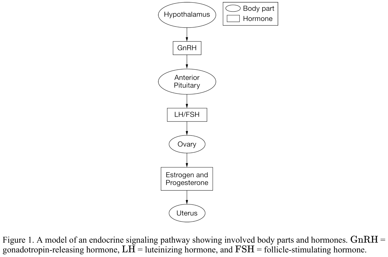
[Figure 1. A model of an endocrine signaling pathway showing involved body parts and hormones. GnRH = gonadotropin-releasing hormone, LH = luteinizing hormone, and FSH = follicle-stimulating hormone.]
Figure 1 shows a model of the endocrine signaling pathway that regulates ovulation.
[See figure]
Q93. Which of the following observations would provide evidence of a positive feedback mechanism in this system?
a) Estrogen from the ovaries inhibits the release of GnRH from the hypothalamus. b) Progesterone from the ovaries stimulates the thickening of the uterine lining. c) Progesterone from the ovaries inhibits the release of LH and FSH from the anterior pituitary. d) Estrogen from the ovaries stimulates the hypothalamus and anterior pituitary to secrete more GnRH, LH, and FSH.
d) Estrogen from the ovaries stimulates the hypothalamus and anterior pituitary to secrete more GnRH, LH, and FSH.
The endocrine system incorporates feedback mechanisms that maintain homeostasis.
Q94. Which of the following demonstrates negative feedback by the endocrine system?
a) During labor, the fetus exerts pressure on the uterine wall, inducing the production of oxytocin, which stimulates uterine wall contraction. The contractions cause the fetus to further push on the wall, increasing the production of oxytocin. b) After a meal, blood glucose levels become elevated, stimulating beta cells of the pancreas to release insulin into the blood. Excess glucose is then converted to glycogen in the liver, reducing blood glucose levels. c) At high elevation, atmospheric oxygen is more scarce. In response to signals that oxygen is low, the brain decreases an individual’s rate of respiration to compensate for the difference. d) A transcription factor binds to the regulatory region of a gene, blocking the binding of another transcription factor required for expression.
b) After a meal, blood glucose levels become elevated, stimulating beta cells of the pancreas to release insulin into the blood. Excess glucose is then converted to glycogen in the liver, reducing blood glucose levels.

Figure 1 is a proposed model of the feedback system controlling erythrocyte (red blood cell) production.
[Figure 1. Model of erythrocyte production control]
Air is less dense at very high elevations, so less oxygen is available than in the denser air at sea level.
[See figure]
Q95. Based on the model in Figure 1, if a person travels from sea level to a high elevation location, which of the following correctly predicts the response to the decreased blood oxygen level?
a) More erythropoietin will be secreted from the kidneys, decreasing production of erythrocytes. b) More erythropoietin will be secreted from the kidneys, increasing production of erythrocytes. c) Less erythropoietin will be secreted from the kidneys, decreasing production of erythrocytes. d) Less erythropoietin will be secreted from the kidneys, increasing production of erythrocytes.
b) More erythropoietin will be secreted from the kidneys, increasing production of erythrocytes.
Ethylene gas is an organic molecule that serves many cell signaling roles in flowering plants.
Q96. Which of the following best explains how a positive feedback mechanism involving ethylene works?
a) Cells of ripening fruit produce ethylene, which activates the ripening response in other fruit cells. b) Low water stress causes cells to produce ethylene, which binds to root cells and initiates cell division. c) Cells damaged by leaf-eating insects produce ethylene, which is released into the air, and repels insects. d) Fertilized ovules produce ethylene, which initiates apoptosis in flower petal cells.
a) Cells of ripening fruit produce ethylene, which activates the ripening response in other fruit cells.
Beta cells, a type of cell found in the pancreas, are responsible for producing, storing, and releasing the hormone (chemical signal) insulin. An increase in blood glucose levels stimulates beta cells to produce insulin, which facilitates the uptake of glucose by body cells and reduces the glucose levels in the blood. As the blood glucose levels decrease, beta cells are no longer stimulated and stop producing insulin.
Q97. Which of the following best explains the feedback mechanism that regulates blood glucose levels?
a) Regulation of blood glucose levels demonstrates positive feedback, because as the blood glucose levels increase, insulin acts to raise blood glucose levels. b) Regulation of blood glucose levels demonstrates negative feedback, because as the blood glucose levels increase, insulin acts to lower blood glucose levels. c) Regulation of blood glucose levels demonstrates positive feedback, because as the production of insulin by beta cells increases, blood glucose levels increase. d) Regulation of blood glucose levels demonstrates negative feedback, because as the production of insulin by beta cells decreases, blood glucose levels decrease.
b) Regulation of blood glucose levels demonstrates negative feedback, because as the blood glucose levels increase, insulin acts to lower blood glucose levels.
Glycogen synthetase kinase 3 beta is a protein kinase that has been implicated in many types of cancer. Depending on the cell type, the gene for glycogen synthetase kinase 3 beta (GSK3β) can act either as an oncogene or as a tumor suppressor.
Q98. Which of the following best predicts how GSK3β mutations can lead to the development of cancer?
a) Cells with inactive GSK3β fail to trigger apoptosis. b) Cells with inactive GSK3β fail to proceed past the G₂/M checkpoint. c) Cells with overactive GSK3β are more likely to repair DNA damage. d) Cells with overactive GSK3β have longer cell cycles.
a) Cells with inactive GSK3β fail to trigger apoptosis.

The relative amounts of DNA present in the nucleus of a cell at four different stages of the life cycle are shown in Figure 1.
[Figure 1.
[See figure]
Q99. Relative amounts of DNA present in the nucleus of a cell]
Based on Figure 1, which of the following statements correctly links a stage of the cell cycle with the event occurring at that stage?
a) Stage I represents the G₂ phase of the cell cycle. b) Synthesis of sufficient DNA for two daughter cells occurs in stage II. c) Stage III includes mitosis. d) The replication of genetic material occurs in stage IV.
b) Synthesis of sufficient DNA for two daughter cells occurs in stage II.
Q100. Which of the following best describes the role of mitosis in the cell cycle?
a) Distributing replicated chromosomes to daughter nuclei b) Dividing the cytoplasm to form four gametes c) Producing organelles and replicating chromosomes d) Exchanging genetic material between homologous chromosomes
a) Distributing replicated chromosomes to daughter nuclei