Week 9 Cell bio
 Call Kai
 Learn
 Practice Test
 Spaced Repetition
 Match
 Flashcards
 Knowt Play
1/78

There's no tags or description
Looks like no tags are added yet.
79 Terms
Kerr et al. 1972: authors, journal and institution
J.F.R. Kerr, A.H. Wyllie and A.R. Currie, Department of Pathology, University of Aberdeen. Published in the British Journal of Cancer (1972) 26, 239.
What does the Greek word “apoptosis” describe?
The “dropping off” or “falling off” of petals from flowers or leaves from trees. The term was suggested by Professor James Cormack of the Department of Greek, University of Aberdeen.
How do the authors want “apoptosis” pronounced?
“Apo’tosis”: stress on the second-to-last syllable, with the second “p” silent, so the second half sounds like “ptosis” (same Greek root, “to fall”; ptosis is the drooping of the upper eyelid).
Kerr et al.’s definition of apoptosis
A mechanism of controlled cell deletion that plays a complementary but opposite role to mitosis in regulating animal cell populations.
Why did the authors give this type of cell death a new name?
Because of its kinetic significance: it regulates cell numbers in many tissues, under both physiological and pathological conditions.
What did Kerr earlier call apoptosis?
Shrinkage necrosis (a name based on its morphology).
Necrobiosis
An older term sometimes used for “physiological cell death”; its morphological features had never been clearly defined.
Councilman (Councilman-like) bodies
An earlier name for apoptotic bodies in the liver. Because it was unclear what Councilman had described in yellow fever, Kerr et al. called them apoptotic bodies instead.
What do apoptosis’s morphological features suggest about it?
That it is an active, inherently programmed (controlled) process, which can be started or inhibited by many environmental stimuli, both physiological and pathological.
What molecular signals of apoptosis were not yet known in 1972?
Caspases. The paper’s evidence is purely morphological (light and electron microscopy).
Cell number homeostasis
The balance of cell division and cell death that lets an organ or organism stay the same size during maturity.
How can apoptosis be identified (journal club handout)?
By its classical morphology, first seen by electron microscopy but also visible by light microscopy; newer methods use fluorescence microscopy and electrophoresis of DNA fragments.
Coagulative necrosis (modern name)
Simply “necrosis.”
What causes coagulative necrosis, according to Kerr et al.?
It seems always to be caused by noxious (harmful) stimuli and probably results from an irreversible disturbance of the cell’s homeostatic mechanisms. Nothing suggests it helps control cell populations.
What does electron microscopy show early in coagulative necrosis?
Signs of degeneration like those seen in in vitro autolysis.
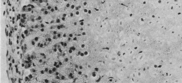
Fig. 3b (right side): what type of cell death is this, and how does it look different from apoptosis?
Coagulative necrosis. It is almost always confluent (a continuous area of dead cells), and the “ghosts” of the dead cells keep their size and shape. Apoptosis affects scattered single cells that condense and break into small, dense bodies.
Which light-microscope feature shows up in both necrosis and apoptosis?
Nuclear pyknosis (noted in the Fig. 3 caption).
Inflammation: coagulative necrosis vs. apoptosis
Coagulative necrosis triggers inflammation; apoptosis does not.
Can one agent cause both necrosis and apoptosis?
Yes. Hepatotoxins, electromagnetic radiation and DMBA can induce either coagulative necrosis or apoptosis (and are also carcinogenic and teratogenic); the authors call the significance of this obscure.
Apoptosis in tissue injury
It is increased in tissue injury, often alongside focal coagulative necrosis. Ischaemia probably explains the dense clusters of apoptotic bodies often seen near coagulative necrosis in tumors.
The two stages of apoptosis seen by electron microscopy
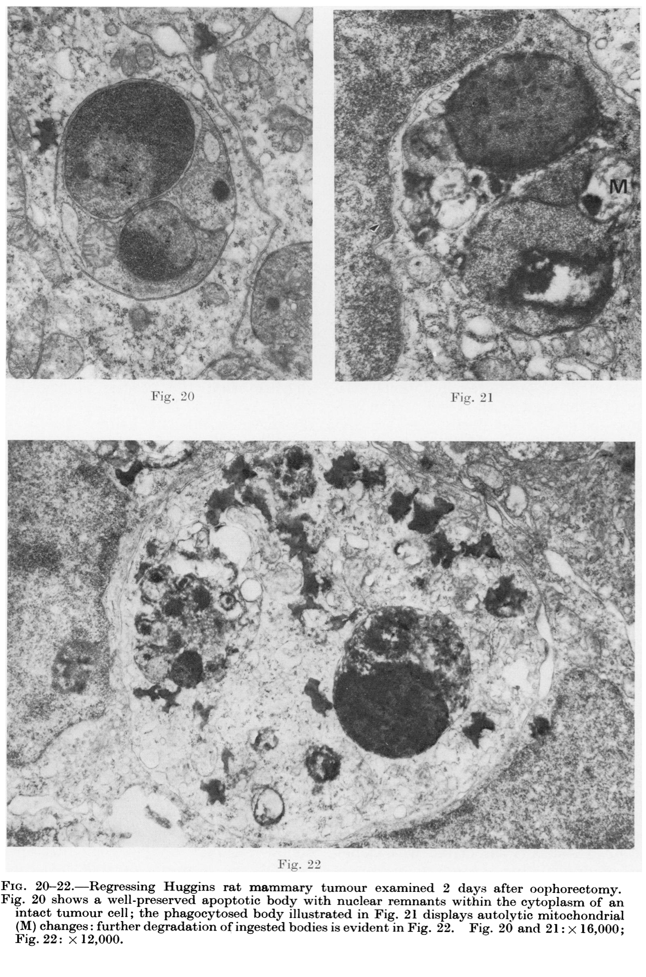
Stage 1: formation of apoptotic bodies (the cell condenses and breaks up into membrane-bound fragments). Stage 2: the bodies are phagocytosed and degraded by other cells (or shed from epithelial surfaces).
Steps of the apoptosis model (Fig. 5), in order
Separation → condensation → fragmentation → phagocytosis → autolysis → lysosomal digestion → residual body.

Which Fig. 5 steps are Stage 1 and which are Stage 2?
Stage 1 (apoptotic body formation): separation, condensation, fragmentation. Stage 2 (phagocytosis and degradation): phagocytosis, autolysis, lysosomal digestion, ending in a residual body.
What happens to the nucleus in apoptosis?
It condenses markedly, its chromatin gathers into dense masses beneath the nuclear envelope, and it breaks into fragments. Dense nuclear remnants end up in some apoptotic bodies but not others.

What happens to the cytoplasm in apoptosis?
It condenses, so the organelles become closely packed (sometimes condensed themselves) but stay intact, both chemically and structurally.
How do apoptotic bodies form?
Protuberances form on the cell surface and separate, producing many membrane-bounded, compact but otherwise well-preserved cell fragments of greatly varying size.
What is inside an apoptotic body?
Whatever was in the protuberance it came from: closely packed intact organelles, sometimes clear vacuoles and dense nuclear fragments. Some small bodies are almost all condensed chromatin; others contain only cytoplasm.
Earliest recognizable stage of apoptosis
The very first events have not been identified. Cells recognizable as apoptotic have already condensed, separated from their neighbors, and have chromatin clumped under the nuclear envelope.
What probably causes the condensation in apoptosis?
Extrusion of water, but the mechanism is unknown. Rough estimates suggest the fragments could form without making new plasma membrane.
What apoptotic bodies look like in the light microscope (H&E)
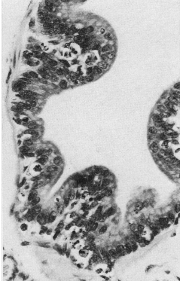
Small, roughly spherical or oval cytoplasmic fragments, some containing pyknotic nuclear remnants. Only the larger bodies in a cluster are visible; smaller ones, especially those without a nuclear fragment, take practice to spot.

Where do extracellular apoptotic bodies end up?
Often in clusters in the space between cells. Smaller ones drift away (in liver and adrenal cortex, into the spaces between parenchymal and sinusoid-lining cells). Bodies from glandular and mucosal epithelium and renal tubules are shed into the lumen; a few enter blood vessels.
Do free extracellular apoptotic bodies show degeneration?
No. They never show ultrastructural signs of degeneration and are probably still metabolically active, though irreversibly committed to destruction.
Why are most apoptotic bodies found inside other cells?
They are phagocytosed rapidly, possibly because the properties of their surface membranes change.
Which cells phagocytose apoptotic bodies?
After birth, under normal conditions: connective tissue cells (sinusoid-lining cells and “histiocytes”). In the embryo and in diseased adult tissue: epithelial cells too. In carcinomas: mostly the tumor cells themselves, more often than histiocytes.
Histiocyte
Another word for macrophage white blood cells.
Parenchymal cells
Cells of the parenchyma: the essential, distinctive tissue of an organ, as opposed to its supporting framework.
Autolysis of a phagocytosed apoptotic body
Inside the phagosome the body degenerates much like ischaemic necrosis or in vitro autolysis: the mitochondrial matrix turns electron-lucent with flocculent densities, organelle and body membranes break down, and ribosomes swell and become indistinct.
When does an apoptotic body first show that its metabolism has stopped?
Only during autolysis inside the phagosome, the first stage with changes that clearly show co-ordinated metabolic activity has stopped.
Do lysosomes cause the autolysis of ingested apoptotic bodies?
No. The evidence suggests lysosomes aren’t involved; the body probably autolyzes because it can’t maintain chemical homeostasis inside the phagosome.
Role of lysosomes after phagocytosis
Lysosomal enzymes (hydrolases) degrade the ingested bodies into electron-dense lysosomal residual bodies. The ingesting cell’s secondary lysosomes fuse with the phagosomes, and the cell also makes new hydrolases.
Figs. 8–9: evidence that secondary lysosomes fuse with phagosomes
Fig. 8: apoptotic bodies sit in phagosomes of a hepatocyte while normal numbers of secondary lysosomes (L) are still present. Fig. 9: residues of degraded bodies are present and the normal secondary lysosomes have disappeared.

Ergastoplasm
The rough ER.
Residual body
The electron-dense lysosomal remnant left after an apoptotic body is digested. Residual bodies soon disappear, possibly by “cell defaecation.”

How long does apoptosis take?
Less than 24 hours: the bodies may form and disappear within 24 hours.
Why is it hard to time apoptosis?
It goes on continuously, and even when stimulated it starts in individual cells of the same tissue at different times.
What does finding only a few apoptotic bodies in a tissue section mean?
That quite extensive cell “drop-out” is happening, just as mitotic figures signal cell proliferation. The bodies are cleared fast, and the light microscope sees only a small fraction of them.
Why is apoptosis well suited to tissue homeostasis?
It removes many cells with little disruption: fragments are quickly cleared by nearby intact cells, there is no inflammation, residual bodies soon disappear, and the cell’s components are reused (economical).
Why can ingested apoptotic bodies be mistaken for autophagic vacuoles?
They look alike in electron micrographs; telling them apart is hard unless the ingested body has a recognizable nuclear remnant.
Autophagy (autophagocytosis)
A cell digesting its own components inside autophagic vacuoles [definition not in manual]. In the paper, it helps shrink individual cells in atrophy and tumor regression (e.g., hepatocyte shrinkage after the liver’s portal blood supply is tied off).
Where does apoptosis occur? (paper summary)
Cell turnover in healthy adult tissues; focal cell elimination in embryonic development; spontaneously in untreated malignant tumors; some therapy-induced tumor regression; atrophy and involution of tissues; and after noxious agents in embryos and adults. It is not confined to vertebrates.
Examples of apoptosis in healthy tissue
Normal secretory-phase human endometrium (Fig. 1); adrenal cortex of a normal 4-day-old rat (Fig. 3a), where temporary ACTH deprivation after birth boosts it; tingible bodies in the germinal centers of lymphoid follicles.

Tingible bodies
Bodies in the germinal centers of lymphoid follicles that have the typical features of apoptotic bodies; many come from cells that recently synthesized DNA.
Is apoptosis just a side effect of rapid cell division?
No. The authors found no increase in apoptosis in regenerating rat liver after partial hepatectomy, and in the embryo apoptosis is focal and highly specific.
Roles of apoptosis in embryonic development
Forming the lumina of tubular structures, shaping limbs, forming the clefts between digits, and removing phylogenetic vestiges. It is precisely controlled, probably by diffusible substances, and depends on the cells’ position and developmental stage.
Ontogenesis
The normal development of an embryo.
Teratogenic
Describes an agent that causes embryo malformation.
Carcinogenic
Describes an agent that causes cancer.
Apoptosis in teratogenesis
Teratogens cause massive apoptosis where they act, and some malformations are fully explained by the focal apoptosis seen within hours; locally, apoptosis greatly exceeds mitosis. Figs. 15–18: rat embryo 24 h after the mother was given the teratogen 7-OHM-12-MBA.

Neoplasm
A new and abnormal growth of cells; cancer.
Apoptosis in malignant tumors
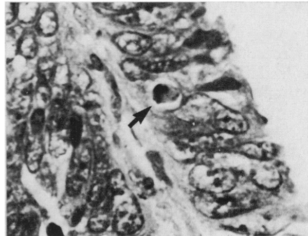
Found in every malignant tumor the authors examined, often in large numbers (e.g., Fig. 4, a human rectal adenocarcinoma). Most bodies are phagocytosed by intact tumor cells, and the balance between apoptosis and mitosis sets how fast the tumor grows.
Apoptosis in therapy-induced tumor regression
Increased in human squamous cell carcinoma after irradiation, and extensive in Huggins rat mammary tumors regressing after oophorectomy (Figs. 20–22). A tumor’s “apoptotic index” a few days into therapy might measure its response.
Atrophy example: rat liver after its portal blood supply is tied off
Within hours, hepatocytes shrink (with more autophagocytosis); then massive apoptotic deletion of liver cells follows, peaking several days later (Figs. 6–10).
Figs. 6–7: what do they show?
A cluster of extracellular apoptotic bodies in atrophying rat liver 3 days after its portal blood supply was blocked: organelles crowded but intact, dense nuclear remnants in some bodies but not others, and histiocytes (H) holding partly degraded bodies.

Hormone withdrawal and apoptosis
Lower ACTH in rats → a shower of apoptotic bodies in the adrenal cortex, prevented by giving ACTH. Orchidectomy → apoptosis in prostate gland epithelium (Fig. 2). Large glucocorticoid doses → deletion of lymphoid cells. Also seen in the involuting human corpus luteum.
ACTH
Adrenocorticotropic hormone, released by the pituitary gland; causes release and production of the stress hormone cortisol.
Example of a hormone causing both proliferation and apoptosis
Thyroid hormone in developing amphibians: it stimulates general body growth and at the same time widespread cell death in the gills and tail.
Steroid hormones and the Müllerian ducts (chick embryos)
Estrogens block the expected Müllerian duct regression in genetic males; androgens promote regression in genetic females.
The authors’ speculation about hyperplasia
Hyperplasia might sometimes come from decreased apoptosis rather than increased mitosis, e.g., focal breast hyperplasia from cells failing to undergo apoptosis when hormone levels fall. They know of no definitive studies supporting it.
Apoptosis in benign tumors
The authors’ impression is that apoptotic bodies are rare in benign neoplasms, but more work is needed.
Ischemic (ischaemic)
Having to do with restriction of blood flow and oxygen deprivation.
Intrinsic “clocks” in apoptosis
In some cases apoptosis is inherently programmed by clocks specific to the cell type: in avian embryo tissue explants, cells died “on schedule.” Diffusible substances from nearby tissue can still delay it, sometimes indefinitely.
The authors’ speculation on how apoptosis triggers work
Triggers and inhibitors might act by stimulating mRNA and protein synthesis. If apoptosis depends on expressing a normally repressed part of the genome, one stereotyped response to many different stimuli would make sense.
Open questions in the paper’s conclusion
Which cells get affected, the role of ageing, how initiators and inhibitors work, the earliest biochemical and morphological events, and the mechanism of cell condensation.
Stain used for the light micrographs (Figs. 1–4)
H. and E. (hematoxylin and eosin).
How the electron micrograph samples (Figs. 6–24) were prepared
Epon-embedded tissue sections stained with uranyl acetate and lead citrate.
Figs. 11–14 (healthy 5-day-old rat adrenal cortex)
Fig. 11: extracellular body with intact mitochondria (M) and lipid globules (L). Fig. 12: dense nuclear fragments (N). Fig. 13: well-preserved body inside a histiocyte (the body’s membrane inside the phagosome membrane). Fig. 14: partly degraded body inside a histiocyte.

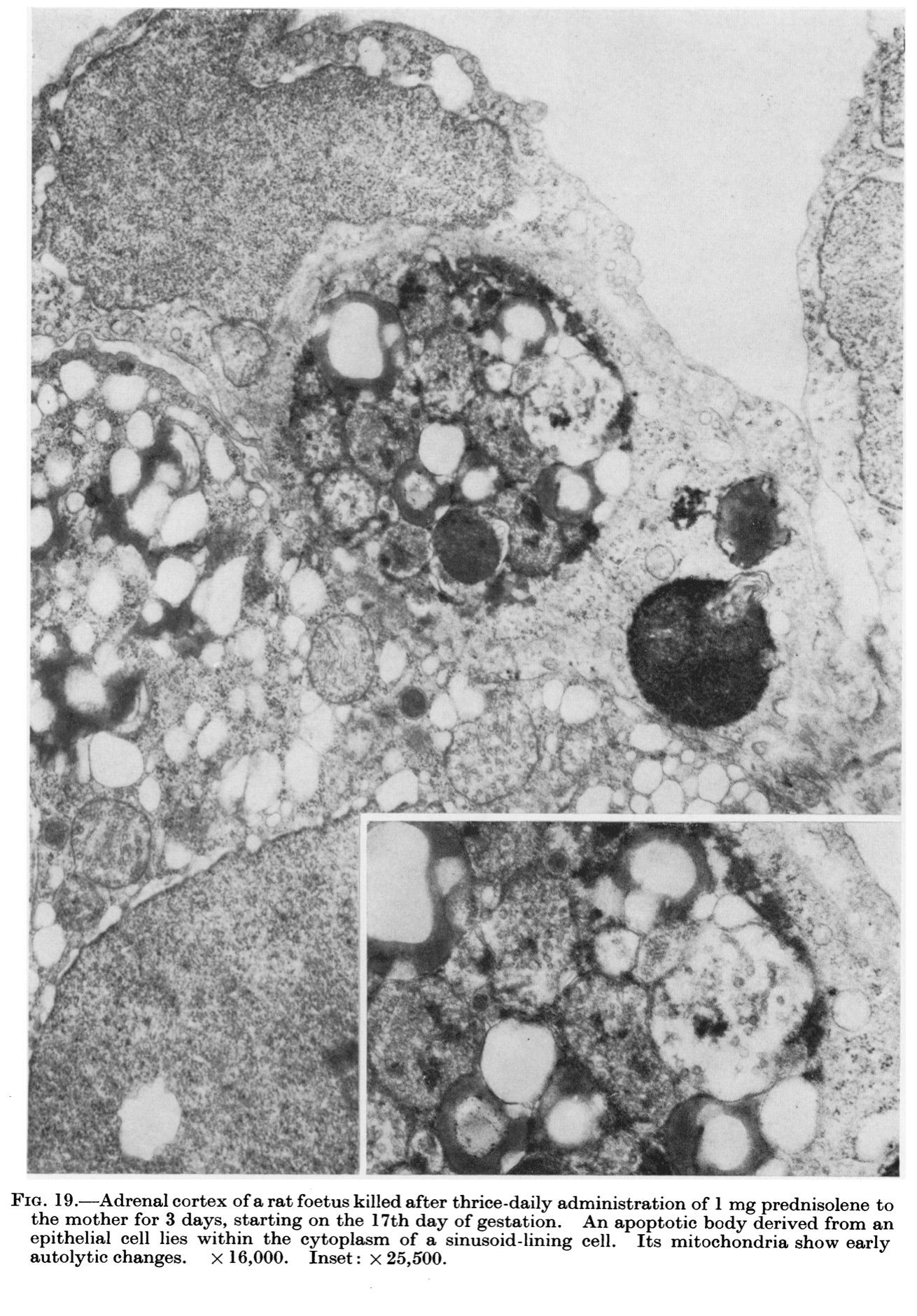
Fig. 19
Adrenal cortex of a rat fetus after the mother was given prednisolone: an apoptotic body from an epithelial cell inside a sinusoid-lining cell, with early autolytic changes in its mitochondria.
Limits of studying apoptosis by microscopy that the paper mentions
Apoptotic bodies are sparse in most tissues, making electron microscopy studies very difficult; the earliest events have not been seen; the timing is hard to pin down; and the light microscope shows only a small fraction of the bodies present.
Size of a mitochondrion (slide 8)
About 0.75–3 µm.
