Introduction to Skin Structure and Function
 Call Kai
 Learn
 Practice Test
 Spaced Repetition
 Match
 Flashcards
 Knowt Play
1/45

There's no tags or description
Looks like no tags are added yet.
46 Terms
What microscopic changes occur as haired skin transitions into a paw pad?
A: The paw-pad side becomes nonhaired, has a much thicker and more ridged epidermis, and contains abundant surface keratin compared with adjacent haired skin. 🔎 What this means: Hair follicles disappear as the tissue enters the pad, while the protective keratinized epidermis becomes much more substantial because the pad experiences high friction and wear.
Where else are melanocytes found, and what melanocyte-to-keratinocyte ratio was given for dogs?
A: Melanocytes are also found in the outer root sheath and hair bulbs, and dogs generally have about one melanocyte for every 10 to 16 keratinocytes. 🔎 What this means: A single melanocyte interacts with many neighboring keratinocytes through its dendritic processes. The lecture also noted that melanogenesis is influenced by genetic, endocrine, and environmental factors.
How do basal keratinocytes ultimately become the corneocytes of the stratum corneum?
A: Mitotically active basal keratinocytes divide, leave the basal layer, move toward the surface while differentiating, and ultimately become dead corneocytes in the stratum corneum. 🔎 What this means: The superficial barrier is not simply loose dead skin; it is the endpoint of an organized differentiation process that produces the corneocyte component of the brick-and-mortar barrier.
What do free nerve endings in the skin detect, and are they easy to see on routine histology?
A: Free nerve endings can function as nociceptors for pain and can also detect temperature, and they are usually not readily seen on routine histologic sections. 🔎 What this means: Not every cutaneous sensory receptor forms a distinctive visible corpuscle on an H&E slide.
Where are Tylotrich pads commonly encountered, and what sensation do their Merkel cells help detect?
A: Tylotrich pads tend to be encountered around the face and distal extremities, and their Merkel cells help detect light or fine touch. 🔎 What this means: The pad is a modified focal area of thicker epidermis, and afferent nerve fibers cup the Merkel cells to carry tactile information to the nervous system.

⭐ 35. On a histologic image of skin, identify the epidermis, dermis, and subcutis.
A: The epidermis is the superficial epithelial layer, the dermis is the connective tissue layer directly beneath it, and the subcutis or hypodermis is the deepest layer and commonly contains adipose tissue. 🔎 What this means: Start at the external surface and work downward. The epidermis is cellular and layered, the dermis contains connective tissue and structures such as follicles or vessels, and the subcutis is the deeper fat-containing region.
She never really went over the subcu layer in histologic images


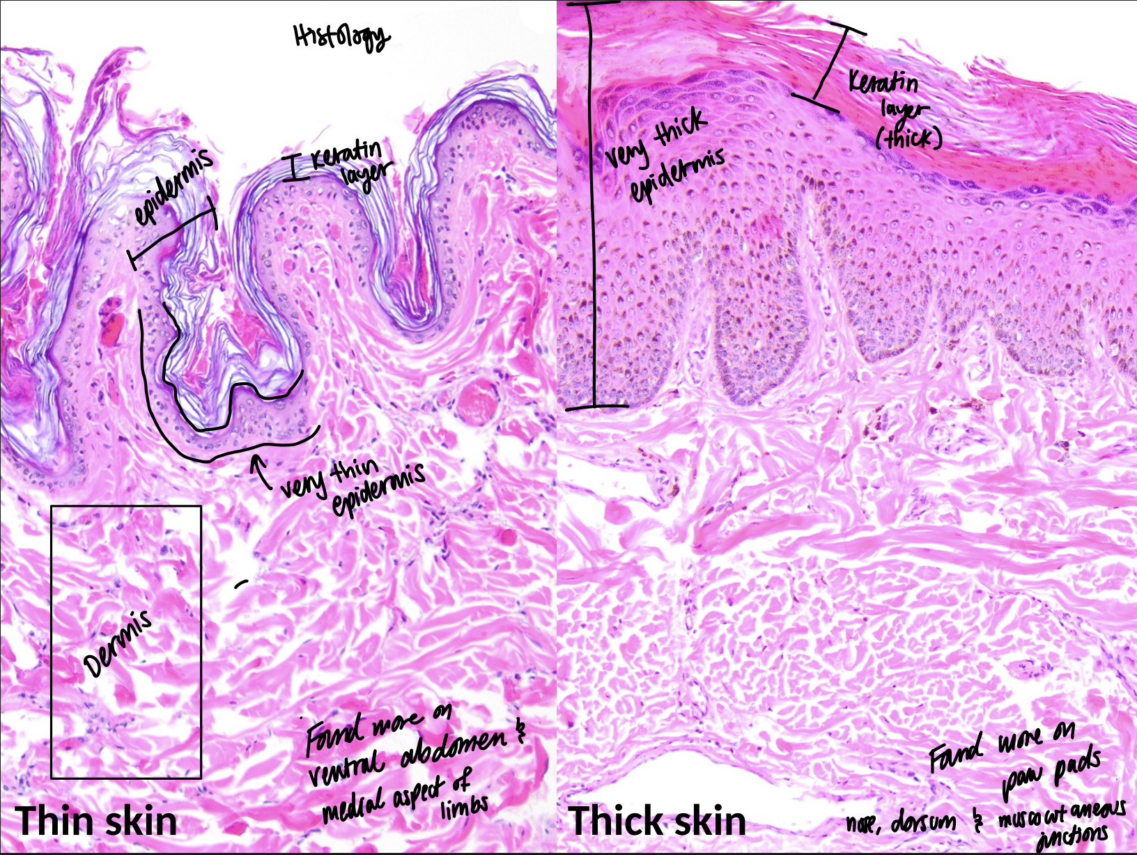
⭐ 36. On a histologic image, how can you identify thick skin versus thin skin?
A: Histologic thick skin has a much thicker epidermis than histologic thin skin. 🔎 What this means: For this identification, judge the thickness of the epidermis rather than the dermis. Thick areas also tend to have more prominent surface keratin and epidermal ridging. 💡 Think: histologic thickness means epidermal thickness.

⭐ 37. On an image of the epidermis, how can you identify a melanocyte?
A: Look for a cell in the basal portion of the epidermis that often has a clear halo and may contain brown melanin pigment. 🔎 What this means: The melanocyte cell body sits near the epidermal-dermal junction. Its thin dendritic processes usually are not easy to see on routine H&E, so location and the clear appearance are important recognition clues.

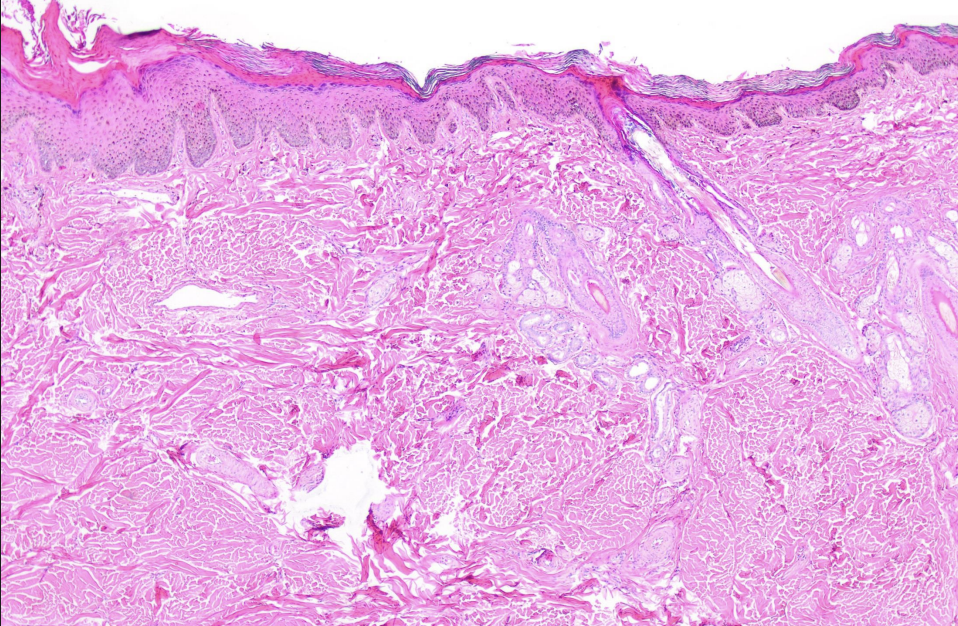
On a histologic image of a haired-to-nonhaired skin junction, how can you distinguish the two sides?
A: Haired skin contains hair follicles and hair shafts in the dermis, whereas nonhaired skin lacks those follicular structures and often has a thicker, more ridged epidermis. 🔎 What this means: At the nasal planum or paw-pad junction, first look for follicles, then compare epidermal thickness and ridging.

On a lip-margin histology image, how can you distinguish keratinized external skin from nonkeratinized oral mucosa?
A: Keratinized external skin has a superficial keratin layer or stratum corneum, whereas nonkeratinized oral mucosa lacks that keratin layer and retains nuclei near the surface. 🔎 What this means: Both are stratified squamous epithelia, so the key visual distinction is whether a true superficial keratinized layer is present.

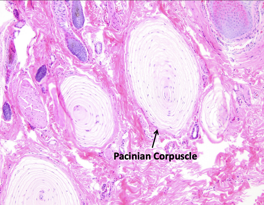
⭐ 40. On histology, how can you identify a Pacinian corpuscle?
A: A Pacinian corpuscle appears as a large laminated or onion-like structure surrounding a central nerve and is located in the mid-to-deep dermis or subcutis. 🔎 What this means: The concentric layers are its strongest visual clue. Pacinian corpuscles detect pressure and vibration. 💡 Think: Pacinian equals pressure plus onion layers.
⭐ 41. On histology, how can you identify a Tylotrich pad containing Merkel cells?
A: A Tylotrich pad appears as a localized thickening of the epidermis with Merkel cells near its base and afferent nerve fibers immediately beneath them. 🔎 What this means: Unlike a Pacinian corpuscle, the important feature is the focal epidermal thickening rather than a large laminated structure in the deep dermis. Merkel cells participate in light or fine touch.

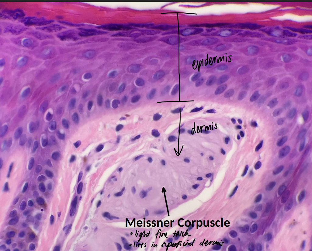
⭐ 42. On histology, how can you identify a Meissner corpuscle?
A: A Meissner corpuscle is a small sensory corpuscle located very superficially in the dermis just beneath the epidermis. 🔎 What this means: Its superficial location helps distinguish it from the much deeper Pacinian corpuscle. Meissner corpuscles participate in light or fine touch.
⭐ 43. If shown the lecture mechanoreceptor diagram, how should you identify a Ruffini corpuscle?
A: Identify the Ruffini corpuscle as a dermal mechanoreceptor associated with detecting stretch. 🔎 What this means: The lecture did not provide a dedicated histologic Ruffini image or a precise depth comparable with the other receptors, so recognize its name, dermal location, and stretch function without inventing additional morphology.

If shown the lecture mechanoreceptor overview, how can you distinguish Merkel, Meissner, Pacinian, and Ruffini receptors by location and function?
A: Merkel receptors are in the epidermis and detect fine touch, Meissner corpuscles are in superficial dermis and detect fine touch, Pacinian corpuscles are deeper in the dermis or subcutis and detect pressure and vibration, and Ruffini corpuscles are dermal receptors associated with stretch. 🔎 What this means: Use depth as your first clue: Merkel is within epidermis, Meissner is just below it, Pacinian is deep, and Ruffini is the stretch receptor.

🟡 LOWER PRIORITY 45. On the frog-skin histology image from this lecture, what structures should you be able to recognize?
A: Recognize the epidermis and dermis, large mucin-producing glands, dense pink poison glands, dermal melanophores, and chromatophores. 🔎 What this means: The frog example demonstrates that non-domestic skin may organize pigment cells and glandular structures differently from typical mammalian skin.

🟡 LOWER PRIORITY 46. If shown the chameleon image under polarized light, what pigment cell type should you recognize?
A: Recognize iridophores, which are a type of chromatophore that can appear strongly reflective or bright under polarized light. 🔎 What this means: This was the lecture's non-domestic example showing how chromatophore structures can interact with light.
