Brain Anatomy, Face Musculature, and Cranial Nerves I BIOL 106 A LA
 Call Kai
 Learn
 Practice Test
 Spaced Repetition
 Match
 Flashcards
 Knowt Play
1/41

There's no tags or description
Looks like no tags are added yet.
42 Terms
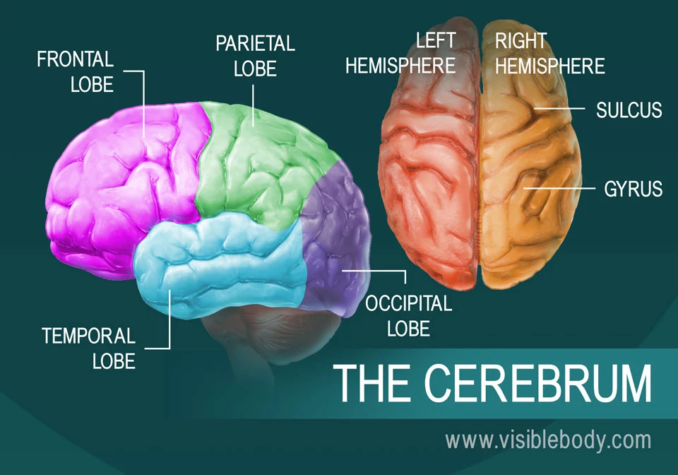
Cerebrum
Largest and topmost part of the brain
Sits above the brainstem and cerebellum
Split into left and right hemispheres
Cerebrum functions
Higher Level Functions
Sensory Processing
Voluntary Movement

Cerebellum
Located in the back of the brain beneath the cerebrum

Cerebellum functions
Movement Coordination
Balance and Posture
Motor Learning
Automate repetitive tasks (ex: riding a bike)

Diencephalon
Located deep in the brain sitting above the brainstem and beneath the cerebral hemisphere
Divided into 4 Structures
Thalamus, Hypothalamus, Epithalamus, Subthalamus
Diencephalon functions
Thalamus: Sensory and Motor Relay
Hypothalamus: Homeostasis and Endocrine Control
Epithalamus: Sleep Wake Cycle
Subthalamus: Muscle Movement Regulation
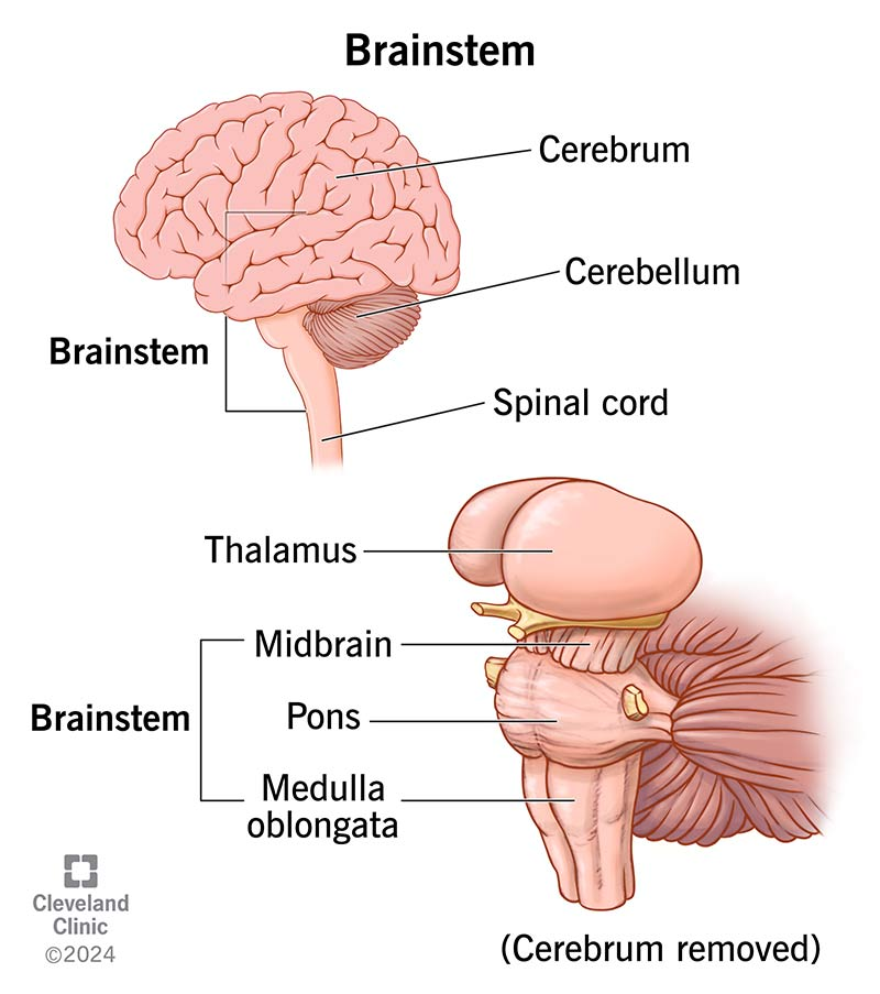
Brain Stem
Stalk-like lower part of the brain
3 Components:
Midbrain
Pons
Medulla Oblongata
Connects the cerebrum and cerebellum to the spinal cord
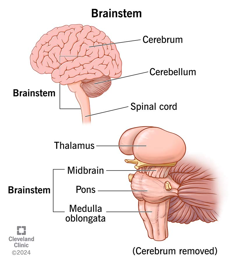
Brain Stem functions
Functions:
Regulates essential involuntary life-support functions aka autonomic functions

Midbrain
Vital relay center for movement, sensory processing and automatic reflexes

Pons
neural relay, respiration regulation, sleep and arousal control, houses nuclei of cranial nerves V-VIII

Medulla Oblongata (Medulla)
Cardiovascular regulation, respiratory control, automatic reflexes, nerve signal relay, houses nuclei for cranial nerves IX-XII
Functional Areas of the Cerebral Cortex
The three types of functional areas are:
Motor areas – control voluntary movement
Sensory areas – conscious awareness of sensation
Association areas – integrate diverse information

Frontal Lobe
Executive control (planning, reasoning, problem-solving, and decision-making)
Managing emotions, personality, and social behavior
Voluntary movement via the primary motor cortex
Speech production via Broca's area

Parietal Lobe
Processing somatosensory data (touch, pressure, temperature, and pain) via the primary somatosensory cortex
Spatial awareness and navigation
Helping identify objects by touch and interpreting symbols or numbers

Temporal Lobe
Auditory processing (hearing and music)
Language comprehension via Wernicke's area
Memory storage and retrieval via the hippocampus
Object and face recognition

Occipital Lobe
Primary visual processing center (interpreting color, light, depth, distance, and movement
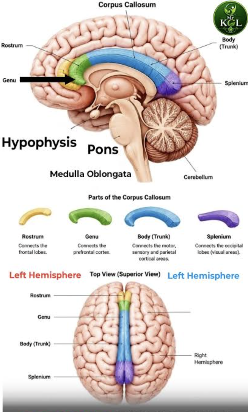
Corpus Callosum
Largest white matter tract in the brain that connects the left and right hemispheres
Interhemispheric Communication: Acts as a bridge, allowing the two sides of the brain to share information and synchronize signals
Coordination: Supports sensory processing (vision, hearing, touch), motor control, and cognitive functions (memory, language, and reasoning)

Temporomandibular Joint (TMJ)
Synovial Sliding Hinge Joint with Articular Disc
Action: depression, elevation, protrusion, retraction and lateral excursion
Connects lower jaw bone (mandible) to temporal bone of skull
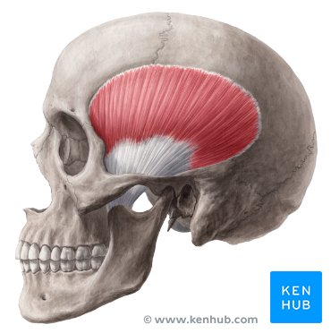
Temporalis Muscle
Responsible for mastication (chewing) by closing the mouth
Origin: Temporal fossa of the skull
Insertion: Coronoid Process of Mandible
Action: Closes mouth and assists in chewing
Innervation: Trigeminal Nerve (Cranial Nerve V)
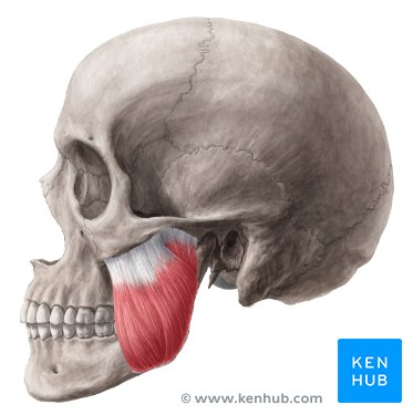
Masseter Muscle
Responsible for mastication (chewing) by elevation and protraction of mandible
Origin: Zygomatic Arch
Insertion: Ramus, Angle of Mandible and Coronoid Process
Action: Elevates and Protrudes Mandible for Chewing
Innervation: Trigeminal Nerve (Cranial Nerve V)
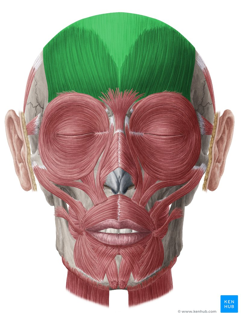
Occipitofrontalis Action
Raises eyebrows

Buccinator Action
Assists with chewing
Puffs out cheeks
“Trumpeter Muscle”

Mentalis Action
Raises and Thrusts Out Lower Lip
“Pouting Muscle”

Depressor Labii Inferioris Action
Pulls Down Button Lip
“Sulking Muscle”

Orbicularis Oculi Action
Open and Closes Eyelids
“Blinking, Winking, Squinting Muscle”

Orbicularis Oris Action
Brings Lips Together
Kissing

Levator Anguli Oris Action
Raises Corners of Mouth
Smiling

Levator Labii Superioris Action
Elevates the Upper Lip

Depressor Anguli Oris Action
Lowers Corners of Mouth
Frowning

Zygomatic Major and Minor Action
Elevates the Upper Lip
Smiling
Olfactory Nerve
Cranial Nerve I
Sensory
Testing: Smell Something with Eyes Closed
Optic Nerve
Cranial Nerve II
Sensory
Vision
Testing: Read something
Oculomotor Nerve
Cranial Nerve III
Motor
Pupillary Reactions
Eye Movement
Testing: Eye Motions (H) / PEARLA
Trochlear Nerve
Cranial Nerve IV
Motor
Eye Movement
Testing: Eye Motions (H)
Trigeminal Nerve
Cranial Nerve V
Sensory and Motor
Muscles of mastication
Facial sensory
Testing: Facial Sensation / Muscles of Mastication
Abducens Nerve
Cranial Nerve VI
Motor
Eye Movement
Testing: Eye Motions (H)
Facial Nerve
Cranial Nerve VII
Sensory and Motor
Facial Expression
Taste
Testing: Facial Expression / Taste on Anterior Tongue
Vestibulocochlear/Auditory Nerve
Cranial Nerve VIII
Sensory
Hearing
Balance
Testing: Hearing / Balance
Glossopharyngeal Nerve
Cranial Nerve IX
Sensory and Motor
Swallowing
Taste
Heart rate regulation
Testing: Say “Ahh” / Gag Reflex / Swallowing / Taste on Posterior Tongue
Vagus Nerve
Cranial Nerve X
Sensory and Motor
Gag Reflex
Testing: Say “Ahh” / Gag Reflex / Heart Rate
Spinal Accessory
Cranial Nerve XI
Motor
Upper trapezius
Sternocleidomastoid muscle
Testing: Shoulder Shrugs
Hypoglossal Nerve
Cranial Nerve XII
Motor
Tongue Movements
Testing: Stick Tongue Out / Move Tongue Side-to-Side