Lecture 4: Patient Monitoring and Induction
 Call Kai
 Learn
 Practice Test
 Spaced Repetition
 Match
 Flashcards
 Knowt Play
1/150

There's no tags or description
Looks like no tags are added yet.
151 Terms
pulse ox on hand with
IV
BP on other arm
limb restrictions for BP
lymph node dissections
AV fistula/graft
lumpectomy
BP set at
3-5 minutes on auto cycle
ASA monitors
EKG
Pulse ox
BP
Temp
Gas analyzer
gas analyzer
CO2
O2/N20/air/inhalational agents
pathway of electricity in heart
Sinus node
AV node
bundle of His
left and right bundle branches
purkinje fibers
SA node
pacemaker, found within right atrium
P wave
regurgitation
blood volume leaks back to previous chamber
stenosis
narrowing causing extra work for previous chamber
p wave
atrial depolarization
PR interval
delay of AV node to allow filling of ventricles
extended = delayed conduction = blockages
QRS interval
ventricular depolarization
T wave
ventricular repolarization
Prolonged PR interval
indicates an abnormal delay at the AV node (delayed conduction, indication of blockage)
Widened QRS complex
Delay in intraventricular conduction, such as bundle branch block
bipolar leads measure
measure potential difference between 2 recording electrodes
bipolar leads comes from
Einthovens triangle

Einthovens triangle
lead II lines up best with heart
what happens when impulse moves from right arm to left leg?
impulse toward a positive; positive upstroke, up swing, upward deflection
this is lead ii
lead II Einthovens triangle
QRS upstroke: positive upstroke down from SA
QRS downstroke: negative downstroke towards purkinje fibers
up swing
impulse toward positive
down swing
impulse away from positive
J point
junction point at S wave and start of ST segment
J point shows
heart attack, ischemia, supply demand issue
what causes differences in action potentials in different parts of heart?
differences in sodium, calcium, potassium (displace in/out myocardial cells during AP)
Calcium blocker has what effect on heart
slows HR because slows repolarization
Lidocaine has what effect on heart
blocks sodium channels (slows influx of na into cells, stabilizing cell membrane), good for v tach
unipolar leads
assess electrical fields in horizontal plane of chest
V1 leads
4th intercostal space to the right of the sternum
V2 leads
4th intercostal space to the left of the sternum
V3
midway between V2 and V4
V4
5th intercostal space, midclavicular line
V5
5th intercostal space, anterior axillary line
V6
5th intercostal space, midaxillary line
1 small box in EKG strip
0.04 sec
0.1 mV
1 big box (5 little boxes) EKG strip
0.20 sec
0.5 mV
rate equation
300 / # of big boxes between R waves
sinus tachycardia
>100 bpm

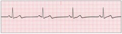
sinus bradycardia
<60 bpm
atrial fibrilation
-irregularly irregular.
-rate varies and depends on the random impulses (not from SA) at random times reaching AV node
-no P wave (SA node is the P wave)

identifying A Fib
-no P waves.
-Baseline shows fibrillatory waves.
in A fib the atria
quiver and dont fully contract so blood only moves passively to ventricles
(blood pools in atria, good treatment is chronic blood thinner) (or cardioversion)
junctional rhythm
normal rate and rhythm but missing P wave
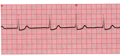
in junctional rhythm the
SA node isnt working so AV node is the main pacemaker
(no SA node so no P wave)
Premature Ventricular Complex (PVC)
no p wave
inverted T wave
wide bizarre QRS
skip beat
extra, abnormal heart beats that start in ventricles instead of SA node (ventricular conduction when ventricles are not filled)
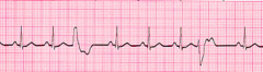
in PVC the electrical signal starts in the
ventricles
ventricular bigeminy
every other beat is a PVC

ventricular trigeminy
every third beat is a PVC

ventricular tachycardia
wide QRS without p waves and very rapid heart rate
100-250 bpm
Pulsatile (NOT SHOCKABLE) vs Pulseless (YES SHOCKABLE)

in ventricular tachycardia the
ventricles contracting very rapidly and not filling with blood
pulsatile v-tach
circulation may or may not be present
emergency-not shockable but could cardiovert
pulseless v tach
no circulation
deadly and shockable
shockable rhythms
pulseless v tach, v fib
Ventricular fibrillation (V-fib)
No QRS complex. Ventricles are quivering and not contracting
shockable

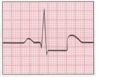
ST segment elevation
ST segment above baseline
myocardial infarction

ST segment depression
ST segment below baseline
myocardial ischemia
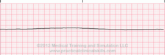
asystole
absence of contractions of the heart
not shockable
transmural ischemia
usually ST elevation
whole tissue lacks blood flow
endocardial ischemia
usually ST depression
first tissue depressed
blood pressure measured in
mmHg
size of BP cuff
too small= high reading
too big = low reading
non invasive blood pressure (NIBP)
oscillometry
korotkoff sounds
oscillometry
automatic BP cuff
measures MAP
MAP
mean arterial blood pressure
dia (+sys-dia/3)
MAP is always more ______
accurate
if BP looks funky double check MAP is more accurate
Korotkoff sounds
listening with stethoscope
measures systolic/diastolic
invasive blood pressure
Direct measurement via catheter in arteries
measures mean/systolic/diastolic
A line transducer location
phlebostatic axis (heart)
transducer too high
BP low
transducer too low
BP high
pulse ox determined by
beers lambert law
Beers Lambert Law
relationship of hemoglobin concentration to intensity of light transmitted through a solution
pulse ox is measured at
red light 660 nm (deoxyhemoglobin)
infrared light 940 nm (oxyhemoglobin_
plethysmography
pulsatile flow waveform
Pulse ox reading below what value is not accurate or reliable?
85%
respiratory gas analysis analyzes
ETCO2
inhalational agents
fresh gases
inspired and expired levels
ETCO2
end tidal CO2
trending decrease in CO2 if
blood isnt circulating to pulmonary circulation
decrease cardiac output
Big immediate decrease in ETCO2
pulmonary embolism
Longer slower decrease in ETCO2
blood pressure low
inhalational agents
isoflurane, sevoflurane, desflurane
fresh gases
O2 air N2O
capnometry
numeric measurement of CO2
capnography
display of CO2 waveform
phase a to B

dead space

phase B to C
expiratory upstroke where alveoli empty

phase C to D
exhalation

point D
where the ETCO2 reading is taken

point D to E
inspiration
temperature probes
esophageal
skin
nasopharyngeal
bladder
acetylcholine
neurotransmitter
acetylcholinesterase
cleaves ACh
anticholinesterases
decrease cleavage of ACh
increased ACh
overcomes NDMR
neuromuscular monitoring measures
measures effects of neuromuscular blockers
common nerves for neuromuscular monitoring
ulnar
facial
posterior tibial
Train of Four (TOF)
4 equali stimuli of 2 Htz
sustained tetany
continuous stimulus of 50 Hz
look for fade for 5 seconds to see if blocked
ulnar nerve monitoring
causes adduction of adductor pollicis (thumb)
black near where pinky meets wrist
facial nerve monitoring
eyelid twitching of orbicularis occuli
black by ear