Learn: MMSC433 Exam 2 Study Guide
 Call Kai
 Learn
 Practice Test
 Spaced Repetition
 Match
 Flashcards
 Knowt Play
1/164

There's no tags or description
Looks like no tags are added yet.
165 Terms
Forward Scatter (FSC) measures what?
cell size (diameter or volume) based on light diffracted along laser path
Side Scatter (SSC) measures whar?
cellular complexity or granularity (internal structures, nucleus, organelles) based on light refracted/reflected at 90 degree angle
What chain do most of hemoglobinopathic abnormalities exist?
beta-chain
Hemoglobinopathy
synthesis of hemoglobin abnormality
sinle amino acid substitution
qualitative/quantitative
inherited OR from new mutation
Qualitative hemoglobinopathies occur as a result of what?
genetic mutations involving globin protein chain
amino acid deletions/substitutions
What do the mutations in qualitative hemoglobinopathies do to the globin chains?
cause structural variations
Examples of qualitative hemoglobinopathy variants?
HbS, HbC, HbM
Quantitative hemoglobinopathies occur as a result of what?
genetic defects that cause reduced synthesis of globin chains
What do the mutations in quantitative hemoglobinopathies do to the globin chains?
nothing they are structurally normal
What are quantitative hemoglobinopathies collectively known as?
thalassemias
Qualitative hemoglobinopathies are most commonly ______ nucleotide point mutations within the globin genes (HBA1, HBA2, HBB) that alter the primary amino acid sequence of the globin chains.
single
Sickle cell disease amino acid substitution
valine replaces glutamic acid at 6th position of B-globin chain
Hemoglobin C disease amino acid substitution
lysine replaces glutamic acid at the 6th position of the B-globin chain
Life-threatening emergency defined by chest pain, fever, tachypnea, and new pulmonary infiltrates on chest X-ray often triggered by fat embolism
Acute Chest Syndrome
sickled cells block blood flow in capillaries, leading to severe recurrent pain in bones, joint, chest, or abdomen
vaso-occlusion
major cause of death in patients -> staph aureus, strep pneumoniae, H. influenzae
bacterial infections
gradual loss of splenic function, presence of howell jolly and pappenheimer bodies
auto splenectomy
howell jolly bodies

Heinz bodies

Pappenheimer bodies

Basophilic stippling

Temporary cessation of RBC production in BM, commonly triggered by parbvo B19 infection, presenting with severe acute anemia
Aplastic crisis
Vaso-occlusion or cerebral infarction (necrosis) /hemorrhage leading to neurological deficits, seizures or silent cerebral infarcts
Stroke
Painful, prolonged penile erection caused by trapped blood flow risking permanent erectile dysfunction if untreated
Priapism
Sickle cell disease (HbSS) key clinical findings
pain crises, hemolysis, infections, auto splenectomy
Sickle cell disease (HbSS) blood smear
sickle cells, target cells, nucleated RBCs, increase reticulocytes
Sickle cell disease (HbSS) heme pattern
HbS > 80%
NO HbA
HbF 1-20%
Sickle cell trait (HbAS) key clinical findings
usually asymptomatic
Sickle cell trait (HbAS) blood smear
usually normal smear
occasional target cells
Sickle cell trait (HbAS) heme pattern
HbA ~60%
HbS ~40%
Target cells

Hemoglobin C disease (HbCC) key clinical findings
mild to moderate hemolytic anemia, possible splenomegaly
Hemoglobin C disease (HbCC) blood smear
target cells, HbC crystals, microspherocytes
Hemoglobin C disease (HbCC) heme pattern
HbC usually >90%
NO HbA
HbC crystals
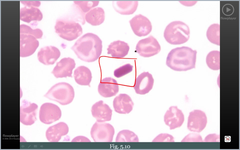
Microspherocytes

Hemoglobin C trait (HbAC) key clinical findings
usually asymptomatic
Hemoglobin C trait (HbAC) blood smear
usually normal occasional target cells
Hemoglobin C trait (HbAC) heme pattern
HbA ~60%
HbC ~40%
Hemoglobin SC disease (HbSC) key clinical findings
pain crises and hemolysis; complications maybe milder or different than HbSS
Hemoglobin SC disease (HbSC) blood smear
target cells, some sickled cells, possible HbC crystals
Hemoglobin SC disease (HbSC) heme pattern
HbS and HbC each ~50%
NO HbA
Alpha thalassemia key clinical findings
ranges from asymptomatic carrier to severe fetal anemia
Alpha thalassemia blood smear
microcytosis, hypochromia, target cells
Alpha thalassemia heme pattern
HbH or Hb Bart's may appear
depends on number of alpha genes affected
Microcytosis

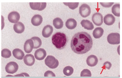
Hypochromia
lack of color
Beta thalassemia major key clinical findings
severe anemia beginning in infancy, growth delay, hepatosplenomegaly, bone changes
Beta thalassemia major blood smear
marked microcytosis, hypochromia, target cells, nucleated RBCs
Nucleated RBCs

Beta thalassemia major heme pattern
HbF markedly increased
HbA absent in B0
HbA reduced in B+
HbA2 often increased
Beta thalassemia minor key clinical findings
usually asymptomatic or mild anemia
Beta thalassemia minor blood smear
microcytosis, hypochromia, target cells, basophilic stippling
Beta thalassemia minor heme pattern
HbA predominant
HbA2 usually above 3.5%
HbF normal or mildly increased
The most common type in healthy adults
95% to 98% of total hemoglobin
consists of two alpha (α) chains and two beta (β) chains.
Hemoglobin A (HbA)
A minor adult type
consists of two alpha (α) and two delta (δ) chains
accounts for about 2% to 3% of total adult hemoglobin
Hemoglobin A2 (HbA2)
two alpha (α) and two gamma (γ) chains
fetal heme
Hemoglobin F (HbF)
abnormal variant responsible for sickle cell disease
Hemoglobin S (HbS)
beta-chain variant that reduces protein solubility and can cause mild chronic hemolytic anemia
Hemoglobin C (HbC)
beta-chain variant common in Southeast Asian populations that results in a reduced rate of synthesis and often mild or no symptoms
Hemoglobin E (HbE)
A abnormal tetramer made of four beta chains (β4), found in types of alpha-thalassemia
Hemoglobin H (HbH)
A tetramer made of four gamma chains, seen in severe alpha-thalassemia conditions
Hemoglobin Barts (γ4)
Measures hemoglobin, hematocrit, RBC count, and RBC indices
CBC test
Low MCV and MCH suggest microcytic, hypochromic anemia, as seen in thalassemia
RBC indices
Identifies sickle cells, target cells, microcytosis, hypochromia, and other abnormalities
Peripheral blood smear
Reticulocyte count is often increased in _______ anemia.
hemolytic
Hemoglobin electrophoresis separates hemoglobins according to their ______.
movement in an electrical field
Separates and quantifies hemoglobin types; commonly used to identify HbA2, HbF, HbS and HbC
HPLC
Screens for HbS but cannot distinguish sickle cell disease from sickle cell trait
Hemoglobin solubility test
Helps distinguish thalassemia from iron deficiency anemia
Iron studies
Identifies mutations or deletions in globin genes
Molecular genetic testing
What factors help assess hemolysis?
serum haptoglobin decreased
bilirubin elevated
LDH elevated
reticulocyte count elevated
HbA 0%
HbS 80-95%
HbC 0%
Sickle cell disease (HbSS)
HbA 55-65%
HbS 35-45%
HbC 0%
Sickle cell trait (HbAS)
HbA 0%
HbS 0%
HbC 35-45%
HbC disease (HbCC)
HbA 55-65%
HbS 0%
HbC 35-45%
HbC trait (HbAC)
HbA 0%
HbS 50%
HbC 50%
HbSC disease
Inherited in a homozygous state
both beta-globin genes are damaged or non-working
HbA ABSENT in B0 REDUCED in B+
HbS only if co-inherited
HbC only if co-inherited
Beta thalassemia major
Inherited in a heterozygous state
only one beta-globin gene is damaged, while the other is normal
HbA usually >90%
HbS & HbC only if co-inherited
HbA2 usually 3.5%
Beta thalassemia minor
Alkaline pH (~8.6) fastest to slowest electrophoresis
HbA > HbF > HbS > HbC
Acid pH (~6.0) fastest to slowest electrophoresis
HbA, HbF > HbS, HbD, HbG > HbC
Acid electrophoresis helps resolve _____ from some variants that overlap with it at alkaline pH
HbS
Sickle cell disease (HbSS) electrophoresis at alkaline
Strong HbS band
Variable HbF
Increased HbA2
No HbA
Sickle cell disease (HbSS) electrophoresis at acid
Strong HbS
HbF present
Sickle cell trait (HbAS) at alkaline
two major bands HbA and HbS
Sickle cell trait (HbAS) at acid
two corresponding bands HbA and HbS
Hemoglobin C disease (HbCC) at alkaline
strong HbC-position band
No HbA
Hemoglobin C disease (HbCC) at acid
strong band in HbC positon distinct from HbS
HbSS -> S predominant
HbAS -> A and S
HbCC -> C predominant
Alkaline pH
HbSS -> S predominant
HbAS -> A and S
HbCC -> C predominant
Acid pH predominance
HbA present in Hb__
AS
HbS present in Hb__
SS and AS
HbC present in Hb__
CC
A positive result for the hemoglobin solubility test suggest Hb_ is present
S
Can the hemoglobin solubility test distinguish sickle cell disease from sickle trait for hemoglobinopathies?
no
The hemoglobin solubility test does not reliably quantify what?
HbS
Thalassemia definition
inherited disorder in which the body produces reduced amounts of one or more globin chains
microcytic hypochromic anemia is a common finding
Abnormal facial bone appearance in Thalassemia major
Prominent cheekbones and forehead
maxillary enlargement and changes in facial structure
Alpha (α)-thalassemia
Reduced or absent production of alpha-globin chains.
Beta (β)-thalassemia
Reduced or absent production of beta-globin chains.