Heart Anatomy, Blood Flow, Valves & Conduction System
 Call Kai
 Learn
 Practice Test
 Spaced Repetition
 Match
 Flashcards
 Knowt Play
1/73

There's no tags or description
Looks like no tags are added yet.
74 Terms
4 chambers of the heart
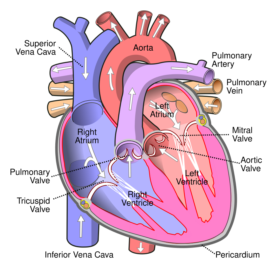
Right atrium (upper right), right ventricle (lower right), left atrium (upper left), and left ventricle (lower left). The right-side chambers contain oxygen-poor blood; the left-side chambers contain oxygen-rich blood.

Blood flow through the heart - complete order
Body → superior/inferior vena cava → right atrium → tricuspid valve → right ventricle → pulmonary semilunar valve → pulmonary trunk/arteries → lungs → pulmonary veins → left atrium → bicuspid (mitral) valve → left ventricle → aortic semilunar valve → aorta → body.
Tricuspid valve
Right AV valve between the right atrium and right ventricle. It has 3 cusps and acts as a gate that prevents blood from flowing backward.
Pulmonary semilunar valve
Between the right ventricle and pulmonary trunk. Blood passes through it as it leaves the right ventricle on the way to the lungs.

Bicuspid (mitral) valve
Left AV valve between the left atrium and left ventricle. It has 2 cusps and prevents backflow toward the left atrium.

Aortic semilunar valve
Between the left ventricle and aorta. Blood passes through it as it leaves the left ventricle for the body.

Function of the heart valves
The 4 valves act as gates. They open and close because of pressure differences and ensure blood flows in only one direction.
Right coronary artery (RCA)
One of the 2 main coronary arteries and one of the first branches off the aorta. It travels in the atrioventricular sulcus on the right side of the heart and gives rise to major right coronary branches.

Marginal artery
Major branch associated with the right coronary artery. Runs along the right margin of the heart and supplies myocardium of the inferior wall of both ventricles.

Posterior interventricular artery (PDA)
Major right coronary branch also called the posterior descending artery. Runs in the posterior interventricular sulcus and supplies the posterior ventricular walls and septum.

Left coronary artery (LCA)
One of the 2 main coronary arteries and one of the first branches off the aorta. It gives rise to the circumflex artery and left anterior descending (LAD) artery.

Circumflex artery
Major branch of the left coronary artery. Runs around the heart in the atrioventricular sulcus and supplies the left atrium and posterolateral left ventricle.
Left anterior descending artery (LAD)
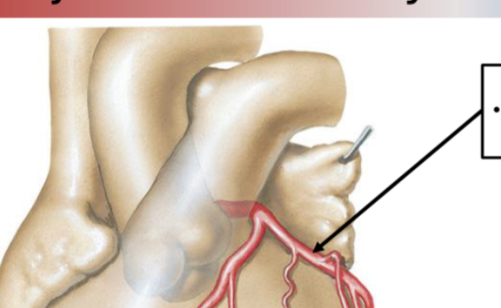
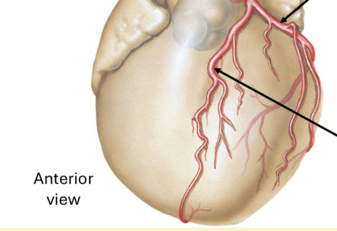
AKA anterior interventricular artery. Runs in the anterior interventricular sulcus and supplies MOST of the interventricular septum and the anterior wall of both ventricles. Costa calls it the "widow maker."
Epicardium
Outermost layer of the heart wall. Same as the visceral pericardium. Made of fibrous connective tissue and fat in the sulci; major coronary vessels are found here.
Myocardium
Middle layer of the heart wall. Made of cardiac muscle and responsible for the muscular contraction of the heart.
Endocardium
Innermost layer of the heart wall. Made of simple squamous epithelium called endothelium; continuous with blood-vessel endothelium and also covers the valves.
Intrinsic vs. extrinsic conduction
Intrinsic = within the heart itself; acts as the pacemaker system, regulates heart rate, and establishes the sequence of chamber contractions. Extrinsic = input from outside the heart through sympathetic and parasympathetic (vagus) nerves; adjusts heart rate to the body's needs.
Intrinsic conduction pathway
SA node → AV node → Bundle of His (AV bundle) → Purkinje fibers → ventricular myocardium contracts.
Parasympathetic extrinsic control
Vagus nerve (CN X) uses acetylcholine. It is inhibitory: decreases heart rate and relaxes/decreases the strength of contractions.
Sympathetic extrinsic control
Sympathetic nerves use norepinephrine. They increase heart rate and increase the strength of contraction.
Aortic auscultation area
Listen at the RIGHT 2nd intercostal space.
Pulmonary auscultation area
Listen at the LEFT 2nd intercostal space.
Tricuspid auscultation area
Listen at the LEFT 5th intercostal space at the sternal margin.
Mitral auscultation area
Listen at the LEFT 5th intercostal space in the midclavicular line.
Location of the heart
Located in the thoracic cavity in the mediastinum, posterior to the sternum. The apex lies at the left 5th intercostal space.
Heart position
The heart sits obliquely: rotated about 45 degrees to the left and tilted inferiorly (caudally).
Anterior wall of heart
Front portion of the heart; includes the right atrium and both ventricles.
Inferior wall of heart
Bottom portion of both ventricles that rests on the diaphragm.
Posterolateral wall of heart
Back portion; mostly the left ventricle and the largest muscular wall in depth and size.
Base of heart
Broad superior surface where the great vessels attach.

Apex of heart
Blunt, cone-shaped region made mainly by the tip of the left ventricle.

Right vs. left side oxygen content
Right atrium and right ventricle contain oxygen-poor blood. Left atrium and left ventricle contain oxygen-rich blood.
Atrioventricular sulcus
Groove separating atria from ventricles. Circles around the heart like a crown and contains the left/right coronary arteries and circumflex artery.

Anterior & posterior interventricular sulci
Grooves separating the right and left ventricles. They contain the anterior and posterior interventricular arteries.

Interatrial septum
Wall separating the right and left atria.

Interventricular septum
Wall separating the right and left ventricles.

Fibrous pericardium
Outer sac enclosing the heart. Tough dense connective tissue that attaches the heart to the diaphragm and base of the great vessels.

Serous pericardium
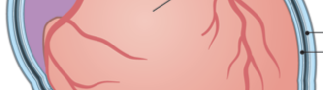
Inner pericardial layer with 2 portions: parietal pericardium lines the inside of the fibrous pericardium; visceral pericardium attaches to the heart surface and is also called epicardium.
Pericardial space
Space between the parietal and visceral pericardium. Contains slippery serous fluid that reduces friction as the heart pumps.
Pericarditis
Inflammation or infection of the pericardium. Costa lists faintness, dizziness, shortness of breath, chest pain, and fever.

Aorta
Great vessel carrying oxygenated blood from the left ventricle to the body.

Pulmonary trunk
Carries deoxygenated blood out of the right ventricle and branches into the right and left pulmonary arteries, which go to the lungs.

Superior vena cava
Carries deoxygenated venous blood from the head, neck, and upper limbs back to the right atrium.

Inferior vena cava
Carries deoxygenated venous blood from the trunk and lower limbs back to the right atrium.

Pulmonary veins
Four vessels total: 2 from each lung. Carry oxygenated blood from the lungs to the left atrium.

Systole vs. diastole
Systole = contraction phase. Diastole = relaxation phase.
Atrial systole
Atria contract and push blood through the open AV valves while the ventricles are in diastole and filling.
Ventricular systole
Ventricles contract; AV valves close. Right ventricle ejects blood into the pulmonary trunk and left ventricle ejects blood into the aorta.
Valve opening/closing during contraction
Atrial systole: AV valves open, semilunar valves closed. Ventricular systole: semilunar valves open, AV valves closed.
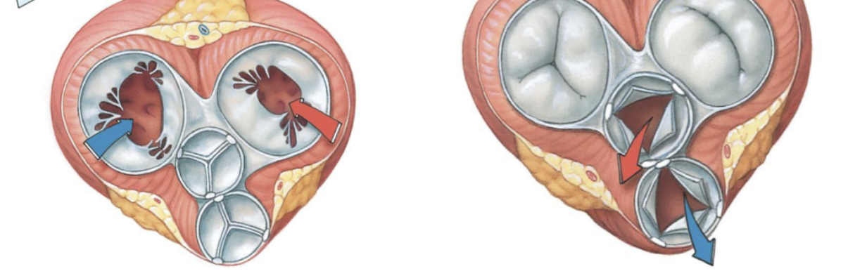
Why is left ventricular myocardium thicker?
The left ventricle must pump blood through the whole systemic circulation, so its myocardium is MUCH thicker. The right ventricle only pumps to the lungs.
Papillary muscles
Finger-like muscular projections from the heart wall that stabilize AV valves and prevent valve inversion (opening in the wrong direction).

Chordae tendineae
Thin "heart strings" connecting papillary muscles to AV valve leaflets. They help prevent the valve leaflets from prolapsing/inverting.

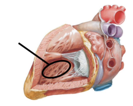
Trabeculae carneae
Crisscrossing ridges in the ventricles that create a mesh-like appearance and help prevent the ventricular walls from sticking together by suction.
Foramen ovale
Fetal opening between the atria that lets blood pass directly from right atrium to left atrium, bypassing the lungs before birth.
Fossa ovalis
After birth the foramen ovale closes, leaving an oval-shaped depression called the fossa ovalis.
Patent foramen ovale
The foramen ovale remains open after birth, allowing venous and arterial blood to mix.
Coronary circulation
Blood supply of the heart itself. Coronary vessels lie in the atrioventricular and interventricular sulci, and branches descend along the cardiac surface.
Coronary sinus
Collects blood from the coronary veins.

Great cardiac vein
Major coronary vein that travels with the LAD.

Middle cardiac vein
Major coronary vein that travels with the posterior interventricular artery.

Small cardiac vein
Major coronary vein that travels with the right coronary artery.

Myocardial infarction (MI)
A heart attack: a coronary artery is blocked by a thrombus (fixed clot) or embolus (moving clot). Cardiac muscle dies, cannot regenerate, and is replaced by scar tissue that cannot contract or conduct impulses.
Atherosclerosis / arteriosclerosis
Fatty deposits build in the vessel wall and narrow the artery. Plaque can rupture and trigger a clot. Costa notes arteriosclerosis when plaque hardens.
Stent
Expandable tube-shaped device inserted into a narrowed vessel to prop it open.

Coronary artery bypass graft (CABG)
A vessel is taken from elsewhere in the body, stitched into the aorta, and directed to the myocardium to bypass impaired coronary flow.

Heart sounds: Lub and Dub
First heart sound, "Lub": closure of AV valves with ventricular contraction. Second heart sound, "Dub": closure of semilunar valves with ventricular relaxation.
Heart auscultation
Listening to heart sounds with a stethoscope. The sounds come from vibrations of valves shutting; used to check for arrhythmias, murmurs, valve defects, etc.
Why are auscultation sites not directly over the valves?
Bone and lungs can muffle the sound. Valve-closure vibrations travel through the heart walls and great vessels, so the stethoscope is placed away from bone where the heart/great vessels contact the anterior thoracic wall.
Heart murmur
Abnormal heart sound associated with valve damage/malformation, turbulent blood flow, and possible regurgitation.
Intrinsic conduction system
Primary conduction system within the heart. Specialized conducting cardiomyocytes carry action potentials and stimulate cardiac muscle contraction; it does NOT need direct nervous-system stimulation to operate.
Sinoatrial (SA) node
Collection of specialized conducting cells in the wall of the right atrium. It is the heart's pacemaker and initiates the heartbeat.

Atrioventricular (AV) node
Receives the message from the SA node in the right atrium and sends it to the Bundle of His.
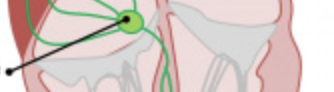
Bundle of His (AV bundle)
Collection of conduction fibers in the interventricular septum that transmits the message toward the Purkinje fibers.
Purkinje fibers
Located in the walls of the ventricles. Conduct the impulse to the myocardium, resulting in ventricular contraction.
