MBM Block 2
 Call Kai
 Learn
Learn Practice Test
 Spaced Repetition
 Match
Match Flashcards
 Knowt Play
1/161

There's no tags or description
Looks like no tags are added yet.
Name | Mastery | Learn | Test | Matching | Spaced | Call with Kai | Chat |
|---|
No analytics yet
Send a link to your students to track their progress
162 Terms
↑ rate of rxn w/o being consumed
↓ Ea
Do NOT change rxn equilibrium
Enzymes
ΔG < 0
Spontaneous
↑ [S] = ↑ rxn velocity until enzyme becomes saturated
x-axis = [S]
y-axis = rxn rate (V)
Michaelis-Menten
Maximum rxn velocity
Enzyme is saturated w/substrate
Vmax
[S] at ½ Vmax
Measure of enzyme affinity for substrate
Km
↑ Km =
LOW affinity
x-axis: 1/[S]
y-axis: 1/V
Lineweaver-Burk Plot
Oxidation-reduction
Add or remove electrons
Ex. dehydrogenase
Oxidoreductase
Transfer functional groups
Ex. kinase
Transferase
Cleave bonds w/ water
Ex. protease
Hydrolase
Cleave or add groups w/o hydrolysis or oxidation
Ex. decarboxylase
Lyase
Rearrange within a molecule
Ex. mutase
Isomerase
Joins two molecules w/ ATP
Ex. synthase
Ligase

Directly competes w/ substrate for enzyme active site
Increasing substrate can overcome inhibition
↑ Km: need more substrate to achieve same velocity
Vmax: unchanged
Competitive Inhibition
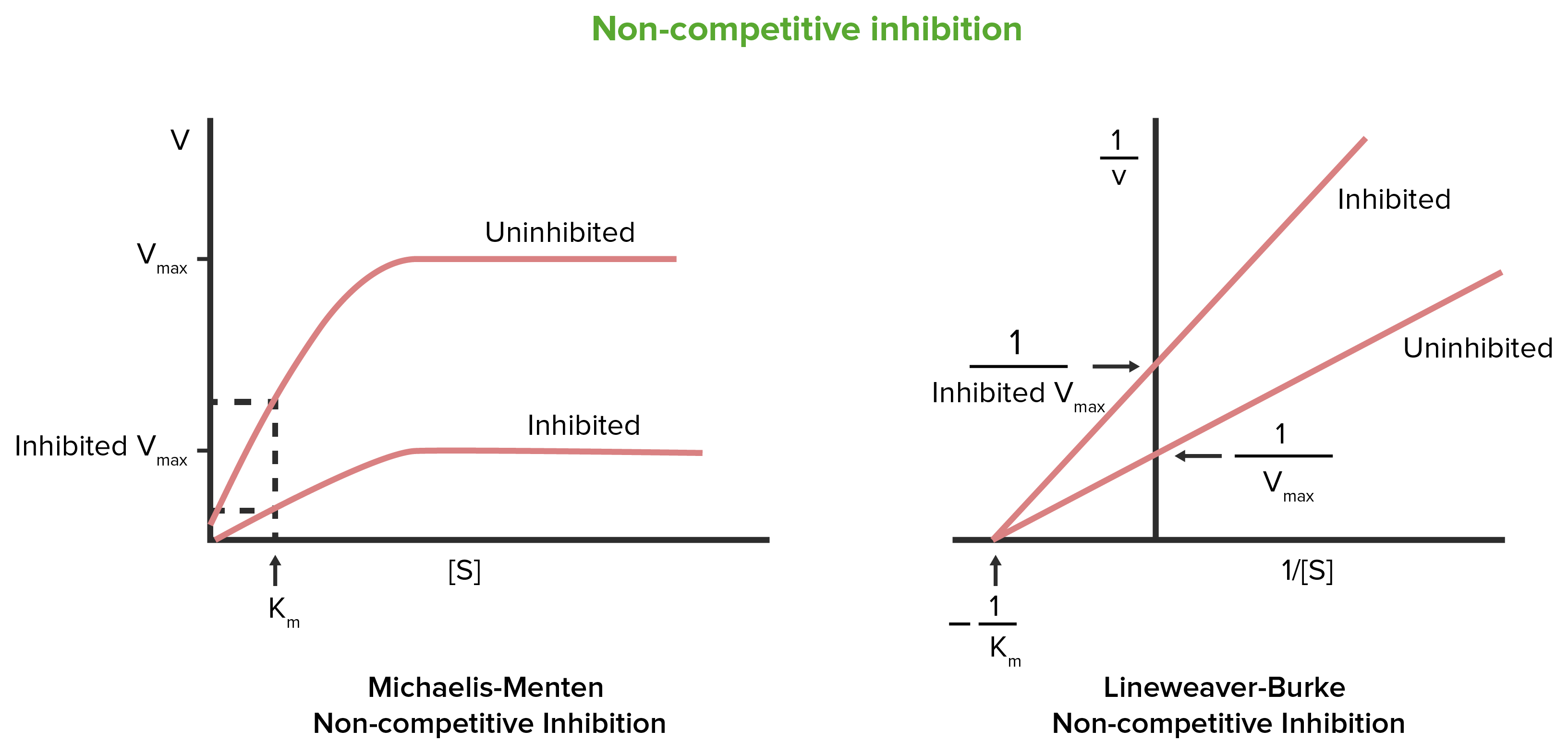
Binds at allosteric site
↓ enzyme activity
↓ Vmax
Km: unchanged
Increasing substrate cannot overcome inhibition
Noncompetitive Inhibition
Irreversibly inhibits enzyme
Ex. allopurinol
Suicide Inhibition
pH on Enzyme Activity
Optimal pH = HIGH activity
Extreme pH can cause denaturation
Temperature on Enzyme Activity
↑ T = ↑ Molecular Movement = ↑ Collisions = ↑ Rxn Rate
HIGH T = Denaturation = ↓ Enzyme Activity
Substrate Concentration of Enzyme Activity
LOW [S] = ↑ substrate = ↑ rxn rate
↑ activity does NOT equal ↑ rxn velocity
Inhibitors on Enzyme Activity
↓ Enzyme Activity
Reversible = inhibitor can dissociate
Irreversible: permanetly inactivates enzyme
Protein component only
Inactive
Apoenzyme
Non-protein component required for enzyme activity
Metal Ions: Mg2+, Zn2+, Fe2+/Fe3+, Cu2+
Cofactor
Organic non-protein molecule that assists an enzyme
Ex. NAD+, FAD+, CoA, TPP
Coenzyme
Coenzyme permanently associated w/enzyme
Ex. heme group of cytochromes, biotin group of acetyl-CoA carboxylase
Prosthetic Group
Apoenzyme + required nonprotein component
Active
Holoenzyme
Irreversible enzyme inhibitor → inhibits bacterial cell-wall synthesis → bacteria can’t maintain/build cell wall
Penicillin
Competitive inhibitors → resemble PABA → interfere with bacterial folate synthesis
Sulfonamides
Competitive inhibitor of folate metabolism → ↓ DNA synthesis/cell division → used as an anticancer drug
Methotrexate
Inhibits cytochrome oxidase
Cyanide
Inhibits enolase
Fluoride
↑ ALT =
Hepatocellular/parenchymal liver injury
↑ ALP =
Obstruction/cholestasis
↑ CK-MM =
Muscle Disease
↑ ALP with increased osteoblastic activity
Bone ALP is heat labile
Bone Disease
↑ PSA =
Prostate Disease/Cancer Marker
↑ Acid Phosphatase =
Prostate Cancer
Which enzyme rises first after a myocardial infarction?
CK-MB
Which enzyme rises after CK-MB in mycardio infarctions?
AST
Which enzyme rises last after a myocardial infarction?
LDH
LDH pattern with myocardial infarction
Flipped LDH pattern
LDH1 > LDH2
Biomarker for myocardial infarction
Cardiac tropinin
CK-MB levels after myocardial infarction
Peak at 24 hrs
Baseline at 2-3 days
Gout =
↑ uric acid → urate crystal deposition → inflammation
Allopurinol
inhibits XO → ↓ uric acid → treats gout
Hemoglobin Structure
4 heme + 4 globin groups
Heme Synthesis Pathway

δ-ALA Dehydratase Defiency
Accumulation of porphyrin precursor ALA within RBCs and hepatocytes
Triggers: alcohol, prolonged fasting
Labs: lactic acidosis, elevated liver levels
Symptoms: neurologic/GI failure
δ-ALA Dehydratase Porphyria
PBG Deaminase Deficiency
Ineffective heme synthesis + accumulation of PBG in cytoplasm
Triggers: drugs
Labs: elevated urine PBG + prophyrins
Symptoms: neuropathic abdominal pain, DARK RED URINE, polyneuropathy, psychological disturbances
Treatment: glucose + hematin to inhibit ALA synthase
Acute Intermediate Prophyria
Uroporphyrinogen decarboxylase deficiency
Triggers: hepatic iron overload, alcohol, sunlight, estrogen therapy, HBV/HCV
Painful, blistering skin lesions on sun-exposed skin
Onset during 4th/5th decade of life
Red to brown urine
Porphyria Cutanea Tarda
Protophorphyngogen oxidase deficiency
In adulthood
Triggers: nongenetic factors - drugs, diet, hormones, stress
Symptoms: abdominal issues, muscle weakness, seizures, mental changes
Photosensitivity
Treatment: liver transplant
Variegate Porphyria
In erythropoietic cells
Enzyme deficiency in RBC
Accumulation of protoporphyrins in RBC
Symptoms: photosensitivity and liver disease
In early childhood
Erythropoietic Protoporphyria
Childhood Symptoms: developmental delays, abdominal issues, irritability
Adult Symptoms: porphyria symptoms, anemia
Interferes with ALA dehydratase and ferrochelatase → decreases heme synthesis
Lead Poisoning
Degradation of Heme
Heme → Biliverden → Bilirubin → Liver via Bilirubin-Albumin Complex → Bilirubin in Liver → Bilirubin Diglucuronide → to blood → Bile
↑ Serum bilirubin levels
Jaundice
Excess RBC breakdown/hemolysis
Prehepatic Jaundice
Problem w/ liver processing/conjugation
Hepatic Jaundice
Obstruction of bile flow
Posthepatic Jaundice
RBC Synthesis > RBC Destruction
Polycythemia
RBC Synthesis < RBC Destruction
Anemia
a globin chains are made on…
Chromosome 16
β globin chains are made on…
Chromosome 11
Hemoglobic Chain Levels
Before Birth =
After Birth =
Before Birth = high γ chains, low β chains
After Birth = low γ chains, high β chains
Sickle Cell Disease
Missense mutation in β-globin gene (Glu → Val)
HbS/HbS (homozygous)
Sickle Cell Trait
HbA/HbS (heterozygous)
No full clinical manifestation
Why do sickle cell symptoms occur after infancy?
Babies have high HbF
↓ HbF = ↑ β-globin production → ↑ HbS
Sickling Cause =
Low O2 + Dehydration → HbS polymerization
Sickling Hemolysis
Sickled RBCs are fragile = destroyed more easily
RBC desctruction → heme breakdown → ↑ bilirubin → jaundice
Sickling Vaso-occlusion
Sickles RBC → occlusion → tissue hypoxia → pain + tissue damage
Chronic hemolytic anemia
Lifelong episodes of pain
↑ infection sisceptibility
Acute chest syndrome
Stroke
Splenic dysfunction
Renal dysfunction
Bone marrow hyperplasia
Sickle Cell Disease Clinical Manifestations
1/4 defective genes
No physical manifestations
a-thalassemia silent carrier
2/4 defective genes
Mild anemia
a-thalassemia trait
3/4 defective genes
Hemolytic anemia
a-thalassemia HbH disease
4/4 defective genes
Fetal death
a-thalassemia hydrops fetalis
1/2 defective genes
Asymptomatic
β-thalassemia minor
2/2 defective genes
Severe disease and deformation
β-thalassemia Cooley Anemia
↑ pH → _____ shift → _____ affinity
LEFT shift → ↑ affinity
↓ temp, ↓ 2.3-BPG
↓ pH → _____ shift → _____ O2 → _____ affinity
RIGHT shift → Hb releases more O2 → ↓ affinity
↑ temp, ↑ 2,3-BPG
Deoxyhemoglobin
Hb w/o O2
Oxyhemoglobin
Hb w/ O2
Methemoglobin
Fe3+ instead of Fe2+
Carboxyhemoglobin
Hb bound to CO
Carbaminohemoglobin
CO2 bound to globin
Sulfhemoglobin
Abnormal Hb associated w/ certain sulfur-containing drugs
Glycohemoglobin
Glucose nonenzymativally attached to Hb
Glucosylation of hemoglobic occurs in…
Diabetes mellitus
Adjacent to cell membrane
Treadmilling: continuously adding at barbed end and dissociation at pointed end
Actin
Actin binding proteins (ABP)
Initiate formation, growth, stabilization
Formin, profilin, Arp2/3, WASP
Binds ATP-actin
Initiates nucleation
Adds actin to barbed end
Forms unbranches chains
Formin
Exchange ADP for ATP
Mutations linked to ALS
Profilin
Forms branches chains
Drives cell movement
Binds at barbed end
Arp2/3
Activated by Rho signaling pathway
Stimulates Arp2/3
Promotes polymerization @ leading edge
WASP
Cell shape, locomotion, intracellular transport, separation of chromosomes during mitosis
Microtubules Function
Microtubules Structure
a- and β-tubulin dimers
13 protofilaments form hollow microtubule
+ and - end
GTP hydrolysis: growth and shrinkage
Kinesin
Move toward + end
Dynein
Move toward (-) end
Generates sliding forces responsible for movement
Cilia Arrangement
9 outer doublets + 2 central microtubules
Provide mechanical strength and structural stability
Help cells withstand physical stress
More stable
Intermediate filament functions
Keratin are in…
Epithelial cells
Vimentin are in…
Mesenchymal cells
Desmin are in…
Muscle