SHS 450 Exam 2
 Call Kai
 Learn
 Practice Test
 Spaced Repetition
 Match
 Flashcards
 Knowt Play
1/92

There's no tags or description
Looks like no tags are added yet.
93 Terms
What does pure tone audiometry tell us?
Pure tone audiometry tells us about the patient’s detection of important frequencies in speech (250-8000 Hz)
Characterizes the degree, configuration, and type of hearing loss
Stimuli are highly artificial
Why is including actual speech in hearing screenings important?
Including speech allows us to characterize a patient’s ability to hear real-world spoken language, because speech stimuli are more ecologically valid than pure tones
What is a VU (volume unit) meter?
In speech audiometry:
a device that measures the intensity (loudness) of an audiologist’s voice to ensure that it’s being presented at the desired intensity level
If it peaks at 0, you are presenting at the desired intensity level
What are the 4 skills included in the hierarchy of hearing?
Detection: Determine the presence/absence of sound
Discrimination: Distinguish between two or more sounds
Recognition: Identify/label a sound
Understanding: Comprehend the meaning of sound
What is the speech detection threshold (SDT)?
The SDT is the lowest intensity level (dB HL) at which the patient can detect speech
Also called Speech Awareness Threshold (SAT)
Stimuli may be words or continuous speech
You don’t have to understand the speech, you just have to recognize that it’s there
Use if the patient cannot reliably repeat back words (e.g., young children)
What is the speech recognition threshold (SRT)?
The SRT is the lowest intensity level (dB HL) at which the patient can correctly repeat words
Stimuli are spondees (two-syllable words with equal stress)
Patient repeats back words
A patient’s SRT is typically 5 to 10 dB higher (more intense) than their SDT
Why would an audiologist choose to run an SDT test instead of an SRT test?
SDT tests can be used for patients who can’t repeat words back, such as young children
What is a spondee?
Stimuli that are two-syllable words with equal stress used in SRT tests

A patient’s SRT threshold is typically ___ to ___ 10 dB higher/more intense than their SDT threshold.
5 to 10 dB
How would one obtain a speech recognition/detection threshold (SRT/SDT)?
Threshold is the lowest intensity at which words can be detected/recognized in at least 2 out of 3 ascending runs
Obtain using the bracketing procedure (down 10 dB, up 5 dB)
Threshold is expressed in dB HL
Threshold is obtained in each ear individually
How does pure tone average (PTA) relate to speech recognition?
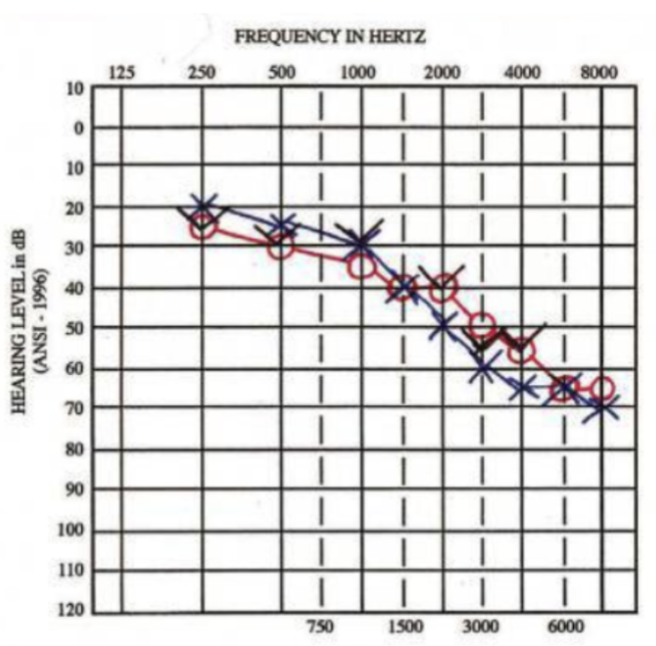
Some use PTA to characterize the degree of hearing loss
PTA is also compared to threshold measures of speech to determine the reliability of pure-tone thresholds

How do the SRT and pure tone average (PTA) relate to one another?
We use the SRT and PTA to judge the reliability of the patient’s pure-tone thresholds
SRT and PTA are expected to be within ±10 dB of each other
Example: PTA of 30 dB HL is expected to have an SRT between 20 and 40 dB HL
A >10 dB difference between SRT and PTA could suggest:
Patient did not understand your instructions
Reliability of pure-tone thresholds is poor
If SRT & PTA do not agree, re-instruct and re-test
If still not in agreement, note the poor reliability in your report

How do the SDT and pure tone average (PTA) relate to one another?
SDT is expected to be within ±10 dB of the lowest pure-tone threshold between 250 to 4000 Hz
Example:
Lowest pure-tone threshold in left ear = 20 dB HL
SDT in left ear expected to be between 10 to 30 dB HL
What are the purposes of word recognition testing?
Estimate the difficulty of understanding speech
Monitor the patient’s hearing ability over time
Assess candidacy for amplification
Provide insight into the potential site of the lesion
Conductive vs. cochlear vs. neural
What is the process of word recognition testing?
The test assesses the patient’s ability to correctly identify monosyllabic spoken words presented above threshold
Typical presentation level: 40 dB sensation level (dB SL)
Example: If SRT is 10 dB HL, present word recognition at 50 dB HL
Audiologist scores the percent of words correctly repeated back

What are two examples of specific word-recognition testing paradigms?
W-22: Developed at Central Institute for the Deaf (CID)
NU-6: Developed at Northwestern University
How should one categorize word-recognition scores?
By percentage level, calculated depending on the number of items presented during the test
90-100%: Excellent—minimal to no difficulty
80-89%: Good—slightly difficult, similar to listening on a telephone
70-79%: Fair—moderate difficulty
60-69%: Poor—Marked difficulty following conversation
<60%: Very poor—Patient may be unable to follow running speech
How can a word-recognition test help an audiologist determine the potential lesion site?
Normal hearing: reaches 100% correct around 40 dB above the SRT
Conductive loss: reaches 100% correct around 40 dB above SRT, but at higher intensity than normal hearing
Sensory (cochlear) loss: does not reach 100% correct even at high intensity levels
Neural (retrocochlear) loss: as intensity increases, performance reaches a point where it starts to decrease
What is a signal-to-noise ratio (SNR)? Which test is it used in?
SNR: Decibel difference between the amplitude of the signal (speech) and competing noise
Positive values: Speech is higher in amplitude than the noise
Negative values: Speech is lower in amplitude than the noise
0 dB: Speech and noise are equal in amplitude
SNRs are used in QuickSIN (Speech in Noise) tests
Results are quantified in terms of “SNR loss”
What is the AzBio test?
Sentences presented in quiet and at one signal-to-noise ratio
Male and female talkers speaking in more conversational style than other tests
Patient repeats back as much of sentence as possible
Audiologist scores number of keywords repeated back correctly
Result is quantified in percent correct
Used as part of evaluation for cochlear implant candidacy
What is the test battery approach?
Each audiologic test provides a piece of information about the patient’s auditory function
No one test provides all the information we need
We therefore use a combination of tests (battery) to make a proper diagnosis
What are the 6 different physiological tests audiologists can perform?
Otoscopy
Tympanometry
Otoacoustic Emissions
Auditory Brainstem Response
Acoustic Reflexes
Audiometry
What does tympanometry test for?
Tympanic membrane mobility
Middle ear pressure
Ear canal volume
What is impedance?
Impedance is the opposition to flow of energy
Impedance of middle ear includes:
Resistance: frictional force due to ligaments holding ossicles together
Mass: weight of tympanic membrane and ossicles
Stiffness: pressure placed on the stapes by the cochlear fluid
What is compliance?
Compliance is the opposite of impedance.
a measure of how easily the eardrum (tympanic membrane) and the middle ear system move and conduct sound vibrations in response to changes in air pressure
What is immittance?
An umbrella term combining the measurement of acoustic impedance (opposition to sound energy flow) and acoustic admittance (ease of sound energy flow) through the middle ear
What does acoustic reflex testing measure?
How the stapedius muscle contracts in response to loud sound
What are 4 examples of barriers to sound transmission in the middle ear?
Otitis media (fluid builds up behind the tympanic membrane)
Tumor in the middle ear space
Torn ligaments of the ossicles
Perforation of the tympanic membrane
What are some effects of Eustachian tube dysfunction?
Build-up of pressure behind the tympanic membrane (because the Eustachian tube’s purpose is to equalize pressure on either side of the tympanic membrane)
Reduced middle ear efficiency
Feelings of aural fullness

What does a tympanometer measure?
Immittance (how easily sound energy moves through the middle ear as a function of changing air pressure in the external ear canal)

How does a tympanometer work?
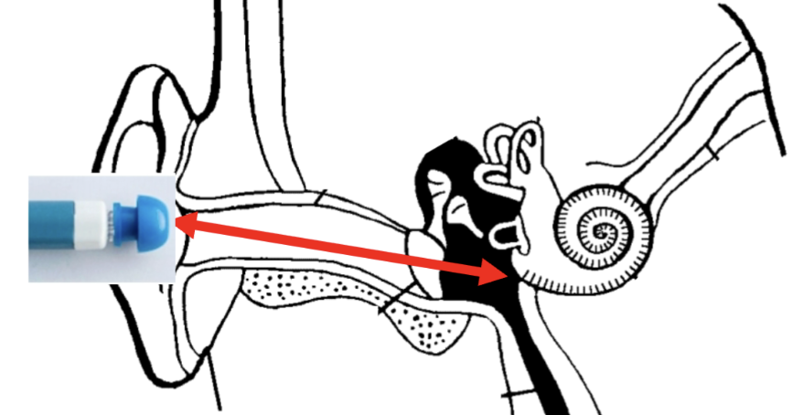
Probe tip is placed into the ear canal to form an air-tight seal
Air pump changes the pressure inside the ear canal
Loudspeaker presents a probe tone of 226 Hz into the ear canal
Microphone measures how much of the 226-Hz probe tone is transmitted through the middle ear

True or false: Tympanometry involves the inner ear
False. Tympanometry only involves the outer and middle ear
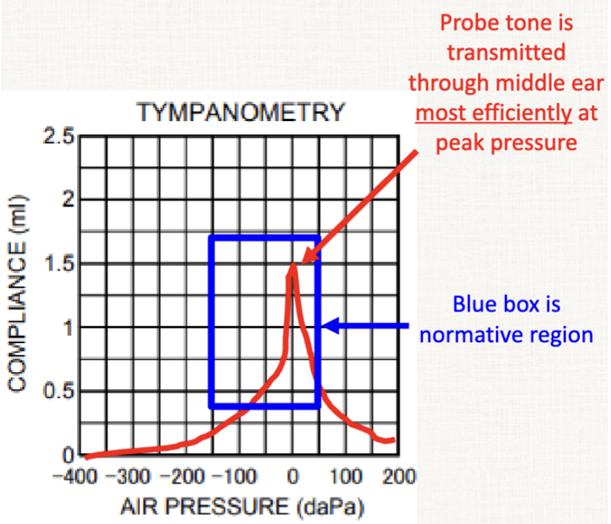
What are the axes of a tympanogram?
x-axis: Air pressure
Expressed in decapascals (daPa)
0 daPa = ambient pressure
y-axis: Compliance
Expressed in mL or mmho (1 mL = 1 mmho)
Larger values indicate more sound is transmitted through middle ear
What is a normal ear canal volume range in adults?
0.9 to 2.0 cc
What type is a Hypercompliant Tympanogram? What does it represent?
Type A-D (D stands for deep)
Compliance is abnormally high
Peak pressure and volume are normal
Causes include ossicular discontinuity and monomeric tympanic membrane
Ossicular Discontinuity: a separation or break in the chain of tiny bones (malleus, incus, and stapes) in the middle ear that stops sound vibrations from traveling properly to the inner ear
This causes the eardrum to become floppy and move with extreme ease, which causes the increase in compliance
Monomeric Tympanic Membrane: a thinned-out area of the eardrum that lacks its middle fibrous layer after a previous perforation heals

What type is a Shallow Tympanogram? What does it represent?
Type A-S (S stands for shallow)
Compliance is abnormally low
Peak pressure and volume are normal
Causes include otosclerosis and scarred tympanic membrane
Otosclerosis: abnormal bone growth in the middle ear that fixates the stapes bone and causes progressive hearing loss

What type is a Flat Tympanogram? What does it represent?
Type B
Causes include:
Otitis media
Cerumen impaction
Tympanic membrane perforation
Pressure equalization tube
We use ear canal volume to differentiate between possible causes
Normal adult volume = 0.9 to 2.0 cc

If a patient has a Type B Tympanogram and an ear canal volume of 0.4 cc, what does this most likely indicate?
Cerumen impaction

If a patient has a Type B Tympanogram and an ear canal volume of 3.0 cc, what does this most likely indicate?
A tympanic membrane perforation or a pressure equalization tube
If a patient has a Type B Tympanogram and an ear canal volume of 1.0 cc, what does this most likely indicate?
Otitis media

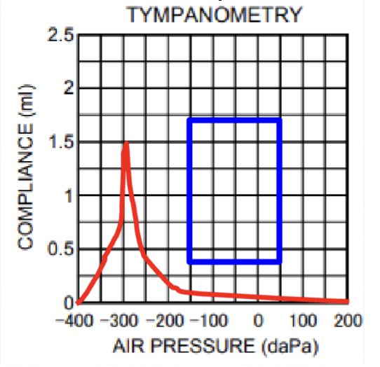
What type is a Negative Middle Ear Pressure Tympanogram? What does it represent?
Type C
Compliance is highest when we make the pressure in the ear canal negative
Peak pressure is abnormally negative
Compliance and volume are normal
Cause is due to Eustachian tube dysfunction
True or false: Tympanometry is not affected by pathology occurring beyond the middle ear
True
Which parts of the auditory system does acoustic reflex testing involve?
Outer ear
Middle ear
Inner ear
Auditory nerve
Brainstem
NOT INVOLVED: Cortex
What is an acoustic reflex?
High-intensity sounds can trigger a reflex that decreases the compliance of the middle ear
Compliance: the ease with which the eardrum and ossicles move
The acoustic reflex may protect the ear from acoustic trauma
Measurement of this reflex is also useful in audiologic diagnosis
What is the ascending acoustic reflex pathway?
Outer, middle, and inner ear
Auditory nerve (cranial nerve VIII)
Cochlear nucleus
Superior olivary complex and facial nerve (CN VII) nucleus
What is the descending acoustic reflex pathway?
Facial nerve (cranial nerve VII)
Stapedius muscle (muscle that contracts the stapes and reduces the compliance of the middle ear system. In other words, contraction of stapedius muscle stiffens ossicular chain, decreasing compliance)

What is the difference between the ipsilateral and the contralateral acoustic reflex pathways?
Ipsilateral Pathway:
Elicitor sound is presented to one ear via an earphone
The stapedius muscle on the same side is contracted
Compliance is measured using a probe
Contralateral Pathway:
Elicitor sound is presented to one ear via an earphone
The stapedius muscle on the opposite side is contracted
Compliance is measured using a probe
When are acoustic reflexes typically present?
Normal hearing
Mild and moderate sensorineural loss
(Rarely) lesions of the auditory cortex
When are acoustic reflexes typically absent?
Conductive hearing loss (ipsilateral reflex)
Severe and profound sensorineural hearing loss
Lesions of the auditory nerve and brainstem
Facial nerve disorders
What are the 2 exceptions you can have within acoustic reflex testing?
Contralateral reflexes can be present if elicitor presented to ear with conductive loss and probe is in unaffected ear (but threshold will be higher than normal)
Contralateral reflexes can be presented if elicitor is presented to unaffected ear and probe is in ear with severe/profound sensorineural hearing loss
How can you find the acoustic reflex threshold?
Start by measuring a tympanogram and obtaining peak pressure and compliance. Perform acoustic reflex testing at peak pressure, where the tympanic membrane is most efficient.
Find the acoustic reflex threshold
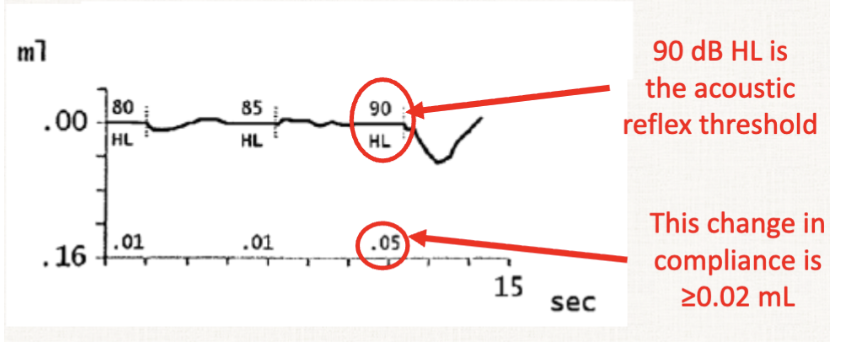
Lowest elicitor intensity that causes a decrease in compliance of ≥0.02 mL
Elicitor sounds may be pure tones (500-4000 Hz) or noise
Threshold measurement involves starting at a lower intensity (e.g., 70 dB HL) and increasing in 5-dB steps
Find lowest intensity that elicits a change of ≥0.02 mL
Ensure that the response is repeatable
Be prepared to stop if the elicitor becomes uncomfortably loud
What are the benefits of acoustic reflex testing?
Acoustic reflexes are useful for determining site of lesion
e.g., middle ear, cochlea, auditory nerve, etc.
Acoustic reflexes can also validate the results of other tests
For example: we expect absent acoustic reflexes in a patient with profound sensorineural hearing loss
What is the normal range of acoustic reflex thresholds?
70 dB to 100 dB HL
What describes the left contralateral acoustic reflex?
Elicitor in left ear, probe in right ear
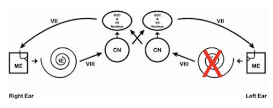
This patient has a severe sensorineural hearing loss in the left ear due to cochlear damage. Which of the following acoustic reflexes would be present in this patient?
Right ipsilateral and Right contralateral

Which of the following acoustic reflexes would be present in this patient?
Right ipsilateral
Right contralateral
Left ipsilateral
Left contralateral

This patient’s acoustic reflex threshold is ______ dB HL.
85
How to find it: causes a decrease of at least 0.02 in compliance (the lowest sound intensity that gives you that change). In this diagram, it’s at 0.3, where we find that characteristic dip
What parts of the ear does Otoacoustic Emissions (OAE) testing involve?
Outer ear
Middle ear
Inner ear
NOT INCLUDED:
Auditory nerve
Brainstem
Cortex
What are Otoacoustic Emissions (OAEs)?
Low-amplitude sounds created by the outer hair cells
A byproduct of the amplification provided by outer hair cells
Outer hair cells create a reverse traveling wave on the basilar membrane that returns to the middle ear and ear canal
OAEs are measured in the ear canal using a sensitive microphone

How are OAEs measured?
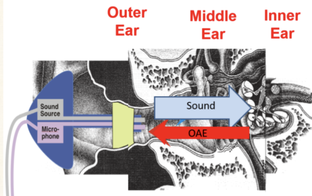
OAEs are measured with a loudspeaker and microphone probe placed in the ear canal
The loudspeaker presents two tones to generate OAEs
The microphone records the OAE returning back to the ear canal
True or false: you can be asleep during an OAE test
True, but both the patient and the room must be quiet because OAEs are low-amplitude sounds (10 dB SPL or less), so noise and vibration from the environment and patient interfere with the measurement
OAEs are absent in what type of hearing loss?
Conductive hearing loss, because it attenuates the incoming stimulus
Lower stimulus amplitude = lower OAE amplitude
Additionally, conductive hearing loss will attenuate the outgoing OAE reaching the microphone
Would a patient with sensorineural hearing loss have present DPOAEs?
It depends on whether that hearing loss is mild or not. We would need more information, but if the patient’s hearing loss is mild, then it is possible that DPOAEs will be present.
Which inner ear structures cause most sensorineural hearing loss?
Outer hair cells
If a patient has absent OAEs and an abnormal tympanogram, they most likely have _________________ hearing loss.
Conductive
If a patient has absent OAEs and a normal tympanogram, they most likely have _________________ hearing loss.
Sensorineural
DPOAE testing is a specific type of OAE testing. What does it involve?
Presenting 2 pure tones (f1 and f2) at the same time to stimulate specific spots in the cochlea
f2 is 1.2 times higher in frequency than f1
f1 is presented at 65 dB SPL
f2 is presented at 55 dB SPL
The inner ear naturally creates a "distortion" sound at 2f₁ - f₂ when hit with those two tones. The probe measures this exact frequency to check specific hair cell function.
What parts of the auditory system are involved in Auditory Brainstem Response testing (ABR)?
Outer Ear
Middle Ear
Inner Ear
Auditory Nerve
Brainstem
NOT INCLUDED:
Cortex
What is an Auditory Brainstem Response (ABR)?
Electrical activity generated by the auditory system in response to sound
Stimulus: short-duration stimuli played through earphones
Measured with electrodes on the scalp and mastoid while patient is asleep or sedated
Used for estimating pure-tone thresholds and for neurodiagnostic purposes
What is the pathway from the auditory nerve to the auditory cortex?
Auditory Nerve, Cochlear Nucleus, Superior Olivary Complex, Lateral Lemniscus, Inferior Colliculus, Thalamus (Medial Geniculate Body), Auditory cortex
AN | CN | SOC | LL | IC | T | AC
A Nice | Child Never | Should Obtain Cyanide| Lest Laurie | Ingest Cyanide | Then | Arrive Coughing |
What are the 4 neural generators of auditory brainstem response (ABR)?
Auditory Nerve
Cochlear Nucleus
Superior Olivary Complex
Lateral Lemniscus

True or false: in ABR testing, each of the 5 waves reflects activity of progressively higher parts of the central auditory system
True
In ABR testing: Wave I represents which part of the brain?
Wave I: Peripheral (distal) portion of the auditory nerve
In ABR testing: Wave II represents which part of the brain?
Wave II: Central (proximal) portion of auditory nerve
In ABR testing: Wave III represents which part of the brain?
Wave III: Cochlear nucleus
In ABR testing: Wave IV represents which part of the brain?
Wave IV: Superior olivary complex
In ABR testing: Wave V represents which part of the brain?
Wave V: Lateral lemniscus
When measuring the auditory brainstem response (ABR), what are the typical steps taken?
Mostly focus on waves 1, 3, and 5
Present the stimulus and measure the ABR
Start around 80 dB HL
Decrease stimulus intensity in steps of 10-20 dB
Measure the latency of Wave V at each intensity
Latency is the time location of the peak of the wave
ABR wave latency increases as stimulus intensity decreases

How can you find the auditory brainstem response (ABR) threshold?
It’s the lowest stimulus intensity that results in a measurable wave V
Obtained in each ear individually, either by air conduction or bone conduction

The auditory brainstem response (ABR) threshold is strongly correlated with what value?
The pure-tone thresholds
We can use ABR thresholds to estimate the degree of hearing loss
This is highly useful in patients who cannot be tested with pure-tone audiometry, such as infants

How can an auditory brainstem response (ABR) test give us information about brain tumors?
Presence of tumor affects brainstem transmission time
We compare the time difference between peaks (interpeak latency)
Tumors cause abnormally long interpeak latencies
Tumors are often unilateral, so we can compare the interpeak latencies in the patient’s affected ear to their unaffected ear

If a person has sensorineural hearing loss, what will their ABR results look like?
They will either have abnormal ABR thresholds, or they will have absent responses
If a person has conductive hearing loss, what will their ABR results look like?
ABR will be present, but threshold and latencies will be outside of normal range
As stimulus intensity decreases, the absolute latency of the auditory brainstem response (ABR):
Increases
In 2022, how many babies were born with permanent hearing loss?
6,000 (1.8 per every 1,000 births)
What percentage of hearing loss cases are due to genetics?
50-60%
What percentage of childhood hearing loss cases are due to maternal infection, environment, or birth complications?
30%
True or false: permanent hearing loss occurs more frequently than any other condition for which we can screen at birth
True

What is the average age at which permanent hearing loss is diagnosed?
35 months
What are the 1-3-6 guidelines for early hearing detection & intervention?
1: Hearing screening should take place no longer than 1 month of age
3: Children who refer on the screening should receive a diagnostic evaluation no later than 3 months of age
6: Children diagnosed with hearing loss should receive intervention no later than 6 months of age
What are 5 examples of risk factors for either congenital, delayed onset, or progressive hearing loss?
Caregiver concern regarding hearing, speech, language or developmental delay
Family history of permanent childhood hearing loss
In utero infections such as herpes, syphilis, and toxoplasmosis
Craniofacial anomalies, including those that involve the pinna, ear canal, ear tags, ear pits, and temporal bone anomalies
Head trauma, especially basal skull/temporal bone fracture that requires hospitalization
True or false: NICU babies are tested using DPOAEs.
False. Healthy newborns are tested with DPOAEs, but babies in the NICU must be tested with ABRs because they face a significantly higher risk of neural and sensory hearing loss