Positive and Negative Selection in T Cell Development
 Call Kai
 Learn
 Practice Test
 Spaced Repetition
 Match
 Flashcards
 Knowt Play
1/48

Earn XP
Description and Tags
Flashcards covering Lecture 14 on T-cell development, positive selection, negative selection, MHC restriction, HLA diversity, AIRE, and bone marrow chimera experiments.
49 Terms
Pre-TCR checkpoint
A developmental checkpoint in the subcapsular region of the thymus where proliferating double-negative thymocytes are assessed for productive rearrangement of their TCR β genes before progressing to immature double-positive cells.
TCR checkpoint
The developmental checkpoint in the thymic cortex where immature double-positive thymocytes are screened following rearrangement of their TCR α genes to confirm assembly of a complete αβ T-cell receptor.
How many rearrangments of VDJ (TCR) does a T cell carry?
ONE
therefore each T cell is specific for only one target
How many TCRs does a T cell carry?
about 30,000 but each TCR is identical bc each T cells only has one rearrangment
TCR α-chain locus - ignore
The genetic locus situated on human chromosome 14 comprising 70–80 Vα gene segments, 61 Jα gene segments, and a single Cα constant region segment.
TCR β-chain locus - ignore
The genetic locus situated on human chromosome 7 comprising 52 Vβ gene segments and two distinct clusters containing Dβ, Jβ, and Cβ segments (Dβ1 with 6 Jβ1 segments and Cβ1; Dβ2 with 7 Jβ2 segments and Cβ2).
Clonal TCR specificity
The organizational rule that each committed T cell carries only one functional V(D)J/VJ rearrangement and is therefore specific for only one target antigen.
Surface TCR abundance
The receptor density on the membrane of a single committed αβ T cell, which carries approximately 30,000 identical T cell receptors.
What does Positive selection test for on a thymocyte regarding its TCR?
Tests whether the TCR will bind MHC
What does Negative selection test for on a thymocyte regarding its TCR?
Tests whether the TCR binds too strongly to self-antigens, leading to thymocyte apoptosis.
where are the peptides located for the TCR to detect
by OUR MHC molecules
Human Leukocyte Antigen (HLA)
The name given to the Major Histocompatibility Complex (MHC) protein family in humans.
what we mean by “OUR” MHC molecules
HLA class I isotypes (3 types)
The three classical MHC class I protein types encoded per chromosome in humans:
HLA-A (with 2980 known allotypes)
HLA-B (3700 allotypes),
HLA-C (2661 allotypes).
HLA class II isotypes
The three classical MHC class II heterodimeric isotypes encoded per chromosome in humans:
HLA-DQ (35 α and 838 β allotypes)
HLA-DP (32 α and 695 β allotypes)
HLA-DR (2 α and 1878 β1 allotypes).
Individual MHC gene complement - how many MHC do we have per chromsomes?
The baseline set of MHC molecules present per person, consisting of 3 MHC-I and 3 MHC-II types encoded per chromosome, giving a total of 6 MHC-I and 6 MHC-II proteins expressed across both chromosomes.
how does the TCR detect MHC?
The requirement that a T cell receptor must simultaneously recognize both the specific foreign peptide antigen and the specific host self-MHC molecule displaying it in order to bind tightly and trigger activation.
does the TCR binds if its the wrong MHC? What is this called?
NO!
MHC restriction The phenomenon whereby T cell receptors (TCRs) only recognize specific peptide-MHC complexes, leading to a failure to bind if a non-matching MHC is present.
Positive selection
The developmental testing process in the thymic cortex providing a survival signal to double-positive thymocytes whose TCRs can bind self-MHC class I or class II molecules with moderate or strong affinity.
what cells do the thymocyte engage with to tests it TCR
Stromal cells - cortical epithelial cells
do stromal cells come from bone marrow or the thymus?
thymus
Death by neglect
The apoptotic death undergone by thymocytes that fail positive selection because their TCRs exhibit weak or no binding to self-MHC class I or class II molecules.
Cortical thymic epithelial cells (cTECs)
Stromal cells of thymic origin located in the thymic cortex that express both MHC-I and MHC-II loaded with self-peptides to mediate positive selection.
Practice peptides
Endogenous self-peptides produced and presented by cortical thymic epithelial cells on self-MHC molecules to test whether a thymocyte's TCR can recognize MHC structure, without testing specific peptide binding.
Single-positive lineage commitment
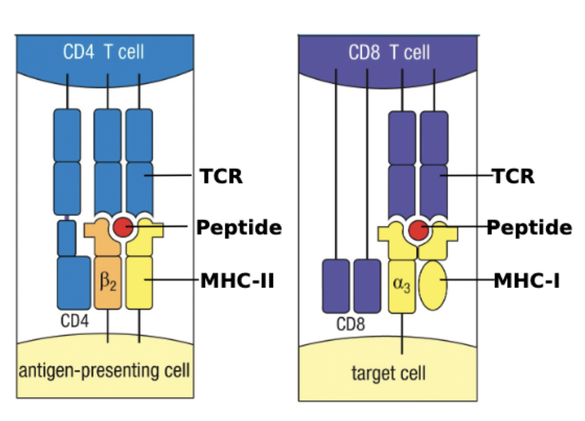
The developmental transition during positive selection where a double-positive thymocyte stops expressing one coreceptor to become either a CD4 or CD8 single-positive cell based on which class of MHC molecule its TCR engaged.
CD4 coreceptor
A surface glycoprotein required to bind MHC class II molecules that is selectively retained by thymocytes whose TCRs engage self-MHC-II during positive selection.
CD8 coreceptor
A surface glycoprotein required to bind MHC class I molecules that is selectively retained by thymocytes whose TCRs engage self-MHC-I during positive selection.
Mediate (immunology)
A term used by immunologists to describe a cell type or factor that directly causes or carries out a biological process.
Thymic stromal cells
Structural, non-immune cells of thymic origin—such as cortical and medullary epithelial cells—that reside permanently in the thymus and do not derive from bone marrow progenitors.
Thymic immune cells
Hematopoietic-derived cells within the thymus—including developing thymocytes, dendritic cells, and macrophages—that originate from bone marrow stem cells.
Bone marrow chimera
An experimental animal constructed by irradiating a host to ablate endogenous bone marrow progenitors and reconstituting it with bone marrow from a genetically distinct donor.
Radiation ablation
The experimental delivery of radiation to kill bone marrow progenitor cells, rendering a recipient mouse unable to generate new immune cells while preserving its radioresistant thymic stroma.
MHCa×b→MHCa bone marrow chimera
An experiment showing that F1 hematopoietic cells expressing both MHCa and MHCb transplanted into an irradiated MHCa recipient develop T cells restricted solely to MHCa, demonstrating that thymic stromal cells mediate positive selection.
Negative selection
The central tolerance process that eliminates developing thymocytes whose TCRs bind self-antigen:self-MHC complexes with high affinity (too tightly), instructing them to die by apoptosis.
OCCURS IN THE CORTICO-MEDULLARY JUNCTION.
Autoimmunity
An immune-mediated pathology that occurs when self-reactive T cells escape tolerance checkpoints and become activated against the body's own tissues.
Clonal deletion
The targeted apoptotic elimination of self-reactive thymocyte clones upon high-affinity interaction with self-antigen presented on MHC molecules during negative selection.
Moderate-affinity survival
The selective survival of thymocytes that bind self-peptide:MHC complexes with moderate affinity, ensuring positive selection survival while preventing gaps or holes in the TCR repertoire against foreign pathogens.
Cortico-medullary junction
The anatomical transition zone between the thymic cortex and medulla where negative selection occurs most prominently as developing thymocytes migrate inward.
Autoimmune regulator (AIRE)
A transcription factor expressed by medullary thymic epithelial cells that enables the expression and presentation of body-wide peripheral tissue antigens in the thymus.
Medullary thymic epithelial cells (mTECs / MECs)
Thymic-origin stromal cells located in the medulla that express AIRE to generate peripheral tissue-specific proteins, directly mediating a minor component of negative selection.
Thymic dendritic cells
Bone marrow-derived antigen-presenting cells residing predominantly at the cortico-medullary junction and medulla that uptake AIRE-derived proteins from apoptotic MECs to mediate negative selection.
Primary mediators of negative selection
Bone marrow-derived antigen-presenting cells—specifically dendritic cells and macrophages—that carry out the majority of negative selection in the thymus.
what happens after negative selection (dendiritic cell)
mature T cells leave the go to the lymphoid to await activation from DCs in an immune response
Minor mediators of negative selection
Medullary thymic epithelial cells (MECs), which can directly present self-antigens to thymocytes to trigger negative selection, though to a lesser extent than bone marrow-derived immune cells.
H-Y antigen
A male-specific self-protein encoded on the Y chromosome used in transgenic TCR models to demonstrate central tolerance and negative selection.
H-Y TCR knock-in mouse
A transgenic mouse engineered with pre-arranged VDJ in the β chain and pre-arranged VJ in the α chain specific for H-Y, bypassing normal recombination checkpoints so nearly all its T cells recognize H-Y.
Female H-Y TCR transgenic phenotype
The experimental observation that female mice lacking a Y chromosome treat H-Y as a foreign antigen, allowing H-Y-specific T cells to survive positive and negative selection and colonize peripheral lymph nodes.
Male H-Y TCR transgenic phenotype
The experimental observation that male mice carrying a Y chromosome treat H-Y as a self-antigen, resulting in the complete deletion of H-Y-specific thymocytes after positive selection and their absence in peripheral lymph nodes.
Thymocyte developmental mortality rate
The percentage of developing thymocytes (98%) that fail selection checkpoints and undergo apoptosis within the thymus.
Thymic cell turnover kinetics
The quantitative cellular metrics of the thymus, maintaining a steady-state pool of 108 thymocytes, receiving 5×10⁵ new progenitors per day, and exporting 106 mature, self-restricted, self-tolerant T cells per day.