Genetics 2250 Topic 4
 Call Kai
 Learn
 Practice Test
 Spaced Repetition
 Match
 Flashcards
 Knowt Play
1/107

There's no tags or description
Looks like no tags are added yet.
108 Terms
nucleotides
A single strand of a DNA molecule is a chain of subunits called nucleotides
Each nucleotide is made of the sugar deoxyribose connected to an inorganic phosphate group and to one of the four nucleobases (a.k.a. nitrogenous bases)
phosphodiester bonds
The phosphodiester bonds that link the nucleotide subunits to each other attach the phosphate group of one nucleotide to the deoxyribose sugar of the preceding nucleotide
Robert Feulgen
The German chemist Robert Feulgen developed a histochemical staining technique in 1924 that detects DNA in cells and tissues.
The Feulgen Reaction is highly specific for DNA because RNA lacks the deoxyribose sugar required for this staining method.
The intensity of staining is proportional to the amount of DNA present, allowing for assessment of DNA content.
Bacteria carry their genetic material in a _
single circular chromosome
bacteria reproduce through _, and they do not use - to apportion their replicated chromosomes to daughter cells or - to produce gametes.
binary fission , mitosis , meiosis
, Frederick Griffith
In 1923, Frederick Griffith (England) distinguished 2 forms of Streptococcus pneumoniae : one with rough colony (R) and one with smooth colony (S). In humans, the virulent S form of S. pneumoniae can cause pneumoniae, whereas the R form is avirulent.
The R form of Griffith’s bacteria lacks an enzyme necessary for synthesis of the —, which helps —
capsular polysaccharide, protect the bacterium from the host’s immune system
griffith’s mouse experiment
Griffith performed an experiment where mice were injected either with the S strain or the R strain.
He noticed that mice infected with the S strain died within a few days after injection, while mice injected with the R strain did not die.
When S cells were heat-inactivated, they failed to cause disease in mice.
When heat inactivated S cells were mixed with live R cells, mice died a few days after injection. Surprisingly, the dead mouse’s blood carried S strains
Something from the heat killed S bacteria must have transformed the living R bacteria into S. What is it?
Avery-MacLeod-McCarty
experiment in 1944 was the first conclusive demonstration of DNA’s role in heredity.
Avery's team employed a method for transforming bacteria in cultures rather than in living mice, by co incubating R cells with heat killed S components in medium
Avery, MacLeod and Maclyn McCarty used a process of elimination to identify the transforming principle
Identical extracts from heat-treated S cells were first treated with hydrolytic enzymes that specifically destroyed protein, RNA, or DNA.
Encapsulated S cells appeared in all of the cultures, except those in which the S strain extract had been treated with DNAse, an enzyme that destroys DNA.

T2 bacteriophage
, a virus that infects the E. coli bacterium, multiply rapidly within their host causing the cells to burst, thus releasing large numbers of new infectious bacteriophages
T2 bacteriophage consists of both protein and DNA, but the role that each substance played in the growth of the bacteriophage was unclear.
Chase & Hershey findings
used radioisotopes to trace the fate of the phage's protein and DNA by taking advantage of their chemical differences. Proteins contain sulfur, but DNA does not. Conversely, DNA contains phosphate, but proteins do not

Maurice Wilkins and Rosalind Franklin
developed methods to produce diffraction patterns of DNA.

Each nucleotide consists of
a deoxyribose sugar, a phosphate and a nitrogenous base (or nucleobase)
Nucleobases include
purines (A, G) and pyrimidines (C, T).
nucleoside
Covalent attachment of a nucleobase to the 1’ carbon of deoxyribose forms a nucleoside.
5’-to-3’ direction
The DNA polymer (or chain) has a direction, i.e. polarity.
By convention, a DNA chain is described in terms of its bases, written with the 5’-to-3’ direction.
Watson and Crick
published the structure of DNA in 1953 in the journal Nature
In their model, two DNA chains spiral around an axis with the sugar-phosphate backbone on the outside and flat pairs of bases meeting in the middle
Watson determined that A-T pairing involves 2 non covalent hydrogen bonds, whereas G-C pairing involves 3 hydrogen bonds
Erwin Chargaff
noticed that in every organism, the ratios of A to T and G to C are not significantly different from 1:1
This means A and T, and G and C, probably pair together
Antiparallel Chains
The two chains of DNA spiral around an axis with the sugar-phosphate backbone on the outside and flat pairs of bases meeting in the middle.
One chain runs 5’ to 3’ upwards, while the other runs in the opposite direction of 5’ to 3’ downward
major and minor grooves
The two backbones are vertically displaced from each other, generating two grooves: The major groove and the minor groove
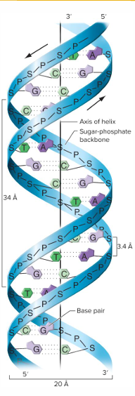
B and Z DNA
B DNA: Majority of naturally occurring DNA, spiraling to the right
Z DNA: Z-form DNA in which the helix spirals to the left (can be observed in vitro)

Linear DNA

Circular DNA

The information content of DNA resides in the sequence of its _
bases
Most genetic information is read from
unwound DNA chains.
unwound DNA chains.
makes the information accessible for fundamental processes to occur, including DNA replication and transcription.
E.g.: RNA polymerase reads DNA on one strand to make RNA
protein binding
Some proteins can recognize and bind to specific base-pair sequences in double-stranded DNA
Some information is « visible » in the major and minor grooves, where certain atoms at the periphery of the nucleobases are exposed
RNA
Ribose, not deoxyribose
Uracil, instead of thymine
Single-stranded, rather than double-stranded
Most RNA molecules are single-stranded, but are sufficiently flexible so that some regions can fold back and form base pairs with other parts of the same molecule
Transfer RNA (tRNA)
molecule is a single-stranded molecule that exhibits significant intracellular base pairing, giving it its characteristic three-dimensional shape.
Model proposed by Watson and Crick
The double helix unwinds to expose the bases in each strand of DNA
Each of the two separated strands acts as a template for the synthesis of the second strand
The newly replicated strands form as complementary bases align opposite to the exposed bases on the two parental strands
Enzymes join the base’s nucleotide to the preceding nucleotide by a phosphodiester bond, linking nucleotides into a continuous strand.
This is called semiconservative replication: a copying in which one strand of each new double helix is conserved from the parent molecule and the other is newly synthesized
semiconservative replication
a copying in which one strand of each new double helix is conserved from the parent molecule and the other is newly synthesized.
conservative model
Unlike the semiconservative model, the conservative model proposes that the parental double helix remains intact while both strands of the doughter helix are newly synthesized
dispersive replication model
In dispersive replication model, both strands of both daughter double helixes contain both original and newly synthesized material
Meselson-Stahl
The Meselson-Stahl experiment used Cesium Chloride gradient density centrifugation and differentially labeled DNA, one with 14N (light) and another one with 15N (heavy), to decipher the mechanism of DNA replication (proved it is semi-conservative).

Kornberg lab
The Kornberg lab purified individual components of the replication machinery in E. coli and was able to elicit the reproduction of genetic information outside a living cell, i.e. in a tube; in vitro
This required: dNTPs (deoxyribonucleotide triphosphates) DNA template (single stranded, DNA must be unwound) Primer with a free 3’-OH group Purified enzymes
The template strand is antiparallel to the newly synthesized DNA strand
DNA polymerase cleaves dATP between the first and second phosphate groups, providing the energy needed to form a phosphodiester bond between the terminal 3’-OH group on the preceding nucleotide to the first phosphate of the dATP substrate with release of pirophosphate as a byproduct
does DNA Polymerase have strict Operating Requirements
yes
DNA replication
Inside a living cell, DNA replication is a highly coordinated, complex, process
It includes 2 stages: Initiation and elongation
Initiation
Initiation involves unwinding of the double helix by DNA helicase which is attracted by the initiator protein at the replication fork. The single strands in the replication bubble serve as templates for DNA synthesis.
The enzyme DNA polymerase III requires a primer to synthesize DNA. In living cells, primers are short RNA fragments called RNA primers synthesized by the enzyme primase.
elongation
During elongation, DNA polymerase III catalyzes polymerization to form phosphodiester bonds such that the DNA under construction grows in the 5’→3’ direction.
On the DNA strand with 3’ end, polymerization is continuous: Leading strand
On the other strand, polymerization is discontinuous: Lagging strand, generating Okazaki fragments (of about 1,000 bases)
Finally, DNA polymerase I and other enzymes replace RNA primers between Okazaki fragments with DNA, then DNA ligase joins fragments into a continuous strand of DNA
dna replication of a circular chromosome
In the circular E. coli chromosome, there is only one origin of replication.
When both forks moving in opposite direction meet at termination region, replication ends
This generates overwound, supercoiled region that are subsequently relaxed by DNA topoisomerase that break, unwind then suture the DNA!

The most important mechanism of generating genetic diversity is the production of
new combinations of already existing alleles —> Meiosis
two ways of getting new combinations during Meiosis
Independent assortment (free, random, segregation of homologous chromosomes)
Crossing over (two homologous chromosomes exchange parts) RECOMBINATION
Tetrads
Some fungi like yeast and Neurospora generate asci carrying all the products of individual meiosis
Tetrad Analysis

How does recombination occur from the molecular genetics perspective? (Meselson and Weigle experiment)
The Meselson and Weigle experiment (1961) proved that genetic recombination happens through the physical breakage and reunion of DNA molecules
E. coli cells were co-infected with two different strains of bacteriophage lambda, one carrying heavy DNA with 13C and 15N and the other carrying light DNA 12C and 14N
The progeny was spun on a Cesium Chloride density gradient, where Ab and aB recombinants were detected
Breakage and reunion of DNA

Crossing over step 1
Step 1: Spo11 protein (meiosis-specific endonuclease) cleaves a phosphodiester bond on both strands of DNA
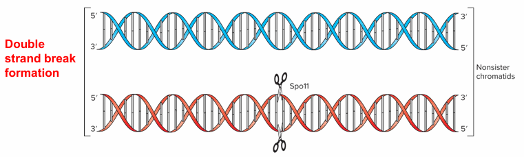
Crossing over step 2
Step 2: An exonuclease degrades the 5’ ends on each side of the break to form 3’ overhang strands

Crossing over step 3
Step 3: Dmc1 protein catalyzes base pairing between the invading strand and its complementary strand. The non-complementary strand is pushed out forming a D-loop, stabilized by Replication Protein A (RPA). A heteroduplex is formed.

crossing over step 4
Step 4 – Crossover pathway: An additional heteroduplex is formed within the additional non-sister chromatid. DNA ligase forms phosphodiester bonds between the adjacent ends to rejoin DNA strands and generate Holliday Junctions, which are interlocked regions of two non-sister chromatids in recombination intermediates

crossing over step 5
Step 5 – Crossover pathway: The two invading strands tend to “zip up” by base pairing and migrate, generating extended heteroduplexes

crossing over step 6
Step 6 – Crossover pathway: Resolvase breaks the two strands at each Holliday Junction and ligase rejoins ends.

DNA Repairof Heteroduplexes
can Result in Gene Conversion
In heteroduplexes, mismatched bases do occur (e.g. in case of SNPs in mother vs father DNA) leading to the induction of a repair mechanism that corrects the mismatch
Depending on the nature of DNA repair, gene conversion can occur.

Homologous recombination
n helps to increase genetic diversity, upon which natural selection can act
site-specific recombination
Some organisms find it useful to have systems for site-specific recombination.
It is a crossing-over that occurs between two specific, short, DNA target sites.
Site-specific recombination is much simpler than homologous recombination at the molecular level
A single protein, called recombinase (e.g. Cre, FLP), is sufficient to catalyze breakage and joining of DNA ends Site-specific recombination can lead to integration of genetic material into a genome – e.g. Phage infection in bacteria
what catylizes site-specific recombination
enzymes called recombinases.
Excision
Excision can allow phage DNA to be removed from bacterial genome

integration

inversion

chromosome exchange

applications of site-specific recombination
Scientists can direct where DNA recombine using recombinases and genetic engineering of DNA sequence. New technologies have been created by Biotech companies, allowing for example high-throughput cloning using recombinases instead of restriction enzymes

CRISPR Cas9 genome editing technology
small guide RNA and a nuclease called Cas9 allow to induce double strand breaks at a specific location of the genome

What are mutations?
Changes in DNA base sequences
A forward mutation
changes a wild-type allele of a gene to a different allele
A reverse mutation
causes a mutant to change back to wild type. Synonymous with reversion.
Substitution
A base at a certain position is replaced by one of the other three bases
Deletion
One or more base(s) removed
Insertion
One or more base(s) added
Spontaneous Mutations Occur at a _Rate
Low
human mutation rate
Mutations can impact on the function of genes, but such events are rare; occurring nearly at an average rate of 2-12 × 10-6 per gene per gamete (but this varies a lot from a given gene to another: less than 10-9 to more than 10-3)
The rate of forward mutation is usually much _ than the rate of reversion.
higher

Sperm drives the majority of new point mutations in humans, exhibiting a mutation rate roughly __higher than that of eggs.
3 to 4 times
why is sperm more prone to mutation than eggs
This is primarily driven by the fundamental differences in how male and female gametes are produced.
Male germ cells undergo continuous, lifelong mitotic divisions to produce sperm. By age 20, a sperm cell has undergone roughly 200 rounds of DNA replication, increasing to over 600 rounds by age 40. Every replication cycle introduces a baseline risk of random copying errors.
Female germ cells complete all their mitotic divisions during fetal development (approximately 22 to 30 replication cycles). The resulting oocytes remain arrested in Meiosis I until ovulation, completely bypassing ongoing replication errors.
The Fluctuation Test
Experiment by Salvador Luria and Max Delbruck (1943): The Fluctuation Test Genetic mutations occur randomly in the absence of selection rather than as an adaptive response to environmental stress
Finding: The number of resistant bacteria fluctuate (i.e. vary) after exposure to antibiotic → Random events
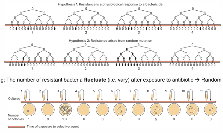
Replica plating experiment
the mutations conferring bacterial resistance occur before the cells encounter the antibiotic that selects for their resistance:
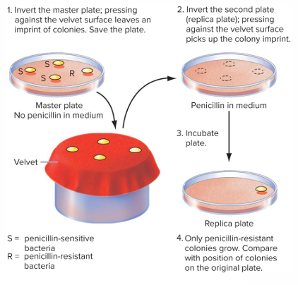
Three competing processes: - - -are at the origin of the emergence of mutations
DNA alteration, DNA repair and DNA replication
DNA alteration
(the damage): DNA is constantly damaged by internal factors (like metabolic byproducts or replication errors) and external factors (like UV light or chemicals). This creates a lesion or change in the DNA structure
DNA repair
(the fix): Cellular repair mechanisms constantly scan the genome to fix these alterations and restore the original sequence.
DNA replication
(the lock-in): If DNA replication occurs before the repair machinery can fix the alteration, the DNA polymerase will read the damaged or incorrect strand and permanent copy it into the new strand. Once replication is complete, the change is permanently locked into the genetic code of the daughter cells as a mutation.
Point mutations result when _
DNA replication wins the race with DNA repair
DNA damage is _ in a cell
quite frequent
depurination
For example, purine base (e.g. Guanine) hydrolysis occurs 1000 times/hour in every human cell. This is called depurination, leading to apurinic site. Apurinic site does not specify a complementary base, consequently DNA replication process introduces a random base causing a mutation with a probability of 75%
Deamination
consists of the removal of an amino group (NH2-) from a nucleobase like cytosine, changing it to Uracil. Following DNA replication, this leads to a transition from C to T.
X-rays
(cosmic radiation) break the sugar-phosphate backbone leading to DNA strand breaks and potentially deletions

UV radiation
causes adjacent Thymines to form dimers, which leads to substitutions when DNA is replicated

Oxidation and Oxidative stress
can damage DNA by generating oxidized Guanine, specifically 8-oxodG (8-oxo-7,8-dihydro2'-deoxyguanosine). During replication, 8-oxodG incorrectly mispairs with Adenine, which ultimately generates a G:C to T:A transversion mutation

Mistakes in DNA Replication
DNA replication machinery can make errors, although rarely.
In vitro, DNA polymerases replicate DNA with an estimated error rate of about 1 mistake every 106 base pairs.
1 in 10^6 is still low, owing to proofreading activity of DNA polymerases.
In addition to their polymerization function, DNA polymerases carry a proofreading/editing function in the form of a 3’→5’ exonuclease activity that becomes active when the polymerase makes a mistake.
The exonuclease removes the wrongly incorporated base so that DNA polymerase adds the correct base.

Base tautomerization
is one of the mechanisms that trigger DNA replication errors by DNA polymerase.
Tautomers
are structural isomers of a molecule that spontaneously interconvert with each other. It exists in a chemical equilibrium.
If by chance a base is in its rare tautomeric form when DNA polymerase is active, __
the wrong base will be incorporated.
Trinucleotide repeat diseases
exemplify the impactful consequences of mutations in humans
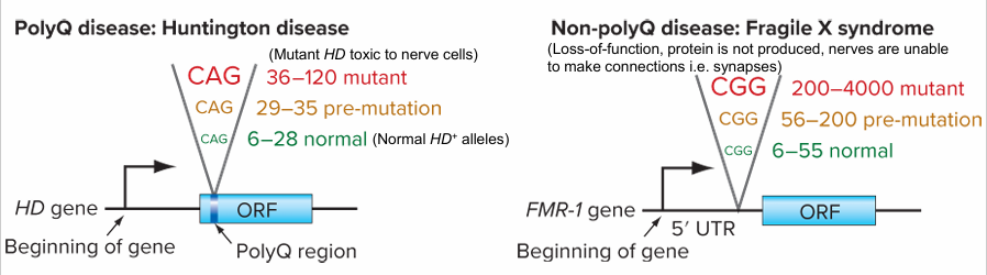
20 known neurodegenerative diseases with triplet repeats, fall into 2 categories
PolyQ (Gln, CAG triplet repeat) diseases: E.g. Huntington disease (coordination issues, cognitive decline, psychiatric problems)
Non-polyQ (CGG, CTG, GCC, GAA) diseases: E.g. fragile X syndrome (CGG triplet repeat, inherited intellectual disability)
mutagen
Any physical or chemical agent that raises the frequency of mutations above the spontaneous rate is called a mutagen
The use of base analogs
These are mutagens that are so similar in chemical structure to the normal nitrogenous bases in DNA that the replication mechanism can incorporate them into DNA in place of the normal bases.
Their incorporation can cause base substitutions on the complementary strand synthesized in the next round of DNA replication.
Alteration of base structure and properties
Some mutagens alter the structure of bases such that they pair inappropriately in the next round of replication.
Agents can alter base structure through hydroxylation, alkylation or deamination.
Alkylation

Deamination

Intercalators
are a class of chemical mutagens composed of flat, planar molecules that can sandwich themselves between successive base pairs, disrupt the machinery of DNA replication, and thereby cause mutation.

Somatic mutations
in genes that help regulate the cell cycle may, for example, lead to cancer. For this reason, most mutagens act as carcinogens (i.e. cancer-causing agents)
The Food and Drug Administration (FDA)
in the U.S. tries to identify carcinogens by using the Ames Test to screen for chemicals that cause mutations in bacterial cells.

DNA Repair Mechanisms
Cells have evolved a variety of enzymatic systems that repair damaged DNA and thereby minimize dramatically the occurrence of mutations
One of the repair mechanisms involve reversion of DNA base damage
For example if ethyl groups are added to guanine, alkyltransferase enzymes can remove them
Similarly, the enzyme photolyase recognizes the thymine dimers produced by exposure to UV light and reverses the damage by splitting the chemical linkage between the thymines.
Other repair systems employ a general strategy of homology-dependent repair in which they first remove a short region from the DNA strand carrying the altered base, and then use the complementary strand as a template to re-synthesize the removed region
base excision repair mechanism