Endocrine OSPE flashcards
 Call Kai
 Learn
Learn Practice Test
 Spaced Repetition
 Match
Match Flashcards
 Knowt Play
1/232

There's no tags or description
Looks like no tags are added yet.
Name | Mastery | Learn | Test | Matching | Spaced | Call with Kai | Chat |
|---|
No analytics yet
Send a link to your students to track their progress
233 Terms

Identify:
Mention histological features:


Identify:
Mention histological features:


Identify:
Describe:
Causes :
Complications :
Mutations :
Clinical features :


Identify:
Describe:
Causes :
Complications :
Mutations :
Clinical features :


Identify:
Describe:
Complications :
Mutations :
Clinical features :


Identify the section:
Mention histological Features:
Identify the section: Ovary
Mention histological Features:
• Stroma: Tunica albuginea covered by germinal epithelium.
• Parenchyma:
1. Cortex:
contain ovarian follicles.
2. Medulla: highly vascular CT. It
contains:
- Blood vessels. - Smooth muscle ibers - Hillus cells

Identify the section:
Mention histological Features:
Mention features of the uterine gland:
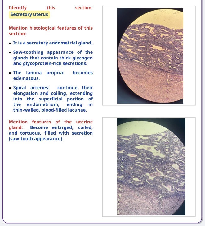

Identify :
Describe :


Identify the section:
Mention histological Features:


Identify :
describe :


Identify :
Describe :


Identify :
Mention histological features :


Identify :
Mention histological features :
Identify :
Steroid secretory cell
Mention its histological features :
1-Polyhedral cell with central rounded nucleus.
2- Have acidophilic cytoplasm because it's contains mitochondria, SER.
3-Contains many lipid droplet.
4-Have fenestrated blood capillaries

Identify the largest pale area? / Identify this picture :
Mention its histological features :
list / mention the cell in these areas:
mention the most numerous / common one / mention its histological feature ?
cell in the periphery and the a count 15% , mention its histological feature ?
Identify the largest pale area? / Identify this picture?
Islet of Langerhans
Mention the histological features?
1. multiple large pale areas scattered between the darkly
stained pancreatic Acini.
2. More numerous at the tail region.
3. Not capsulated.
4. surrounded by reticular CT.
5. formed of cell masses and cords
6. secreting cells separated with fenestrated blood
capillaries.
list / mention the cell in these areas?
Alpha-Beta-C cells-Delta-E cells - F cells - G cells - Ganglion
mention the most numerous / common one / mention its histological feature ?
( Beta )
cell in the periphery and the a count 15% , mention its histological feature ?
( Alpha )
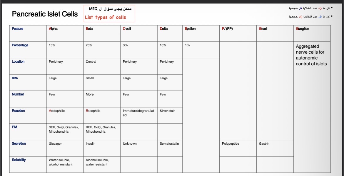

Identify :
Mention histological features :
Identify this picture?
EM of Islet of Langerhans
2 - Mention the histological features?
-Beta cells
• more numerous 70%
• Smaller.
• Central.
• basophilic granular cytoplasm.
• Granules are water resistant.
• Cytoplasm contains RER,Golgi,mitochondria,
• They release insulin hormone

Identify the marked structure
Mention its site
Mentions its Relations
Mention its blood supply
Mention its parts
Pituitary gland( Hypophysis cerebri)
Located in the (cranial cavity) within the (pituitary fossa or sella turcica) on
(superior surface of the sphenoid body)
Relations
-Antero-superior: optic chiasma
-Inferiorly: sphenoidal air sinuses
-Superiorly: the diaphragma sellae
-Laterally On each side: Cavernous sinus
*Cavernous sinus contents:
1-Internal carotid artery
2-Cranial nerves : 3rd,4th,5th v1 , 5th v 2 , 6th
Blood Supply:
-Arteries:
•Superior hypophyseal artery
•Inferior hypophyseal artery
(branches from internal carotid arteries ICA)
-Veins:
•hypophyseal veins
(drains into Cavernous sinuses)
Parts of Pituitary Gland:
-Adenohypophysis (Anterior lobe):
•Pars distalis
•Pars intermedia
•Pars tuberalis
-Neurohypophysis (Posterior lobe):
•Pars nervosa
•Infundibular stem
•Median eminence

Identify :
Mention histological features:
Identify: EM of thyroid follicle:
Histological features:
- Lined with follicular F and parafollicular C cells resting on a
basement membrane BM and surrounding a lumen filled with
homogeneous colloid Co.
- Follicular cells F: cuboidal, joined by junctional complex and
show central rounded pale nuclei, short apical microvilli Mv,
numerous basal rER, mitochondria Mi , supranuclear Golgi G,
many lysosomes L & apical vesicles .
- Parafollicular C cell: is present on the BM but not exposed to
lumen. The cytoplasm contains numerous small electron-dense
secretory granules , well developed RER , mitochondria ,Golgi
- A fenestrated capillary Cap is present close to the
follicular BM

Identify :
Mention histological features:
Identify marked structures x,y
Identify : Parathyroid gland
Histological features:
- Formed of glandular tissue G intermixed with adipose tissue A.
- Glandular tissue G is formed of
two cell types: chief (principal) cells and oxyphil cells.
- Cells are arranged as clusters or anastomosing cords surrounded by fenestrated blood capillaries
X = glandular tissue
Y= adipose tissue

Identify :
Mention histological features:
Identify marked structures x,y
Define: Parathyroid gland
Histological features:
- Chief (principal) cells P are more numerous, small in size, polyhedral in shape with central rounded nuclei and pale clear or slightly acidophilic cytoplasm contain
RER, mitochondria, Golgi, small secretory granules
They secrete PTH.
- Oxyphil cells O are less in number, larger in size and clustered. They are rounded or oval cells with deeply acidophilic cytoplasm packed with mitochondria.
These cells are of unknown function.
- Many fenestrated capillaries C are present between the cells
X= chief cells
Y= oxyphil cells

Identify :
mention histological features:
Identify : SEM of primary follicle
Histologyical features:
1- central oocyte surrounded by zona pellucida(ZP).
2- granulosa cells(GC)

Identify :
Mention histological features :
Identify: Ovarian Cortex
Histological features:
1- Cortex cover by Tunica Abuginea(TA)
contains collagen fiber covered by germinal epithelium(E).
2- Priomrdial follicles(PF).
3- primary oocyte(O1) surrounded by zona pellucida(ZP).
It’s divided into: Unilaminar and Multillminar

Identify :
Mention histological features :
Identify : Ovary
Histological Features:
Stroma: tunica albuginea covered by germinal epithelium.
Parenchyma:
1- Cortex: contains ovarian follicles.
2- Medulla: highly vascular CT
Contains
1- blood vessels.
2-smooth muscle fibers.
3-Hillus cells

Identify :
Mention histological features :
Identify marked structure x,y,z


Identify :
Mention histological features :
Identify marked structure x,y,z


Identify :
Mention histological features :
Identify: Mature Graafian Follicle
Histological features:
1-Secondary oocyte(O2)
2-Follicular Antrum(FA): contains fluid (liquor folliculi)
3-The Cumulus oophorus.
4-Corona Radiata (CR)
5-Zona Pellucida (ZP)
6-Zona Granulosa (ZG)
7-Theca Folliculi (TF) has 2 layers:
A-Theca Interna (TI) consists of rounded cells.
B-Theca Externa (TE) consists of collagen fibers

Identify :
Mention histological features :
Identify: Corpus Luteum
Histological features:
1- Granulosa lutein cells[G](steroid secreting cells):
Large, pale , polygonal cells and 80%of corpus luteum.
2- Theca lutein cells[T]:
Small, dark and 20% of corpus luteum.
3- Blood clot [B]
4- Septa [S]

Identify :
Mention histological features :
Identify: Fallopian Tube
Histological Features:
The layers from inside to outside:
1-The mucosa: highly folded
A-Two cell types of simple Columnar epithelium(E):
non-ciliated peg cells and ciliated cells.
B-The lamina propria containing:
Mast cells and lymphoid cells.
2-The musculosa: inner circular and outer longitudinal.
3-The Serosa:CT covered by simple squamous epithelium, it contains large blood vessels

Identify :
Mention histological features :
Identify : Proliferative uterus.
What are the histological features of his section?
S: stratum functionalis.
B: stratum basalis.
My: myometrium
G: straight tubular form of the endometrium glands.
The glands G at this are empty , straight and narrow
As the glands , stroma and vessels proliferate , the endometrium gradually becomes thicker [2-3mm]

Identify :
Mention histological features :
Mention the features of the glands :
endometrium reaches its maximum thickness [5mm]
![<p>endometrium reaches its maximum thickness [5mm]</p>](https://assets.knowt.com/user-attachments/f614ae8d-cee8-4f0a-b9e0-d59d41f5ffb0.jpg)

Identify :
Mention histological features :
Identify : Uterine cervix
What are the histological features of his section?
EC: endocervical canal, it is lined by a single layer f columnar mucus secreting epithelial cells.
V: vaginal part of the cervix, called ecto - exocervix, it is lined by thick stratified squamous epithelium, the cells of the ectocervix usually have clear cytoplasms due to their high glycogen content.
J is Transformation zone, junction between endo and ectocerviacl epithelium
•the main bulk of the cervix is composed of tough, collagenous tissue containing a little smooth muscles.
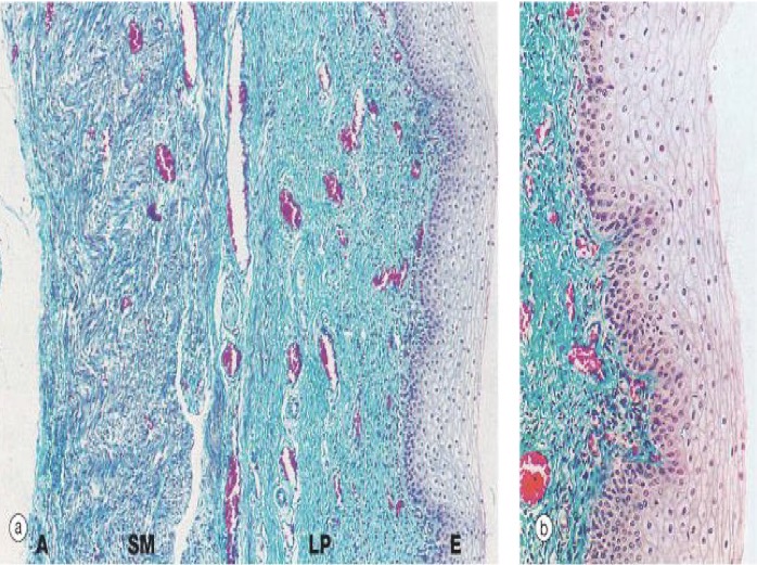
Identify :
Mention histological features :
Identify : Vagina
What are the histological features of his section?
. 1/Mucosa , and composed of:
1. Stratified squamous non-keratinized epithelium
2. Muscularis, and composed of:
1. Inner circular fibers and Outer longitudinal smooth muscle layer
3. Adventitia

Identify :
Mention histological features :


Identify :
Mention histological features :
Mention the functions of the cell :
It’s meiotic not mitotic


Identify :
Mention histological features :
Identify: Testes
Histologiacal Features:
1- Interstitial Tissue (I) :
Loos CT with leydig clles
Fibroblast, mast cell, lymphatic, nerves, macrophages
fenestrated blood capillaries
undifferentiated mesenchymal and Myoid cells
2- seminiferous tubules (S) :Lined by
1- Stratified epithelium
( 1-spermatogonia
2- primary spermatocytes
3-secondary spermatocytes
4-spermatide
5-spermatoza)
2-Sertoli cel

identify the marked structures


identify the marked structures

How many lobules divide the testis?
What divides the testis into lobules?
How many seminiferous tubules are present in each lobule?
What are the coverings of the testis?
What is the arterial supply of the testis?
What is the venous drainage of the testis?
Where does the right testicular vein drain?
Where does the left testicular vein drain?
200–300 lobules.
Incomplete fibrous septa.
Two to four coiled seminiferous tubules.
Tunica vaginalis, tunica albuginea, and tunica vasculosa.
Testicular artery.
Pampiniform venous plexus.
Inferior vena cava (IVC).
Left renal vein.
What are the contents of the scrotum?
Enumerate the layers of the scrotum from superficial to deep.
What is the arterial supply of the scrotum?
What is the nerve supply of the anterior one-third of the scrotum?
What is the nerve supply of the posterior two-thirds of the scrotum?
What is the nerve supply of the dartos muscle?
Where does lymph from the scrotum drain?
Testes, epididymis, and lower parts of the spermatic cords.
Skin, dartos muscle, external spermatic fascia, cremasteric muscle and fascia, and internal spermatic fascia.
Superficial external pudendal artery, deep external pudendal artery, internal pudendal artery, and cremasteric artery.
Ilioinguinal nerve and genital branch of the genitofemoral nerve.
Posterior scrotal nerves and posterior cutaneous nerve of the thigh.
Sympathetic fibers.
Superficial inguinal lymph nodes.
What are the main parts of the penis?
Enumerate the arterial supply of the penis.
What are the supporting ligaments of the penis?
Root and body.
Dorsal arteries of the penis, deep arteries of the penis, and arteries of the bulb.
Suspensory ligament of the penis and fundiform ligament of the penis.
What are the surfaces of the prostate?
What is the relation of the anterior surface of the prostate?
What is the relation of the posterior surface of the prostate?
Which muscle is related to the inferolateral surfaces?
Enumerate all the lobes of the prostate.
What are the capsules of the prostate?
What structures are found within the prostate?
Anterior surface, posterior surface, and two inferolateral surfaces.
Behind the pubic symphysis; the anterior surface is pierced by the urethra.
In front of the rectum; the posterior surface is pierced by the ejaculatory ducts.
Levator ani muscle.
Anterior lobe, posterior lobe, median (middle) lobe, and right and left lateral lobes.
True capsule and false capsule.
Prostatic urethra, ejaculatory ducts, and prostatic utricle.
Where are the seminal vesicles located?
What is their relation to the base of the urinary bladder?
What is their relation to the termination of the vas deferens?
What is their relation to the rectum?
At the base of the urinary bladder.
They lie at the base of the urinary bladder.
They lie lateral to the termination of the vas deferens.
They lie in front of the rectum.
Where does the vas deferens begin?
What structure does it ascend within?
How does the vas deferens end?
Which duct joins the vas deferens to form the ejaculatory duct?
What is the arterial supply of the vas deferens?
What is the origin of arteries ?
At the inferior pole of the testis.
The spermatic cord.
It joins the duct of the seminal vesicle to form the ejaculatory duct.
The duct of the seminal vesicle.
Artery to the vas deferens.
Arise from superior vesical and sometime from inferior
Where does the spermatic cord begin?
Where does the spermatic cord end?
What is the relation of the deep inguinal ring to the inferior epigastric vessels?
What are the coverings of the spermatic cord?
What are the three arteries contained in the spermatic cord?
What are the three nerves contained in the spermatic cord?
What are the other contents of the spermatic cord?
In which part of the spermatic cord is the vas deferens located?
At the deep inguinal ring, lateral to the inferior epigastric vessels.
In the scrotum, at the posterior border of the testis.
The deep inguinal ring lies lateral to the inferior epigastric vessels.
Internal spermatic fascia, cremasteric muscle and fascia, and external spermatic fascia.
Testicular artery, cremasteric artery, and artery to the vas deferens.
Genital branch of the genitofemoral nerve, cremasteric nerve, and sympathetic fibers.
Pampiniform venous plexus, lymphatics, and vestige of the processus vaginalis.
The posterior part.
What are the parts of the epididymis?
Arrange the parts of the epididymis from above downward.
Head, body, and tail.
Head → Body → Tail.

Identify the marked structures


Identify the marked structures


Identify the marked structures


Identify the marked structures


Identify the marked structures


Identify the marked structures

What is the shape of the breast?
Where is the breast located?
What are the upper and lower limits of the base of the breast?
What are the medial and lateral limits of the base of the breast?
What is the axillary tail of the breast?
How many lobes does the mammary gland contain?
What are Cooper’s ligaments?
What is the function of Cooper’s ligaments?
What structures lie deep to the breast?
What is the arterial supply of the breast?
What is the lymphatic drainage of the breast?
What percentage of breast lymph drains into the axillary lymph nodes?
Where does the nipple lie in young adults of both sexes?
Hemispherical in shape; its covering skin contains the nipple and areola.
In the superficial fascia of the pectoral region at the front and side of the thorax.
From the 2nd rib above to the 6th costal cartilage below.
From the lateral border of the sternum medially to the mid-axillary line laterally.
A tongue-like prolongation from the superolateral quadrant of the gland.
16–20 lobes.
Fibrous septa that support and separate the lobes of the breast, extending from the overlying skin to the deep fascia covering pectoralis major.
To suspend and anchor the breast and maintain its shape.
Pectoral fascia, pectoralis major, pectoralis minor, thoracic wall (ribs and intercostal muscles), serratus anterior laterally, and external abdominal oblique aponeurosis inferiorly.
Mammary branches from the 2nd part of the axillary artery, perforating branches of the internal mammary artery, and perforating branches of the intercostal arteries.
Axillary lymph nodes, parasternal lymph nodes, posterior mediastinal lymph nodes, subdiaphragmatic lymphatic plexus, and lymph nodes in the rectus sheath.
More than 75%.
The 4th intercostal space.
What are the four parts of the uterine tube from medial to lateral?
What is the narrowest part of the uterine tube?
What is the most expanded part of the uterine tube?
Where does fertilization occur?
Which part of the uterine tube contains fimbriae?
What is the arterial supply of the uterine tube?
Which artery supplies the lateral one-fourth of the uterine tube?
Which artery supplies the medial three-fourths of the uterine tube?
What is the venous drainage of the uterine tube?
What is the lymphatic drainage of the uterine tube?
Intramural part, isthmus, ampulla, and infundibulum.
Isthmus.
Ampulla.
Ampulla.
Infundibulum.
Ovarian artery and uterine artery.
Ovarian artery.
Uterine artery.
Uterine veins, which drain into the internal iliac vein, and ovarian veins.
Internal iliac lymph nodes and para-aortic lymph nodes.
Where is the ovary located?
What is the depression in which the ovary lies called?
How is the anterior border of the ovary attached?
What is the condition of the posterior border of the ovary?
What structures are related to the superior pole of the ovary?
How is the inferior pole of the ovary attached?
Which structure does the medial surface of the ovary face?
What is related to the lateral surface of the ovary?
What is the arterial supply of the ovary?
At what vertebral level does the ovarian artery arise from the aorta?
Where does the right ovarian vein drain?
Where does the left ovarian vein drain?
What is the lymphatic drainage of the ovary?
In the lateral wall of the lesser pelvis.
Ovarian fossa.
Attached to the posterior surface of the broad ligament by the mesovarium.
Free.
Fimbriae of the fallopian tube and suspensory ligament of the ovary.
By the ovarian ligament.
The uterus.
Ovarian fossa.
Ovarian artery.
L2.
Inferior vena cava (IVC).
Left renal vein.
Para-aortic lymph nodes.
What are the three ligaments of the ovary?
Which ligament attaches the ovary to the posterior surface of the broad ligament?
Which ligament connects the ovary to the uterus?
Which ligament suspends the ovary and is related to its superior pole?
Mesovarium, suspensory ligament of the ovary, and ovarian ligament.
Mesovarium.
Ovarian ligament.
Suspensory ligament of the ovary.
What is the embryological origin of the round ligament?
Through which canal does the round ligament pass?
Where does the round ligament extend?
What is the function of the round ligament?
It is an embryological remnant of the gubernaculum.
Inguinal canal.
To the mons pubis.
It helps maintain the uterus in an anteverted and anteflexed position.
Enumerate the three ligaments of the cervix mentioned in the lab.
What are the alternative names of the cardinal ligament?
Name the ligament that supports the cervix laterally.
Name the ligament that extends toward the sacrum.
Name the ligament that extends toward the pubis.
Cardinal ligament, uterosacral ligament, and pubocervical ligament.
Transverse cervical ligament and Mackenrodt’s ligament.
Cardinal ligament.
Uterosacral ligament.
Pubocervical ligament.
What is the vagina?
From where to where does the vagina extend?
What is the direction of the vagina?
At what angle does the vagina ascend relative to the longitudinal axis of the cervix?
What is the anterior relation of the upper part of the vagina?
What is the anterior relation of the middle part of the vagina?
What is the anterior relation of the lower part of the vagina?
What is the posterior relation of the upper part of the vagina?
What is the posterior relation of the middle part of the vagina?
What separates the lower part of the posterior vaginal wall from the anal canal?
What are the lateral relations of the middle part of the vagina?
What are the lateral relations of the lower part of the vagina?
What is the arterial supply of the vagina?
What is the venous drainage of the vagina?
What is the nerve supply of the superior part of the vagina?
Which parasympathetic nerves supply the superior part of the vagina?
What is the nerve supply of the inferior part of the vagina?
What is the lymphatic drainage of the upper part of the vagina?
What is the lymphatic drainage of the middle part of the vagina?
What is the lymphatic drainage of the lower part of the vagina?
A fibromuscular tube in the pelvis.
From the vaginal vestibule to the cervix.
It ascends upwards and backwards.
At an angle of 90° to the longitudinal axis of the cervix; the lecture associates this with the angle of anteversion.
The cervix of the uterus pierces the upper anterior wall.
Base of the urinary bladder.
Urethra.
Peritoneum of the pouch of Douglas.
Rectum.
Perineal body.
Anterior fibers of the levator ani muscle.
Sphincter vaginae (bulbospongiosus) muscle and Bartholin gland (greater vestibular gland).
Branches of the internal iliac artery: vaginal artery, uterine artery, middle rectal artery, and internal pudendal artery.
Vaginal venous plexus → vaginal veins → internal iliac veins.
Inferior hypogastric plexus, including sympathetic fibers.
Pelvic splanchnic nerves (S2, S3, S4).
Pudendal nerve (somatic).
External iliac lymph nodes.
Internal iliac lymph nodes.
Inguinal lymph nodes.
What are the anterior relations of the body of the uterus?
What is the anterior relation of the supravaginal cervix?
What is the anterior relation of the vaginal cervix?
What are the posterior relations of the body and supravaginal cervix?
What is the posterior relation of the vaginal cervix?
What structures are related to the uterine cornu?
What are the lateral relations of the body of the uterus?
What is the lateral relation of the supravaginal cervix?
Which artery crosses the ureter near the supravaginal cervix?
What is the lateral relation of the vaginal cervix?
Superior surface of the urinary bladder and uterovesical pouch.
Base of the urinary bladder.
Anterior vaginal fornix.
Pouch of Douglas, loops of sigmoid colon, and upper two-thirds of the rectum.
Posterior vaginal fornix.
Uterine tube, round ligament, and ovarian ligament.
Broad ligament and uterine artery.
Ureter, approximately 1 inch lateral to the cervix; it is crossed by the uterine artery.
Uterine artery.
Lateral vaginal fornix.
What is the arterial supply of the uterus?
What is the origin of the uterine artery?
What is the course of the uterine artery?
With which artery does the uterine artery anastomose?
What is the venous drainage of the uterus?
What is the nerve supply of the uterus?
Which nerves provide sympathetic supply to the uterus according to the lab?
Which nerves provide parasympathetic supply to the uterus?
What are the lymphatic drainage pathways of the uterus?
Name the lymph nodes that receive lymph from the uterus.
Uterine artery.
Internal iliac artery.
It has a tortuous course.
Ovarian artery.
Uterine vein → internal iliac vein.
Inferior hypogastric plexus and pelvic splanchnic nerves.
Sympathetic fibers from the inferior hypogastric plexus.
Pelvic splanchnic nerves (parasympathetic).
The lymphatic drainage reaches aortic, internal iliac, external iliac, rectal, and sacral lymph nodes.
Aortic nodes, internal iliac nodes, external iliac nodes, rectal nodes, and sacral nodes.
Enumerate the factors maintaining the position of the uterus.
Which ligament is also called the transverse cervical ligament?
Which ligament helps maintain the uterus in an anteverted and anteflexed position?
Which muscles of the pelvic floor help maintain uterine position?
Cardinal ligament, uterosacral ligament, pubocervical ligament, pelvic floor muscles, perineal body, deep perineal muscles, and round ligament.
Cardinal ligament (Mackenrodt’s ligament).
Round ligament.
Pelvic floor muscles and deep perineal muscles.

Identified the marked structures in numbers
Mention boundaries of pelvic inlet
Mention boundaries of pelvic outlet
1: Identify marked structures
1- Symphysis Pubis
2- Sacrum
3- Sacroiliac Joint
4 Hip Bone
2: Mention Boundaries Of Pelvic Inlet
▪ Posteriorly: by sacralpromontory.
▪ Anteriorly: by upper margin of the pubic symphysis.
▪ On either side: ilio-pectineal line
3: Mention Boundries Of Pelvic Outlet
▪ Anteriorly: lower margin of symphysis pubis.
▪ Posteriorly: coccyx.
▪ On each side: by ischiopubic ramus, ischial tuberosity,
and sacrotuberous ligament.

identify the marked muscle
Mention muscles of pelvic floor
Mention nerve supply and action if the muscles
1: Identify the muscle
Levator ani of inferior wall of pelvic
2: Mention muscles of pelvic floor
levator ani and coccygeus muscles
3: Mention nerve supply and action of the muscles forming pelvic floor
Nerve supply:
a) (b) perineal branch of sacral nerve (S4).
perineal branch of the pudendal nerve.
Action:
1-Support: Support pelvic organs and resist
abdominal pressure.
2-Control: Act as a sphincter to maintain bowel
control (both sexes) and support the vagina
(females)

Identify the specimen shown in the image.
What disease does it cause?
What is the approximate size of the female?
Describe the body shape.
Describe the dorsal surface.
Describe the ventral surface.
Name the mouthparts.
How many pairs of legs does the female have?
Describe the anterior two pairs of legs.
Describe the posterior two pairs of legs.
Female Sarcoptes scabiei.
Scabies.
Approximately 0.4 mm.
Hemispherical.
Convex dorsally.
Flat ventrally.
Hypostome, chelicerae, and pedipalps.
Four pairs.
The anterior two pairs have suckers (short hair).
The posterior two pairs have long bristles.

Identify the parasite associated with the skin lesion.
What disease does it cause?
What does the parasite form in the skin?
What may happen when the tunnels unite?
What may happen when the tunnels rupture?
Sarcoptes scabiei.
Scabies.
Tunnels in the skin.
The tunnels may unite together.
Rupture may lead to formation of a large ulcer, according to the lab notes.

Identify the specimen shown in the image.
What disease is associated with this parasite?
How many pairs of legs does the larva have?
How does its number of leg pairs differ from that of the adult?
What structures are present on the posterior pair of legs according to the lab?
Sarcoptes scabiei larva.
Scabies.
Three pairs of legs.
The larva has three pairs, whereas the adult has four pairs.
Two bristles on the posterior pair of legs.

Identify the specimen shown in the image.
What disease does it cause?
What is the approximate size of the male?
Describe the body shape.
Describe the dorsal surface.
Describe the ventral surface.
Name the mouthparts.
How many pairs of legs does the male have?
What structures are present on the anterior two pairs of legs?
Describe the posterior legs according to the lab diagram.
Male Sarcoptes scabiei.
Scabies.
Approximately 0.2 mm.
Hemispherical.
Convex dorsally.
Flat ventrally.
Hypostome, chelicerae, and pedipalps.
Four pairs.
Suckers (short hair).
The diagram indicates two suckers and two bristles on the posterior legs.

Identify the specimen shown in the image.
What disease is associated with this parasite?
How many pairs of legs does the larva have?
How does its number of leg pairs differ from that of the adult?
What structures are present on the posterior pair of legs according to the lab?
Sarcoptes scabiei larva.
Scabies.
Three pairs of legs.
The larva has three pairs, whereas the adult has four pairs.
Two bristles on the posterior pair of legs.

Identify the specimen shown in the image.
What disease does it cause?
What is the approximate size of the male?
Describe the body shape.
Describe the dorsal surface.
Describe the ventral surface.
Name the mouthparts.
How many pairs of legs does the male have?
What structures are present on the anterior two pairs of legs?
Describe the posterior legs according to the lab diagram.
Male Sarcoptes scabiei.
Scabies.
Approximately 0.2 mm.
Hemispherical.
Convex dorsally.
Flat ventrally.
Hypostome, chelicerae, and pedipalps.
Four pairs.
Suckers (short hair).
The diagram indicates two suckers and two bristles on the posterior legs.

Identify the specimen shown in the image.
What disease does it cause?
What is the approximate size of the female?
Describe the body shape.
Describe the dorsal surface.
Describe the ventral surface.
Name the mouthparts.
How many pairs of legs does the female have?
Describe the anterior two pairs of legs.
Describe the posterior two pairs of legs.
Female Sarcoptes scabiei.
Scabies.
Approximately 0.4 mm.
Hemispherical.
Convex dorsally.
Flat ventrally.
Hypostome, chelicerae, and pedipalps.
Four pairs.
The anterior two pairs have suckers (short hair).
The posterior two pairs have long bristles.


Identify the finding in the photograph.
Name the parasite associated with it.
What are the attached structures called?
What disease does this parasite cause?
Nits attached to pubic hair. ( in this picture eyelashes nor pubic hair )
Phthirus pubis.
Nits (eggs).
Pediculosis pubis.

Identify the structure shown attached to the hair shaft.
To which parasite does it belong?
What is the common name for these eggs?
What is the disease associated with the parasite?
Egg.
Phthirus pubis.
Nits.
Pediculosis pubis.

Identify the specimen shown in the image.
What disease does it cause?
Describe the head.
Describe the thorax.
How many pairs of legs are present?
Describe the legs.
How many abdominal segments are described in the lab?
How many lateral abdominal processes are present in the female?
Describe the posterior end of the female abdomen.
State its habitat.
Female Phthirus pubis.
Pediculosis pubis.
Short; it lies in a depression on the thorax.
Formed of three fused segments; one respiratory spiracle is described.
Three pairs.
Claw-like.
Five segments, each has a respiratory spiracle while the uppermost one has 3 respiratory spiracles
Four lateral processes.
Notched.
Pubic region, axillae, and eyelashes.

Identify the specimen shown in the image.
What disease does it cause?
Describe the head.
Describe the thorax.
How many pairs of legs are present?
Describe the legs.
How many abdominal segments are described in the lab?
How many lateral abdominal processes are present in the male?
Describe the posterior end of the male abdomen.
State its habitat.
Male Phthirus pubis.
Pediculosis pubis.
Short; it lies in a depression on the thorax.
Formed of three fused segments; one respiratory spiracle is described.
Three pairs.
Claw-like.
Five segments, each has a respiratory spiracle while the
uppermost one has 3 respiratory spiracles
Two lateral processes.
Bulging.
Pubic region, axillae, and eyelashes.

Identify the specimen shown in the image.
What disease does it cause?
Describe the head.
Describe the thorax.
How many pairs of legs are present?
Describe the legs.
How many abdominal segments are described in the lab?
How many lateral abdominal processes are present in the female?
Describe the posterior end of the female abdomen.
State its habitat.
Female Phthirus pubis.
Pediculosis pubis.
Short; it lies in a depression on the thorax.
Formed of three fused segments; one respiratory spiracle is described.
Three pairs.
Claw-like.
Five segments, each has a respiratory spiracle while the uppermost one has 3 respiratory spiracles
Four lateral processes.
Notched.
Pubic region, axillae, and eyelashes.
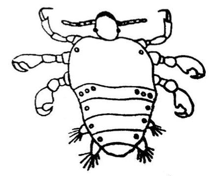
Identify the specimen shown in the image.
What disease does it cause?
Describe the head.
Describe the thorax.
How many pairs of legs are present?
Describe the legs.
How many abdominal segments are described in the lab?
How many lateral abdominal processes are present in the male?
Describe the posterior end of the male abdomen.
State its habitat.
Male Phthirus pubis.
Pediculosis pubis.
Short; it lies in a depression on the thorax.
Formed of three fused segments; one respiratory spiracle is described.
Three pairs.
Claw-like.
Five segments, each has a respiratory spiracle while the uppermost one has 3 respiratory spiracles
Two lateral processes.
Bulging.
Pubic region, axillae, and eyelashes.

Identify each organism


Identify the organism shown in the image.
Describe its size and shape.
State its habitat.
How many nuclei does it have, and where is the nucleus located?
Describe its cytostome.
How many flagella does it have?
Describe its undulating membrane.
Describe its axostyle.
Describe its parabasal body.
Trichomonas tenax trophozoite.
15 × 8 µm; oval to pear-shaped.
Oral cavity.
One nucleus at the anterior end.
Small and anterolateral.
Four flagella: three free anterior flagella and one marginal flagellum.
The undulating membrane ends at the posterior end.
One slender rod.
Thin.

Identify the organism shown in the image.
Describe its size and shape.
State its habitat.
How many nuclei does it have, and where is the nucleus located?
Describe its cytostome.
How many flagella does it have?
Describe its undulating membrane.
Describe its axostyle.
Is the parabasal body present or absent?
Trichomonas hominis trophozoite.
15 × 8 µm; oval to pear-shaped.
Large intestine.
One nucleus at the anterior end.
Large and anterolateral.
Four flagella: three free anterior flagella and one marginal flagellum.
The undulating membrane emerges free at the posterior end.
One thick rod with a sharp posterior end.
Absent.

Identify the organism shown in the image.
Describe its size and shape.
State its habitat.
How many nuclei does it have, and where is the nucleus located?
Describe its cytostome.
How many flagella does it have?
Describe its undulating membrane.
Describe its axostyle.
Describe its parabasal body.
Trichomonas tenax trophozoite.
15 × 8 µm; oval to pear-shaped.
Oral cavity.
One nucleus at the anterior end.
Small and anterolateral.
Four flagella: three free anterior flagella and one marginal flagellum.
The undulating membrane ends at the posterior end.
One slender rod.
Thin.

Identify the organism shown in the image.
Describe its size and shape.
State its habitat.
How many nuclei does it have, and where is the nucleus located?
Describe its cytostome.
How many flagella does it have?
Describe its undulating membrane.
Describe its axostyle.
Is the parabasal body present or absent?
Trichomonas hominis trophozoite.
15 × 8 µm; oval to pear-shaped.
Large intestine.
One nucleus at the anterior end.
Large and anterolateral.
Four flagella: three free anterior flagella and one marginal flagellum.
The undulating membrane emerges free at the posterior end.
One thick rod with a sharp posterior end.
Absent.

Name the two culture media used for the clinical diagnosis of vaginal trichomoniasis.
Which organization is mentioned in relation to these media?
What inoculation period is mentioned?
How long may it take to establish that a specimen is negative?
Which medium allows more prolific growth over a shorter period?
Identify the medium shown in the left image.
Identify the medium shown in the right image.
Diamond’s medium and Kupferberg’s medium.
Food and Drug Administration (FDA).
72 hours.
Up to 7 days.
Diamond’s medium.
Diamond’s medium.
Kupferberg’s medium.

1. Identify the organism shown in the image.
2. Describe its size and shape.
3. State its habitat.
4. What is its diagnostic stage?
5. How many nuclei does it have, and where is the nucleus located?
6. How many flagella does it have?
7. Describe the flagella.
8. Describe the undulating membrane.
9. Describe the axostyle.
10. Describe the parabasal body.
11. Describe the cytostome.
12. What disease does it cause?
13. What specimen is collected for diagnosis?
14. How is the specimen examined?
15. Which stain is mentioned in the lab?
Is the diagnostic stage a trophozoite or a cyst?
1. Trichomonas vaginalis trophozoites
2. 15 × 8 µm; oval to pear-shaped.
3. Vagina or urethra.
4. Trophozoite.
5. One nucleus at the anterior end.
6. Four flagella.
7. Three free anterior flagella and one marginal flagellum.
8. It reaches approximately half the body.
9. One very slender rod.
10. Thick.
11. Very small and anterolateral.
12. Trichomoniasis.
13. Vaginal or urethral discharge.
14. Wet preparation and direct microscopic examination to detect motile trophozoites between pus cells.
15. Giemsa stain.
16. Trophozoite


What specimen is collected to diagnose Trichomonas vaginalis?
From which two sites can the specimen be collected?
How is the specimen examined?
What preparation is used?
Which form of the parasite is detected?
Where are the trophozoites seen?
Why is direct examination of the specimen important according to the lab?
Which stain is mentioned in the lab?
Genital discharge.
Vagina or urethra.
Direct microscopic examination.
Wet preparation.
Motile trophozoites.
Between pus cells.
Because of the small number of trophozoites.
Giemsa stain.
List the parasites transmitted by sexual contact
Which parasite causes trichomoniasis?
Which parasite causes pediculosis pubis?
Which parasite causes scabies?
Trichomonas vaginalis, Phthirus pubis, and Sarcoptes scabiei.
Trichomonas vaginalis.
Phthirus pubis.
Sarcoptes scabiei.

Identify the test shown in the picture.
What is the purpose of Fehling’s test?
What is the principle of Fehling’s test?
Why is the sample heated during the test?
What does a brick-red precipitate indicate?
Is Fehling’s test specific for glucose?
Why can Fehling’s test give a false-positive result?
Can a diabetic patient have a negative Fehling’s test?
What is the renal threshold for glucose ?
What is one disadvantage of Fehling’s test?
Fehling’s test.
To detect reducing substances in urine, including glucose.
Reducing sugars reduce the reagent, producing a coloured precipitate.
Heating helps the reaction occur.
A positive test for reducing substances in urine.
No. It is a non-specific test.
Other reducing sugars can also give a positive result.
Yes. If blood glucose is below the renal threshold, urine glucose may be absent.
Approximately 180 mg/dL.
It cannot specifically distinguish glucose from other reducing substances.

Identify the instrument shown in the picture.
Which method is used to estimate plasma glucose?
What does GOD-POD stand for?
What is the principle of the GOD-POD method?
Which enzyme oxidizes glucose in this method?
Which enzyme participates in the colour-producing reaction?
What colour develops in the final reaction?
At what wavelength is absorbance measured?
Which instrument measures absorbance?
What are the three tubes used in the procedure?
What is the purpose of the blank tube?
What is the purpose of the standard tube?
What is the purpose of the test tube?
What is the relationship between colour intensity and glucose concentration?
What is the formula for calculating glucose concentration?
The test absorbance is 0.60, standard absorbance is 0.40, and standard glucose concentration is 100 mg/dL. Calculate the glucose concentration.
How long does the final pink colour remain stable at room temperature according to the lecture?
Spectrophotometer.
Glucose oxidase–peroxidase (GOD-POD) method.
Glucose Oxidase–Peroxidase.
Glucose is oxidized by glucose oxidase, and the subsequent peroxidase reaction produces a coloured compound.
Glucose oxidase (GOD).
Peroxidase (POD).
Pink.
505 nm.
Spectrophotometer.
Blank (B), Standard (S), and Test (T).
To provide the blank reference when measuring absorbance.
To provide a known glucose concentration for comparison.
To measure the glucose concentration in the patient’s sample.
Directly proportional.
Glucose (mg/dL) = (Test absorbance ÷ Standard absorbance) × Standard glucose concentration.
(0.60 ÷ 0.40) × 100 = 150 mg/dL.
One hour.

Identify the test shown in the picture.
What does OGTT stand for?
What is the definition of OGTT?
What are the indications for performing OGTT?
What samples are collected before giving the glucose load?
What is the glucose load used in the test?
In how much water is the glucose load dissolved?
At what intervals are blood and urine samples collected?
How are the OGTT results presented?
What is the normal fasting plasma glucose range shown in the table?
What is the fasting plasma glucose level in diabetic individuals?
How does the normal glucose curve behave after the glucose load?
What does the blue line (Patient #1) represent?
What does the green line (Patient #2) represent?
What does the red line (Patient #3) represent?
What is the interpretation of Patient #1’s glucose curve?
What is the interpretation of Patient #2’s glucose curve?
What is the interpretation of Patient #3’s glucose curve?
At what time does the normal glucose curve reach its peak?
When does the normal glucose curve return to normal?
What does the green curve indicate about the 2-hour glucose reading?
Why is Patient #3 classified as diabetic according to the slide?
What happens to urine glucose in normal subjects?
What happens to urine glucose in diabetic individuals?
Oral Glucose Tolerance Test (OGTT).
Oral Glucose Tolerance Test.
A test that measures the body’s capacity to tolerate an ingested or injected standard dose of glucose.
To diagnose gestational diabetes mellitus (GDM), impaired glucose tolerance (IGT), and diabetes mellitus. It is requested when a patient has glucosuria or when random/fasting glucose results are suggestive but not fully diagnostic of diabetes.
Fasting venous blood and urine samples.
0.75 g/kg body weight.
250–300 mL of water.
Every 30 minutes for 2 hours. For GDM: fasting, 1 hour, and 2 hours postprandially.
By plotting the results against time.
70–100 mg/dL.
≥126 mg/dL.
It starts with normal fasting glucose, peaks at 1 hour, and returns to normal after 2 hours.
Normal glucose tolerance.
Impaired glucose tolerance (IGT), or prediabetes.
Diabetes mellitus.
Normal fasting glucose, a peak at 1 hour, and return to normal after 2 hours.
Normal fasting glucose, but the 2-hour glucose reading is greater than 140 mg/dL, indicating glucose intolerance (prediabetes).
Fasting plasma glucose exceeds 126 mg/dL, and glucose levels remain elevated throughout the test.
At 1 hour.
After 2 hours.
The 2-hour glucose reading is greater than 140 mg/dL, indicating impaired glucose tolerance.
The fasting plasma glucose exceeds 126 mg/dL, and glucose levels remain elevated throughout the test.
None; urine glucose is below the renal threshold.
Glucosuria is present because glucose exceeds the renal threshold.
How long should a patient fast before fasting plasma glucose testing?
What is a post-prandial glucose sample?
What is a random glucose sample?
Which sample is preferred for glucose estimation?
Why is sodium fluoride added to the sample?
Which anticoagulants are mentioned in the lecture?
What is the normal fasting plasma glucose range ?
8–10 hours.
A sample collected after a meal.
A sample collected regardless of meal timing.
Plasma.
It inhibits glycolysis and prevents falsely low glucose results.
Potassium oxalate and sodium fluoride.
70-100 mg/dL.
A patient has fasting plasma glucose of 130 mg/dL. Does this meet the diabetes threshold?
A patient’s 2-hour OGTT glucose is 210 mg/dL. What does this indicate?
A patient has HbA1c of 6.8%. Does this meet the diabetes threshold?
A patient has random glucose of 230 mg/dL and classic symptoms. What is the likely diagnosis?
What is the fasting plasma glucose threshold for diabetes?
What is the HbA1c threshold for diabetes?
What random glucose level supports diagnosis when classic symptoms are present?
Yes. ≥126 mg/dL.
It meets the diabetes threshold of ≥200 mg/dL.
Yes. HbA1c ≥6.5%.
Diabetes mellitus.
≥126 mg/dL after at least 8 hours of fasting.
≥6.5%.
≥200 mg/dL.
What is glucosuria?
At what blood glucose level can glucosuria occur according to the lecture?
Name four causes of glucosuria.
Which urine test detects reducing substances?
Why is Benedict’s or Fehling’s test non-specific?
The presence of glucose in urine.
When blood glucose exceeds approximately 180 mg/dL.
Diabetes mellitus, gestational diabetes, renal threshold defect, and alimentary glucosuria.
Benedict’s or Fehling’s test.
Other reducing sugars can also produce a positive result.
At what gestational age is GDM screening usually performed according to the lecture?
Which glucose tolerance test is used?
What is the fasting glucose threshold for GDM?
What is the 1-hour glucose threshold for GDM?
What is the 2-hour glucose threshold for GDM?
How many abnormal values are required for diagnosis according to the lecture?
Name three hormones that contribute to insulin resistance during pregnancy.
What is the long-term risk after GDM?
24–28 weeks of gestation.
A 75-g OGTT.
≥92 mg/dL.
≥180 mg/dL.
≥153 mg/dL.
One or more values meeting or exceeding the thresholds.
Estrogen, progesterone, and placental lactogen.
Increased risk of developing type 2 diabetes later in life.
What does HbA1c reflect?
Over approximately how long does HbA1c reflect average glucose?
What is the normal HbA1c category?
A patient has HbA1c of 6.1%. What category does this fall into?
What HbA1c level meets the diabetes threshold?
Name two advantages of HbA1c.
Name two factors that can affect HbA1c independently of blood glucose.
Why should HbA1c be interpreted cautiously in anemia?
Name disadvantages of HbA1c.
Average blood glucose.
Approximately 2–3 months; the lecture describes it as about 2 months.
Below 5.7%.
Prediabetes range: 5.7–6.4%.
≥6.5%.
Fasting is not required, and the test has good preanalytical stability.
Anemia or hemoglobinopathies, and altered red blood cell turnover.
Anemia can alter red blood cell lifespan and affect HbA1c independently of actual glucose levels.
Lower sensitivity.
Greater cost.
Limited availability.
Imperfect correlation with true average glucose.
Indirect measure (affected by non-glucose factors).
What does HOMA-IR assess?
What is the HOMA-IR formula when glucose is measured in mg/dL?
A patient has fasting glucose of 90 mg/dL and insulin of 9 μU/L. Calculate HOMA-IR.
What does a high HOMA-IR suggest?
What does HOMA-IR below 1.0 suggest according to the lecture?
Insulin resistance.
HOMA-IR = [Fasting glucose (mg/dL) × Fasting insulin (μU/L)] ÷ 405.
(90 × 9) ÷ 405 = 2.0.
Insulin resistance.
Greater insulin sensitivity.
A patient with uncontrolled diabetes develops ketones in the urine. What serious condition should be suspected?
Name the three ketone bodies.
Which method is used to detect urine ketones?
What is the normal blood pH range?
What happens to pH and bicarbonate in DKA?
What is the normal serum potassium range given in the lecture?
Why can serum potassium be high in DKA?
Why can urea and creatinine increase in DKA?
Why can osmolarity increase in DKA?
Diabetic ketoacidosis.
Beta-hydroxybutyric acid, acetoacetate, and acetone.
Strip method.
7.35–7.45.
Both decrease, indicating metabolic acidosis.
3.5–5.0 mmol/L.
Insulin deficiency reduces potassium uptake into cells.
Dehydration due to excessive urination can impair renal function.
Renal impairment caused by dehydration.
What is the normal serum insulin level in the lecture?
What is the normal serum C-peptide level?
What happens to C-peptide in type 1 diabetes?
What happens to C-peptide when endogenous insulin production is preserved?
What is the main clinical use of C-peptide measurement?
5–15 μU/L.
0.9–4.2 ng/mL.
It is usually low due to reduced endogenous insulin production.
It is present because the pancreas produces endogenous insulin.
To assess endogenous insulin production and help distinguish it from injected insulin.
What is microalbuminuria?
What is the microalbuminuria range in the lecture?
A diabetic patient has urinary albumin excretion of 100 mg/day. How is this classified?
Which diabetic complication is associated with microalbuminuria?
What does microalbuminuria indicate about future kidney health?
The presence of moderately increased albumin in urine.
30–300 mg/day.
Microalbuminuria.
Diabetic nephropathy.
It may indicate early kidney damage and increased risk of progression to renal disease.
What is impaired glucose tolerance (IGT)?
What is another name for IGT?
What is the fasting blood glucose range in IGT?
What is the 2-hour postprandial blood glucose range in IGT?
What is the HbA1c range in IGT?
What lifestyle changes may help people with IGT return their glucose levels to normal?
How are blood glucose levels in IGT compared with normal and diabetic levels?
A transition phase between normal glucose tolerance and diabetes.
Pre-diabetes.
100–125 mg/dL.
140–199 mg/dL.
5.7%–6.4%.
Weight loss and exercise.
Higher than normal but below the diabetic range.

Identify:
Ex of organism :
How to take Specimens :
why we used this media :
Identify: Mc Coy cells
Ex of organism : Chlamydia Trachomatis
How to take Specimens : endocervical , urethral , rectal swab
why we used this media : Chlamydia can't grow on artificial culture
media ,It requires living host cells (Mc Coy cells or Hela cells)
Identify :
Comment:
Can give false negative results in :
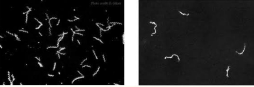
Identify : Dark-field microscopy of Treponema Pallidum
Comment: T. pallidum appears as white spiral bodies, motile like spring against a
black background
Can give false negative results in : low number of organisms,Antimicrobial
treatment

Identify:
Example of organism:
Media on which the organism grows:
On microscope:
Environment required by the organism:
Identify: Chocolate agar
Example of organism: Neisseria gonorrhoeae
Media on which the organism grows:
1. Chocolate agar
2. Thayer-Martin medium
On microscope: Gram-negative diplococci.
Environment required by the organism: An
environment containing 5–10% CO₂.