ABD: CTL Pathology 42% - Liver
 Call Kai
 Learn
Learn Practice Test
 Spaced Repetition
 Match
Match Flashcards
 Knowt Play
1/152

There's no tags or description
Looks like no tags are added yet.
Name | Mastery | Learn | Test | Matching | Spaced | Call with Kai | Chat |
|---|
No analytics yet
Send a link to your students to track their progress
153 Terms
What abnormality causes focal developmental lesions of the liver and is associated with congenital hepatic fibrosis, autosomal dominant polycystic disease, and cholangiocarcinoma?
a. schistosomiasis
b. biliary hamartomas
c. candiadiasis
d. Wilson disease
b
Which of the following is commonly associated with contaminated water and causes debris/occlusion within intrahepatic portal venous system?
a. candidiasis
b. schistosomiasis
c. pyogenic abscess
d. amebic abscess
b
A liver mass with a hypoechoic rim should create a strong suspicion of:
a. portal HTN
b. a liver adenoma
c. a hepatic lipoma
d. malignancy
d
Steatosis refers to:
a. chronic hepatitis infection
b. fatty liver disease
c. glycogen storage disease of the liver
d. pneumocystis carinii
b
Which of the following describes a non-infectious cause for hepatitis?
a. anabolic steroids
b. virus
c. fungus
d. bacteria
a
Which of the following correctly describes how contrast enhanced ultrasound can be used to differentiate benign and malignant masses?
a. benign nodules should demonstrate hypervascular enhancement while malignant tumors usually demonstrate hypovascular enhancement
b. significant contrast uptake occurs in benign nodules, while no contrast uptake is seen in malignant nodules
c. significant contrast uptake occurs in malignant nodules, while no contrast uptake is seen in benign nodules
d. benign nodules usually demonstrate isovascular enhancement, while malignant tumors usually demonstrate hypervascular enhancement
d
In a patient with cirrhosis, the _______ lobe of the liver can enlarge to nearly half the size of the RT lobe.
a. left
b. caudate
c. quadrate
d. none of the above, the entire liver shrinks in size with cirrhosis
b
________ can lead to air within the portal venous system and ______ can lead to air within the biliary tree.
a. an ERCP, ulcerative colitis
b. ulcerative colitis, ERCP
c. ulcerative colitis, appendicitis
d. diverticulitis, ulcerative colitis
b
Contrast enhanced ultrasound is used to determine the outcome of tumor ablation in the liver. If the procedure was a success, what are the expected findings on the image?
a. contrast uptake occurs during the arterial phase
b. no contrast enters the lesion during the cardiac cycle
c. contrast uptake occurs during the venous phase
d. consistent contrast uptake occurs during the arterial and venous phase
b
If a liver transplant patient has a piggyback anastomosis, how does this affect your evaluation?
a. each hepatic vein is connected to the native IVC separately, so there will be three anastomosis sites in the IVC that must be evaluated
b. there is a single anastomosis site in the IVC that must be evaluated
c. there will be two anastomosis sites in the MPV that must be evaluated
d. the hepatic artery anastomosis will not be able to be visualized because it is located deep within the liver tissue
b
Which of the following is most likely to demonstrate a simple cyst with gravity dependent debris in the RT lobe of the liver?
a. chronic hydatid infection
b. early echinococcal infection
c. chronic FNH
d. early schistosomiasis infection
b
In patients with cirrhosis, what effect will deep inspiration have on the portal vein?
a. the PV diameter will increased by 50%
b. there will be minimal change in the PV diameter
c. the PV diameter will double in size
d. the PV will collapse at the deepest point of inspiration
b
Which of the following describes the appearance of the liver in a patient in the later stages of cirrhosis caused by viral hepatitis?
a. multiple nodules usually 1-5 cm in size
b. significant hepatomegaly with diffuse decrease in hepatic echotexture
c. multiple nodules usually <1 cm in size
d. diffuse coarse liver echotexture with no nodule formation
a
Which of the following describes the most common sonographic appearance of a hepatoma?
a. solid mass that may be more or less echogenic that liver parenchyma
b. usually causes multiple diffuse solid mass formation
c. most commonly demonstrates diffuse coarse liver texture in the affected lobe
d. echogenic mass with a central scar
a
Compensatory enlargement of the caudate lobe occurs with which of the following?
a. cirrhosis and Budd Chiari syndrome
b. cirrhosis and acute hepatitis
c. fatty infiltration and glycogen storage disease
d. Wilson disease and glycogen storage disease
a
While scanning the liver in a patient with chronic cirrhosis, you notice a tubular vessel connecting to the splenic vein just prior to its confluence with the SMV. Flow within the vessel is very low velocity, continuous and moving in a cephalic direction. Which of the following explains the ultrasound findings?
a. a dilated SMV is present with flow reversal which is a common complication of cirrhosis
b. a dilated coronary vein is present which is a common complication of cirrhosis
c. a dilated umbilical vein is present which is a common complication of cirrhosis
d. the ductus venosus is dilated with antegrade flow which is a common complication of cirrhosis
b
Which of the following is the most common cause for intrahepatic cholestasis?
a. stone in the distal CBD
b. pancreatic head mass
c. hepatitis
d. fatty infiltration of the liver
c
If a liver transplant patient has an interposition anastomosis, how does this affect your evaluation?
a. each hepatic vein is connected to the native IVC separately, so there will be three anastomosis sites in the IVC that must be evaluated
b. there will be two anastomosis sites in the IVC that must be evaluated
c. there will be two anastomosis sites in the MPV that must be evaluated
d. the hepatic artery anastomosis will not be able to be visualized because it is located deep within the liver tissue
b
What is the most common cause of a hepatic abscess in the US?
a. cholangitis and cholecystitis
b. appendicitis and diverticulitis
c. osteomyelitis and endocarditis
d. E. coli from digestive tract
a
Which of the following findings will indicate hepatomegaly is present?
a. when the right lobe is rounded inferiorly and extends below the lower pole of the right kidney, the liver is considered enlarged
b. hepatomegaly causes rounding of the superior liver segments
c. the liver is considered enlarged when the AP measurement exceeds 15.5 cm
d. when the right lobe is rounded inferiorly and extends past the upper pole of the right kidney, the liver is considered enlarged
a
A _______ abscess is caused by bacteria and most commonly forms within the right lobe of the liver. A _______ abscess is caused by a parasite and usually forms in the right dome of the liver causing elevation of the diaphragm.
a. amebic, pyogenic
b. parasitic, fungal
c. pyogenic, amebic
d. fungal, pyogenic
c
Which of the following describes the Doppler appearance of liver transplant rejection?
a. mottled appearance of the liver parenchyma with ascites
b. nodular formations within the liver parenchyma
c. low resistance flow in the PV
d. high resistance flow in the hepatic artery
d
What is a potentially life threatening complication of portal HTN?
a. pulmonary embolism
b. aortic embolism
c. ruptured gastroesophageal varices
d. ascites formation
c
The most common benign liver tumor is:
a. Klatskin tumor
b. cavernous hemangioma
c. hepatitis
d. polycystic liver disease
b
About 50% of all primary tumors that metastasize to the liver are drained through the:
a. PV
b. hepatic vein
c. aorta
d. IVC
a
_________ abscesses are most commonly found in countries outside the US. ________ abscesses are most commonly seen in immunosuppressed patients.
a. pyogenic, amebic
b. amebic, fungal
c. fungal, amebic
d. fungal, pyogenic
b
The most frequently involved organ in metastatic disease is the:
a. GI tract
b. liver
c. pancreas
d. spleen
b
The US exam of a patient’s liver demonstrates a patent umbilical vein. The most common cause for this recanalization is:
a. portal HTN
b. hepatitis
c. ascites
d. abscess
a

A 40 y/o female patient presents with increasing RUQ pain over the last month, beginning upon returning from a trip to Indonesia. The findings on her exam are most consistent with:
a. echinococcal cyst
b. schistosomiasis cyst
c. lymphatic cyst
d. fungal abscess
a

Which waveform represents hepatic vein flow in a patient with advanced cirrhosis?
b

Which of the following liver abnormalities is commonly associated with the recanalization of the vessel within the structure indicated by the purple arrows?
a. acute hepatitis
b. cirrhosis
c. fatty infiltration
d. hydatid disease
b

Longitudinal images are presented of the anterior abdomen just superior to the umbilicus. What is the most common cause of the abnormality seen on the images?
a. portal HTN
b. polycythemia vera
c. malignant HTN
d. steatosis
a
Which of the following correctly describes how to determine if a peripheral mass is intrahepatic or extrahepatic?
a. assess the location of the mass in relation to the stomach
b. assess the liver capsule and look for indentations that indicate extrahepatic origin or outward bulging that indicates intrahepatic origin
c. use PW doppler to obtain the RI of the lesion and compare it to the RI of the normal liver tissue
d. evaluate the biliary tree for the presence of dilatation which is only seen with an extrahepatic mass
b
Liver transplant evaluation requires PW doppler evaluation of all of the following except:
a. IVC anastomosis
b. hepatic arterial anastomosis
c. portal anastomosis
d. biliary anastomosis
d

Which of the following organs can be involved in this multi-organ syndrome and can also display the same abnormality seen here in the liver?
a. thyroid and thymus
b. peritoneum and mesentery
c. kidneys and ovaries
d. spleen and adrenal glands
c
The hormone changes with pregnancy have been associated with the enlargement of what type of liver mass?
a. granuloma
b. schistosomiasis
c. hemangioma
d. hepatoma
c
Hepatic artery hypertrophy is a common finding with:
a. celiac axis occlusion
b. cholecystitis
c. splenic vein thrombosis
d. cirrhosis
d
What usually causes an echinococcal cyst in the liver?
a. hemangioma
b. ingestion of raw oysters
c. hepatitis
d. ingestion of raw pork
d
Which of the following liver tumors is usually treated by surgical resection due to risk of hemorrhage and malignant transformation?
a. hemangioma
b. adenoma
c. pyogenic abscess
d. lipoma
b
A patient presents with new onset of jaundice 2 weeks after a liver transplant. This clinical finding is most suggestive of what complication?
a. portal vein stenosis
b. hepatic artery stenosis
c. biliary stricture
d. hepatic vein stenosis
c
The diameter of the _______ will be relatively unaffected by hepatic congestion.
a. MPV
b. LHV
c. RHV
d. hepatic artery
d
The most definitive sonographic sign of cirrhosis is:
a. hepatoma formation
b. portal HTN
c. associated hepatomegaly
d. decreased liver size with coards texture and nodule formation
d
What is the most common primary cancer to metastasize to the liver?
a. lung
b. melanoma
c. ovary
d. GI tract
d
A cyst within a cyst (daughter cyst) is a classic sign of:
a. polycystic liver disease
b. hydatid cyst or fungal abscess
c. echinococcal cyst or pyogenic abscess
d. hydatid cyst or echinococcal cyst
d
A patient presents with lower extremity edema and increased abdominal girth 2 weeks after a liver transplant. These findings are most suggestive of what complication?
a. biliary stricture
b. hepatic vein stenosis
c. hepatic artery stenosis
d. cholangitis
b

Which of the following statements is true regarding the displayed image?
a. the patient’s liver is increased in echogenicity indicating acute viral hepatitis
b. bilirubin, AST, and ALT levels will be elevated
c. the patient has pleural effusion
d. the patient’s liver is decreased in echogenicity indicating cirrhosis
b
The primary finding that differentiates liver mass formation from steatosis is:
a. steatosis will demonstrate normal vessel course through affected areas, while mass formation displaces vasculature
b. steatosis will cause wall thickening of the portal system, while mass formation invades the wall
c. steatosis causes an irregular liver surface contour, while mass formation does not affect the surface contour of the liver
d. steatosis will cause dilated hepatic veins, while mass formation cause narrowed hepatic veins
a
Benign liver masses will usually demonstrate US contrast enhancement during the arterial and portal venous phases of imaging. Malignant masses will demonstrate:
a. contrast enhancement during the arterial and portal venous phases of imaging
b. no contrast enhancement
c. contrast enhancement during portal venous phase of imaging
d. contrast enhancement during the arterial phase of imaging
d
Which of the following statements is true regarding the sonographic appearance of fatty infiltration?
a. as fatty infiltration increases in the liver, the VH branches become more visible
b. as fatty infiltration increases in the liver, all walls of all liver vascularity becomes increasingly echogenic
c. as fatty infiltration increases in the liver, the PV branches become more difficult to visualize
d. as fatty infiltration increases in the liver, the walls of the PV branches become increasingly echogenic
c
The most common parasitic infection in humans is:
a. hydatid disease
b. pneumocystis carinii
c. hepatitis
d. schistosomiasis
d
Shear wave elastography can be used to assess the liver. Which of the following correctly describes the result of an abnormal exam?
a. red color superimposed over liver parenchyma indicates liver fibrosis
b. lower values indicate liver fibrosis
c. blue color superimposed over liver parenchyma indicates liver fibrosis
d. higher values indicate liver fibrosis
d
The most common cause of spread of hepatitis in the US is:
a. liver transplants
b. hereditary infection
c. IV drug users
d. blood infusions
c
Which of the following is true regarding a liver hemangioma?
a. most hemangiomas demonstrate some level of posterior shadowing
b. fluid and debris levels are commonly present in a hemangioma
c. color doppler is a valuable tool in evaluating this highly vascular mass
d. a hemangioma is composed of an abnormal concentration of vascular tissues within the liver
d
A 10 y/o presents for an abdominal US with a palpable RUQ mass. He has no history of liver disease and alpha-fetoprotein levels are normal. You identify a 9 cm mass in the RT lobe that demonstrates punctate calcifications and a central scar. These findings are most suggestive of?
a. metastasis
b. hepatoma
c. fibrolamellar carcinoma
d. hepatoblastoma
c
A patient presents with abdominal pain for 3 months following a missionary trip to India. Lab values demonstrate normal LFTs. The US exam demonstrates a 3 cm cyst with septations in the right lobe of the liver. These findings are most consistent with:
a. candidiasis
b. schistosomiasis
c. hydatid disease
d. histoplasmosis infection
c
What condition is associated with prominent portal walls?
a. hemangioma
b. cirrhosis
c. fatty infiltration
d. acute viral hepatitis
d

Which of the following statements is true regarding the displayed US image?
a. the PV is a normal diameter but appears to be filled with thrombus related to chronic portal HTN
b. the PV is at top normal diameter at 17 mm. the PV is considered abnormally dilated at a diameter 18 mm or greater
c. the PV is abnormally dilated at 17 mm and is filled wtih mildly echogenic thrombus
d. the PV is dilated at 17 mm but does not demonstrate color doppler signals due to the perpendicular incidence with the probe
c

Which of the following statements is true regarding the doppler evaluation of the liver vasculature displayed?
a. the hepatic artery and PV are demonstrated with normal flow directions
b. the hepatic artery is hepatopetal and the PV is hepatofugal
c. the hepatic artery is hepatofugal and the PV is hepatopetal
d. the PV flow is normal but the hepatic artery demonstrates increased resistance
b

When reviewing a prior US exam for an upcoming follow up exam for the same patient, you come across an image labeled “sandwich sign?” What are they referring to?
a. lymphadenopathy surround the PV
b. the head of the pancreas is abnormally compressed between the liver and great vessels
c. extrinsic compression of the IVC by a pancreatic tumor
d. the layering of the stomach wall at the head of the pancreas
a
The US-LIRADS is used to assess patients at risk for:
a. liver metastasis
b. lymphoma
c. HCC
d. cirrhosis
c
What type of liver infection/abscess is commonly associated with immunocompromised patients?
a. amebic
b. schistosomiasis
c. candidiasis
d. pyogenic
c
A patient presents with a history of hep C and you identify a solid liver mass with a hypoechoic halo. This finding is most consistent with:
a. hepatic adenoma
b. FNH
c. hepatic metastasis
d. HCC
d
Triglyceride accumulation in the liver leads to _______, while copper accumulation in the liver leads to _______.
a. steatosis, hemochromatosis
b. steatosis, Wilson disease
c. amyloidosis, hemochromatosis
d. amyloidosis, steatosis
b
If a patient presents with a history of right breast cancer and you identify a solid liver mass with a hypoechoic halo, what is the most likely diagnosis of the finding?
a. HCC
b. hepatic metastasis
c. FNH
d. hepatic adenoma
b
Liver hemangiomas are a common benign neoplasm that will usually appear on an US of a normal liver as:
a. complex liver mass with irregular borders
b. cystic mass within the liver
c. solid hypoechoic mass with posterior shadowing
d. well-defined hyperechoic liver mass
d
A vascular condition of the liver seen in women who take oral contraceptives is:
a. portal venous gas
b. Budd Chiari syndrome
c. portal aneurysm
d. hereditary telangiectasia
b
Which of the following statements is true regarding the image displayed?
a. the liver is demonstrated in the transverse plane at the level of the caudate lobe and has a normal echotexture. There is an enlarged lymph node seen anterior to the aorta and adjacent to the IVC
b. the liver is demonstrated in the transverse plane at the level of the caudate lobe and has a normal echotexture
c. there is a mass identified in the lateral left lobe of the liver adjacent to the IVC
d. there is a mass identified in the caudate lobe of the liver adjacent to the IVC
d
If tardus parvus waveforms are identified in the intrahepatic artery waveforms of a transplant, this finding is __________. If biphasic waveforms are identified in the right and left hepatic arteries, the finding is _________.
a. suspicious for hepatic artery stenosis, suspicious for organ rejection
b. normal, suspicious for organ rejection
c. suspicious for organ rejection, suspicious for hepatic artery stenosis
d. suspicious for hepatic artery stenosis, normal
a
A TIPS shunt is placed into the liver circulation to alleviate portal HTN. What two vessels are usually connected?
a. RPV and LPV
b. hepatic artery and PV
c. RPV and RHV
d. RHV and LHV
c
Liver elastography is used for:
a. staging of secondary malignancy
b. staging of primary malignancy
c. staging of fibrosis
d. staging of steatosis
c
Fulminant hepatitis is most commonly caused by:
a. acetaminophen abuse
b. alcohol abuse
c. viral infection
d. fungal infection
a
What liver complication is associated with cholangitis, diverticulitis, and osteomyelitis?
a. metastasis
b. pyogenic abscess
c. steatosis
d. fungal infection
b
How can a hepatic adenoma be differentiated from FNH on contrast enhanced ultrasound?
a. FNH lesions demonstrate no contrast uptake while adenomas demonstrate significant contrast uptake
b. FNH lesions usually demonstrate centrifugal enhancement while adenomas demonstrate centripetal enhancement
c. FNH lesions demonstrate significant contrast uptake while adenomas demonstrate no contrast uptake
d. FNH lesions usually demonstrate centripetal enhancement while adenomas demonstrate centrifugal enhancement
b
A mass that presents with a bull’s eye appearance within the liver tissue is most likely ________.
a. fatty sparing
b. lymphoma
c. metastasis
d. FNH
c
You are scanning through the liver and note the caudate lobe is nearly the same size as the right lobe. The surface of the liver appears lobulated or nodular. Which of the following is the most likely diagnosis of these findings?
a. FNH
b. cirrhosis
c. hepatitis
d. HCC
b
_______ should be suspected in an infant with a large hepatic hemangioma and unexplained thrombocytopenia.
a. Budd Chiari syndrome
b. Kasabach-Merritt syndrome
c. cirrhosis
d. fragile X syndrome
b
Which of the following is a potential collateral pathway formed in patients with cirrhosis?
a. blood moves from the splenic vein to the coronary vein
b. blood moves from the emissary veins to the umbilical vein
c. blood moves from the gastric vein to the splenic vein
d. blood moves from the superior rectal vein to the middle rectal vein
d
Which of the following Doppler characteristics of the hepatic arteries will be seen in a normal liver transplant?
a. diastolic flow reversal
b. sharp systolic upstroke
c. prolonged acceleration time
d. increased RI
b
Which of the following is true regarding a liver hemangioma?
a. color doppler is not usually helpful in evaluating this highly vascular mass
b. the hemangioma is composed of an abnormal concentration of smooth muscle and Kupffer cells within the liver
c. most hemangiomas demonstrate some level of posterior shadowing
d. fluid and debris levels are commonly present in a hemangioma
a
What is the most common liver malignancy identified on ultrasound?
a. adenoma
b. metastatic tumor
c. hepatoma
d. Klatskin tumor
b
Which of the following is not an expected finding with fatty infiltration of the liver?
a. increased attenuation
b. brighter parenchymal echoes
c. cystic degeneration
d. degraded visualization of vessels
c
Which of the following is true regarding a transjugular intrahepatic shunt?
a. the shunt connects the dilated umbilical vein to the IVC
b. air is retained within the shunt after placement which limits the US evaluation for the first 3-5 days
c. a harvest branch of the hepatic vein is normally used as the shunt
d. the shunt is advanced into the liver through a puncture of the groin and common iliac vein
b
Which of the following describes the sonographic appearance of hepatic candidiasis?
a. cystic mass with gravity dependent debris
b. complex cystic mass that typically forms near the porta hepatis
c. mass formation is described as a wheel within a wheel pattern
d. a large solid mass replaces the caudate lobe and displaces the IVC
c
High-intensity transient signals (HITS) are seen on the Doppler evaluation of a patient with:
a. hyperfunctioning adenoma
b. critical renal artery stenosis
c. significant aortic stenosis
d. portal venous gas
d
A patient presents with a history of AIDS and recent pneumocystis carinii infection. What changes to the liver do you expect to see on the ultrasound exam?
a. diffuse echogenic foci throughout the liver tissue
b. formation of multiple abscesses within the liver tissue
c. gas-producing abscess near the porta hepatis
d. diffuse increase in liver tissue echogenicity with ascites
a
A liver ultrasound demonstrates a hyperechoic mass with smooth borders and posterior enhancement in the posterior right lobe. These findings are most suggestive of:
a. FFS
b. HCC
c. hepatic adenoma
d. cavernous hemangioma
d
__________ is the most common cause for micronodular cirrhosis and ___________ is the most common cause for macronodular cirrhosis.
a. analgesic consumption, alcohol consumption
b. alcohol consumption, chronic viral hepatitis
c. alcohol consumption, analgesic consumption
d. chronic viral hepatitis, alcohol consumption
b
If discovered in early stages, which of the following effects on the liver can be reversed?
a. fatty infiltration
b. cirrhosis
c. metastasis
d. hepatoma
a

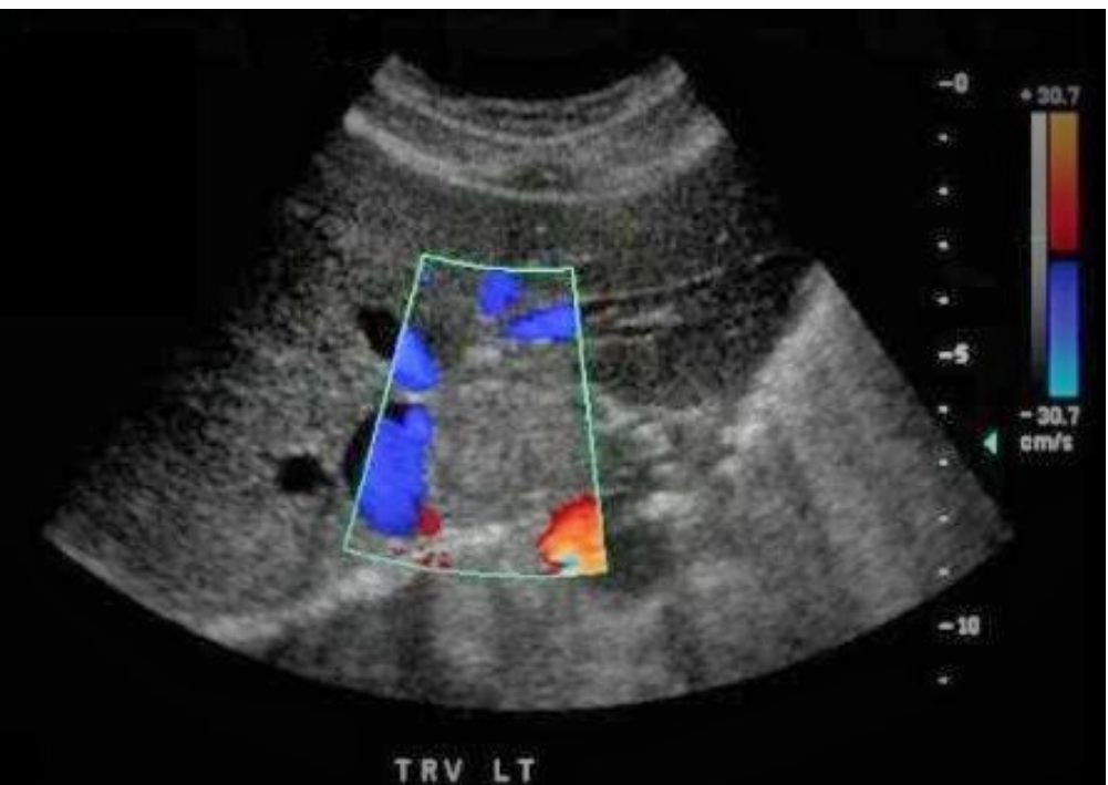
A 50 y/o female presents with nausea, vomiting, and increased levels of AFP in lab testing. Which of the following pathologies is demonstrated on the image displayed?
a. choledochal cysts
b. hepatoblastoma
c. Klatskin tumor
d. Krukenberg tumor
c
While performing US on the liver, a transverse view reveals a hyperechoic triangular structure in the inferior left lobe. The hyperechoic structure demonstrates an anechoic center that fills with color when doppler is applied. Which of the following best describes the finding identified on the image?
a. metastasis
b. recanalized coronary vein
c. FNH
d. recanalized umbilical vein
d
Which metabolic infiltrative disorder is associated with hepatic adenoma formation?
a. Wilson disease
b. hemochromatosis
c. amyloidosis
d. Von Gierke disease
d
Which of the following doppler parameters is most commonly used when evaluating the common hepatic artery in a patient with a liver transplant?
a. resistive index
b. peak systolic velocity
c. pulsatility index
d. S/D ratio
a
Hypoalbuminemia associated with cirrhosis leads to:
a. liver capsule contraction and surface irregularities
b. gallbladder and bowel wall thickening
c. hepatomegaly
d. nodule formation
b
__________ usually presents as multiple target lesion in the liver.
a. Klatskin tumor
b. metastasis
c. fungal abscess
d. hydatid disease
b
___________ is the most common malignant liver mass found in children.
a. Mets from the brain
b. hepatoma
c. mets from nephroblastoma
d. hepatoblastoma
d
Which of the following vessels will be abnormally dilated with significant portal HTN?
a. MPV only
b. HVs only
c. MPV and left gastric vein
d. HVs, MPV, and left gastric vein
c
Periportal cuffing is a sign of:
a. intrahepatic biliary tree obstruction
b. extrahepatic biliary tree obstruction
c. acute viral hepatitis
d. Caroli disease
c
A patient is scheduled for abdominal US every six months due to their history of cirrhosis. What is the primary reason for these serial exams?
a. increased risk of tumor invasion of the PV from metastatic lesion
b. evaluate for the development of an abscess
c. evaluate for pleural effusion
d. increased risk for portal thrombosis
d

What lobe of the liver is the mass located?
a. mass is within the IVC, not the liver
b. caudate
c. left
d. right
b
A 75 y/o female presents with RUQ pain. She was diagnosed with cirrhosis 4 years ago. Lab values demonstrate increased levels of AFP, ALP, AST, and ALT. The US exam demonstrates a heterogeneous liver texture that is decreased in size. The left lobe contains a new round hypoechoic mass with increased vascularity. These findings are most suggestive of:
a. metastatic lesion
b. hydatid cyst
c. FNH
d. HCC
d