13: General and Special Senses
 Call Kai
 Learn
 Practice Test
 Spaced Repetition
 Match
 Flashcards
 Knowt Play
1/80

There's no tags or description
Looks like no tags are added yet.
81 Terms
Two classes of Senses
General and Special
General Sense
Distributed throughout the body with receptor cells in the structures of other organs - temperature, pain, touch, stretch, and pressure
Special Sense
has a specific organ devoted to it - gustation, olfaction, vision, equilibrium and audition
Types of Receptors
Chemoreceptors, thermoreceptors, photoreceptors, mechanoreceptors, and nociceptors
General: Skin Receptors
Somatosensation pressure, and vibration; thermoreceptors detect temperature; and nocireceptors detect tissue damage

Heat Receptor/nociceptor
first arrow on left

Cold receptor/nociceptor
middle arrrow

Merkel cell-axon complex
arrow on far right

Pacinan Corpuscle
arrow bottom right - Deep pressure and vibration is transduced

Meissner Corpuscles
Top arrow on sode - Light touch is transduced
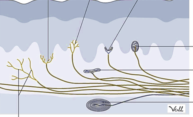
Ruffini corpuscles
middle arrow on side - Stretching of the skin is transduced

Hair follicle receptor - wrapped in a plexus of nerve endings that detect the movement of hair at the surface of the skin
General: Proprioceptors
found in the joints and muscles - They monitor the stretching of tendons, muscles, and the components of joints.

In skeletal muscle tissue, stretch receptors are called…
Muscle spindles

In tendons, stretch receptors are called…
golgi tendon organs
What nerves innervate the tounge and gustation
Facial nerve (anterior 2/3) and Glossopharyngeal nerve (posterior 1/3)

Filiform papillae are not involved in the sense of taste and contain no taste buds, but help with texture and friction when licking or tasting food

Fungiform Papillae - containing few taste buds that are scattered

Foliate Papillae - contain taste buds that are only important during infancy

Circumvallate papillae - contain many taste buds
Taste Buds composition
barrel-shaped collections of cells that contain Gustatory Cells, supporting cells, and basal cells

Gustatory Cells
white part - Chemoreceptors that respond to the five basic tastes: salty, sour, sweet, bitter, and umami (replaced 7-10 days)

Basal Cells
the blobs - Immature cells, replace other two cell types
Olfactory receptor neurons
Made of chemoreceptors and bipolar neurons - supporting cells and basal cells
Where are Olfactory receptor neurons located?
Olfactory epithelium and contains bipolar sensory neurons
What can be recognized by olfactory receptor cells?
thousands of chemical stimuli

arrow on bottom
olfactory epithelium - contains bipolar sensory neurons - pass over the olfactory epithelial region and dissolve into the mucus and bind to proteins that keep them dissolved

arrow on top -
Olfactory Bulb - axons carry information to several brain regions
How many muscles are in the eyes?
6
eyebrows, eyelids, and eyelashes help what?
to protect the eye from abrasions by blocking particles that may land on the surface of the eye
superior rectus, medial rectus, inferior rectus, and lateral rectus
When each of these muscles contracts, the eye moves toward the contracting muscle. Ex- when the superior rectus contracts, the eye looks up.
superior oblique
contraction of the superior oblique rotates the eye medially
inferior oblique
When it contracts, it laterally rotates the eye
levator palpebrae superioris
elevating and retracting the upper eyelid
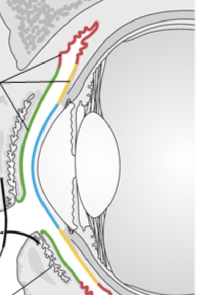
Conjunctiva
top arrow - Covers eyes anterior surface and internal eyelid surface
conjunctivitis
"pink eye" - The eye appears pink because of the dilation of the blood vessels that are found in the conjunctiva

Lacrimal System
lacrimal glands, canaliculi, lacrimal sac, and nasolacrimal duct (bottom arrow is the duct)

Tarsal glands
sebaceous gland found within the eyelids that produce an oily secretion to keep tears in the eye and help prevent tear evaporation.
outermost layer is called and contents
fibrous tunic - Sclera and Cornea
Cornea
allows light to enter
middle layer is called and contents
Vascular Tunic - choroid, ciliary body, suspensory ligaments, and iris
Choroid
highly vascularized connective tissue that provides a blood supply to the eye
What do the Ciliary body and suspensory ligaments do?
bend the lens, allowing it to focus light on the back of the eye
Iris
the colored part of the eye that is a smooth muscle that opens or closes the pupil
pupil
hole at the center of the eye that allows light to enter
innermost layer is called and contents
Retina
retina
contains the nervous tissue responsible for photoreception
What part of the eye is filled with Aqueous humor?
Anterior and Posterior Chamber
What part of the eye is filled with Vitreous humor?
Vitreous Chamber
What are the cells responsible for detecting light?
photoreceptors (rods and cones)

Rods
green things - function in dim light - don’t provide sharp vision or color vision, more numerous than cones

Cones
blue things - Operate best in bright light, provide high acuity color vision
Optic Disc
Contains no rods or cones; axons exit the eye - blind spot

fovea centralis
divit - The exact center of the retina that contains only cones; maximal visual acuity
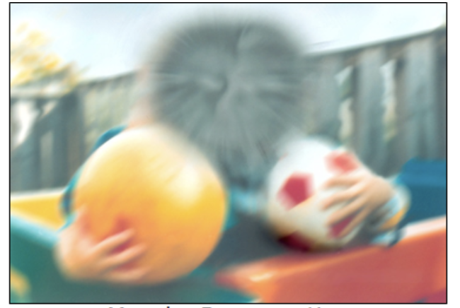
Macular Degeneration - Area around fovea degenerates

Cataracts - cloudy lens

Glaucoma - high pressure in eye hurts optic nerve
External Ear f
Auricle (pinna), External auditory canal, Ceremonious glands, and tympanic membrane
ceruminous glands
produce cerumen or earwax that cleans and waterproofs the canal, impeeding microorganism growth
tympanic membrane
or ear drum, which vibrates after it is struck by sound waves
Middle Ear Structures
Malleus, Incus, Stapes, Auditory tube, Stapedius muscle, roound and oval windows, tympanic cavity, and Tensor tympani muscle

Name the Ossicles in order from left to right
Malleus, Incus, and Stapes
auditory tube
Connects air-filled middle ear to the nasoppharynx to equilibrate air pressure across the tympanic membrane - normally closed but will open during swallowing or yawning
Otitis Media
Infection of the middle ear - trapped fluid and mucus behind the eardrum

stapedius and tensor tympani
(Tensor - red ~ Stap - Gray) contract by reflex during loud sounds. Their action stiffens the ossicle chain, preventing overstimulation and damage of the delicate cells of the inner ear
Inner Ear Structures
Vestibule, Semicircular Canals, Cochlea, and Vestibular and Cochlear Nerves
Vestibule
Has two parts called the utricle and saccule, which detect linear acceleration and head position - helps sense equilibrium
Semicircular Canals
Detect rotational movements and help sense equilibrium
Cochlea
Detects sound waves
Oval Window (Middle Ear)
located at the beginning of a fluid-filled tube within the cochlea called the scala vestibuli
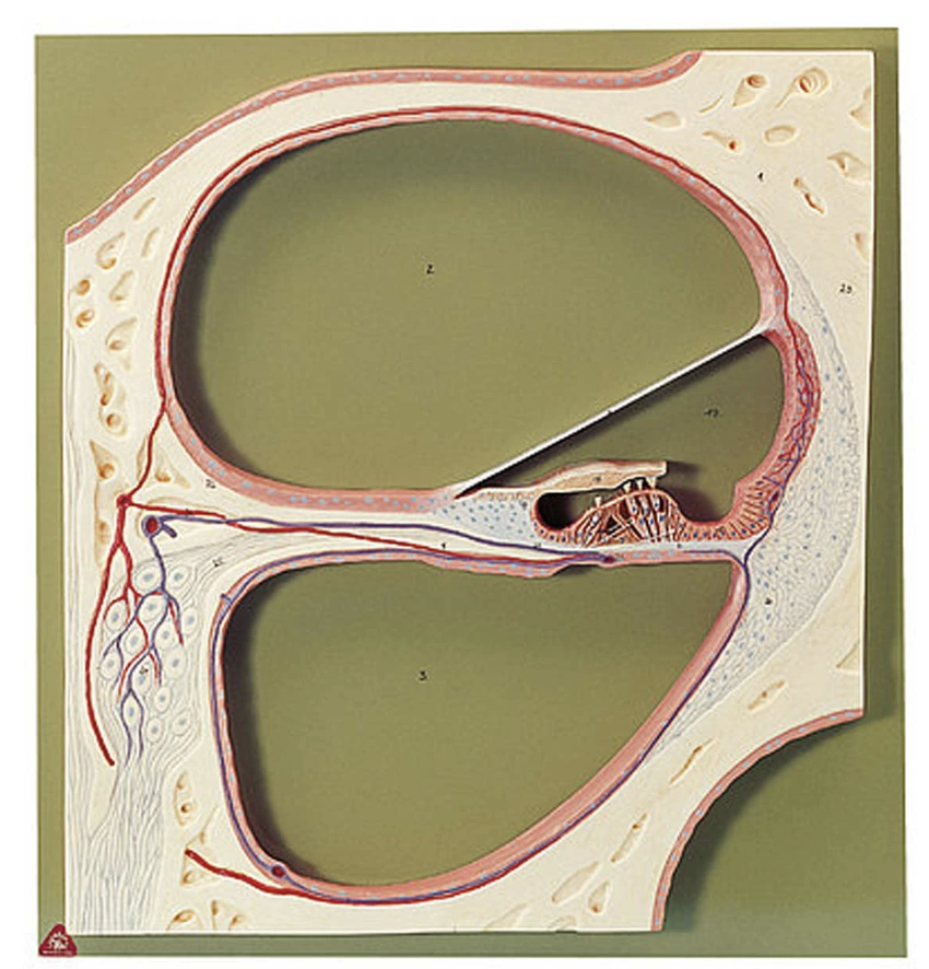
scala vestibuli
top space - containing perilymph (Cushions and protects inner-ear structures and transmit sound)

cochlear duct
middle scala - contains the sound-transducing neurons and Endolymph (clear fluid that helps w hearing and balance)

scala tympani
bottom scala - containing perilymph (Cushions and protects inner-ear structures and transmit sound)
round window
Where the scala tympani ends, which is covered by a membrane that contains the fluid within the scala

basilar membrane
middle bar - As the fluid waves move through the scala vestibuli and scala tympani,it moves depending on the frequency of the waves. Higher frequency waves move closer to the base of the cochlea. Lower frequency waves move near the tip of the cochlea.
hair cells and tectorial membrana
The stereocilia extend up from the hair cells to the overlying tectorial membrane. When the pressure waves move the basilar membrane, the tectorial membrane slides across the stereocilia. This bends the stereocilia, causing hair cells to send messages to sensory neurons within the vestibulocochlear nerve, which in turn, relays the information to the brain.
Gustation - Sensory Cell, Type of Receptor, and Cranial Nerves
Gustatory Cell, Chemoreceptor, and CN VII and CN IX (7 and 9)
Olfaction - Sensory Cell, Type of Receptor, and Cranial Nerves
Olfactory Neuron, Chemoreceptor, and CN I (1)
Vision - Sensory Cell, Type of Receptor, and Cranial Nerves
Rods and Cones, Photoreceptor, and CN II (2)
Audition - Sensory Cell, Type of Receptor, and Cranial Nerves
Cochlear Hair Cells, Mechanoreceptor, and CN VIII (8)
Equilibrium - Sensory Cell, Type of Receptor, and Cranial Nerves
Hair Cells, Mechanoreceptor, and VIII (CN 8)