Neuropharm Exam 2
 Call Kai
 Learn
Learn Practice Test
 Spaced Repetition
 Match
Match Flashcards
 Knowt Play
1/162

There's no tags or description
Looks like no tags are added yet.
Name | Mastery | Learn | Test | Matching | Spaced | Call with Kai | Chat |
|---|
No analytics yet
Send a link to your students to track their progress
163 Terms
K+ Channel Functions
Play important roles in determining neuronal excitability
Shape action potential
Set resting potential
Contribute to excitability of dendrites and cell body, as well as axon
Neuronal excitability
Property or characteristic(s) of neurons that dictate how easy or likely it is for a neuron to fire an action potential.
Contributors:
Sensitivity to neurotransmitters
Difference between membrane potential and threshold
Neuron Summation
Threshold depolarization must be reached for AP to fire
Whether threshold is reached is determined by:
Number & size of excitatory inputs
Number and size of inhibitory inputs
Difference between resting pot’l and threshold

Post-synaptic potentials

6TM K+ Channels
– Voltage gated K+ channels
– Ca2+ activated

2TM K+ Channels
– Inward Rectifying
– Includes KATP Channels

K+ Channel Classes
K+ channels can also be divided into fast vs slow opening channels
Some VGK+C will open quickly in response to depolarization
Others are delayed → for example, the VGK+C that mediates the falling phase of the AP
Fast Opening Channels (KA)
Blocked by 4-AP

Kv Family
Delayed rectifiers: members of Kv1-3, 7, 10 families
Shape repolarization and hyperpolarizaton after action potential → IK(V)
Localize to axons → blocked by TEA

A-type channels (Kv4 family)
Open briefly in response to depolarization after period of hyperpolarization → fast opening and inactivating
Regulate action potential frequency (think of summation)
Blocked by 4-AP

Ca2+ Gated K+ Channels (KCa Family)
Two types → Large (BK) and small (SK) conductance channels
Regulate rate of action potential firing

Bk Channels
These channels mediate long after hyperpolarization (AHP),
Increase interspike interval (decrease AP firing rate)
Also regulate repolarization in dendrites

Sk Channels
These channels mediate long after hyperpolarization (AHP)
Regulate rate of action potential firing, shape EPSP
Blocked by bee toxin apamin (small peptide, crosses BBB)

KIR Structure

Inward Rectifying K+ Channels (KIR)
Establish resting membrane potential
Inactivate when membrane depolarizes
Blocked by TEA, spermine

KIR3 Family (GIRKs)
KIR regulated by metabotropic neurotransmitter receptors (muscarinic acetylcholine, GABAB…)
Kir Block
Block by Endogenous Polyamines
Measure current through Kir channels
Inward rectification of Kir2.1 current in cell-attached patch vs inside-out patch.

Spermine biding
Binds acidic residues in Kir pore to block it

KATP Channels
Consist of KIR subunits (pore forming) and sulfonylurea (SUR) subunits
Open in response to low cellular ATP, lowers membrane potential (hyperpolarizes)
Protect neurons by reducing activity when energy reserves are low
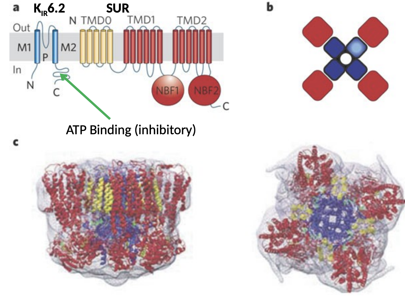
KATP Channel Antagonists
In periphery → drugs that bind SUR subunit and reduce KATP channel opening increase insulin secretion, treating Type II diabetes and hypoglycemia.
Includes Glibenclamide and Repaglinide.

KATP Channel Openers
Can be neuroprotective (Iptakalim?)
Used as antihypertensives as they relax smooth muscle of vasculature
May also treat hair loss

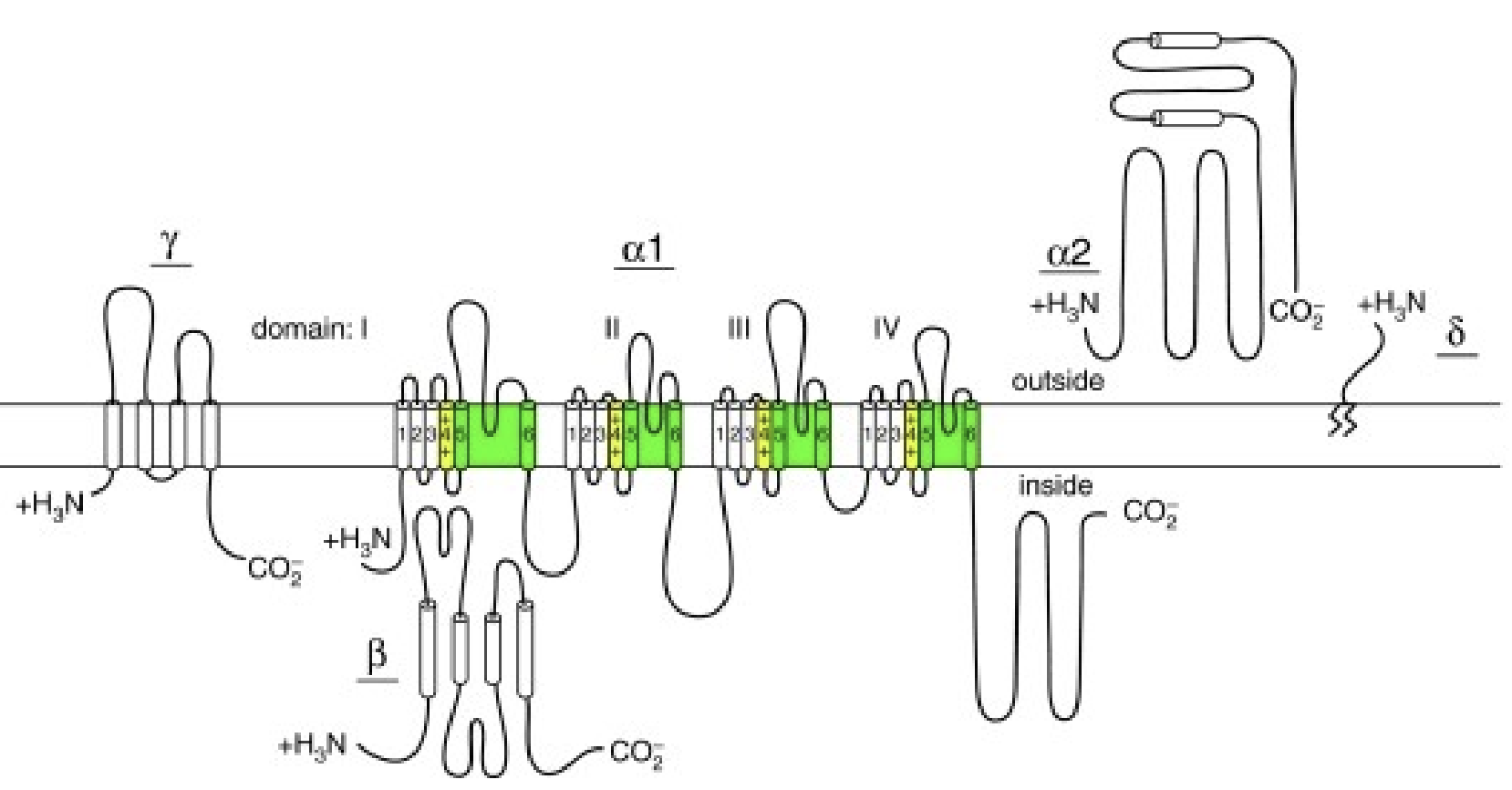
VGIC Structures

VGCC Subunits
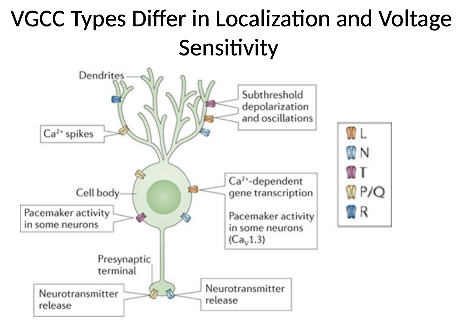
VGCC Localization and Sensitivity
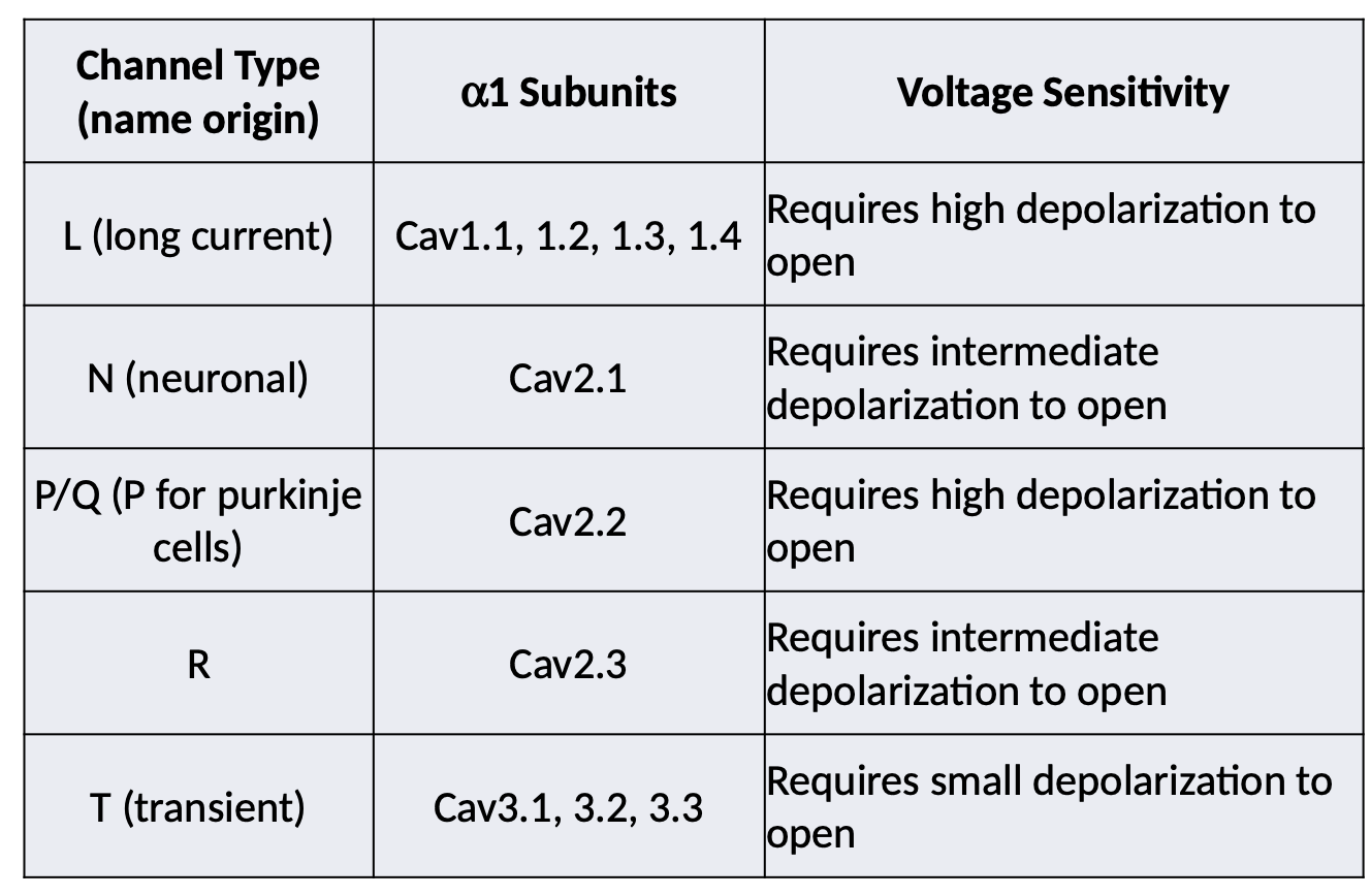
VGCC Types
Dihydropyridines
Large family of L type channel blockers
Some are clinically used antihypertensives

Phenylalkylamines
L type channel blockers
Some are clinically used antihypertensives
Verapamil may also treat cluster headache

w-Conotoxins
Cone snails use small peptide toxins to paralyze their prey
This toxin’s GVIA blocks N VGCCs
N type channels are involved in pain sensation → w-Ctx is analgesic (intrathecal)
1000X more potent than morphine

w-Agatoxin
Small peptide P type channel blocker

T Type Channels
So called “transient” channels as they open at small depolarization and inactivate quickly upon further depolarization
Set “pacemaker” current in thalamic neurons
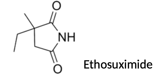
Block of thesechannels (Cav3.1-3) by ethosuximide prevents absence seizures
Ca2+ in Neurons
Normally, intracellular [Ca2+] ~10-100nM, extracellular is ~1-3mM
Huge gradient → massive influx if channels open
Strong depolarization
Second messenger → many proteins are regulated by Ca2+
Ca2+ signaling

Ca2+ Binding Domains
Many proteins sense calcium through Ca2+ binding domains
Calcium binding induces conformational change
Change activity of channel/enzyme
Change in protein-protein interactions
C2 domain, EF Hands
EF Hand Domain
Common Ca2+ domain found in many Ca2+ sensing proteins
Example protein → Calmodulin

C2 Domains
b-sheet Ca2+ binding domain
Synaptotagmin → Ca2+ sensor for neurotransmitter release
Presynaptic Ca2+ Regulation
VG Ca2+C are near active zone in neurons of CNS
Induce large local rise in Ca2+ concentration
Ca2+ is quickly removed into intracellular buffers like ER, or pumped into extracellular space

Vesicular neurotransmitter release
Docking → vesicle is brought close to target membrane
Priming → molecular machinery for fusion is “primed”
Fusion → upon Ca2+ influx, vesicle fuses
Endocytosis → vesicle is retrieved
Recycling → vesicle is refilled with neurotransmitter

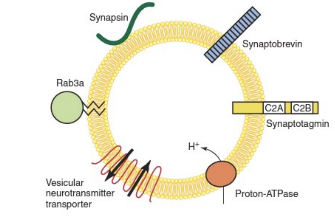
Vesicle Proteins
SNAREs
On vesicular membrane (v-SNAREs)
On target membrane (t-SNAREs)
Bring 2 membranes together to fuse
Synaptotagmin
Ca2+ sensor for vesicular neurotransmitter release
Fusing Proteins

a-Latrotoxin
Forms Ca2+ Channel, Inducing Massive Exocytosis

Botulinum Toxins
Initially taken into cells by endocytosis, then exit vesicles into cytoplasm
These toxins catalyze proteolysis (cleavage) of SNARE complex
Prevents vesicular neurotransmitter release, at NMJ this causes paralysis
Highly toxic → estimated human median lethal dose (LD-50) of 1.3–2.1 ng/kg intravenously or intramuscularly and 10–13 ng/kg when inhaled
NT Release Disruption

Botulinum toxin uses (Botox)
Strabismus (eye muscle disorder)
Chronic migraine
Hyperhidrosis (excess sweating)
Cosmetic applications, especially wrinkle reduction
Cholinergic projections

Acetylcholine
In CNS it modulates → sleep, attention, memory/cognition, and pleasure/reward
In PNS
Excitatory NT of neuromuscular junction (nicotinic)
Several autonomic roles (especially parasympathetic)
Acetylcholine Synthesis
Acetate (from acetyl-CoA) plus choline = acetylcholine
You need to be able to draw acetylcholine

Acetylcholine Metabolism
Acetylcholine is synthesized in the cytosol of presynaptic terminal by choline acetyltransferase
Loaded into vesicles by a transporter, VAChT
Released into synaptic cleft
Broken down into acetate and choline by acetylcholinesterase
Choline is recycled by Na+/Choline transporter

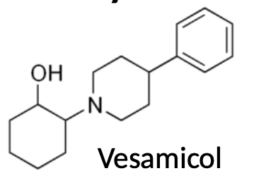
Vesamicol
Vesicular acetylcholine transporter (VAChT) inhibitor
Hemicholinium
Choline transporter (ChT) inhibitor

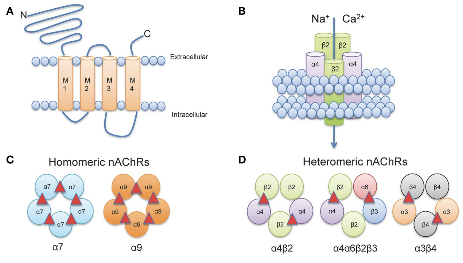
nAChR Channel structure
One receptor binds at least 2 molecules of ACh to open
nAChRs have hydrophobic ring in pore
Leucine residue highly conserved in M2 pore lining helix
Forces cations to shed hydration before entering cell

Homomeric vs Heteromeric
nAChR Channel Properties
Cation channels that pass Na+, K+ → some may pass Ca2+
nAChRs desensitize → pass less current after prolonged ligand binding
Intrinsic property of nAChRs
Can be modulated by posttranslational modifications to channel

Nicotine
Acetylcholine Receptor Agonist
Tobacco was brought to new world by Spanish & Portuguese
Popularized by French ambassador to Portugal, Jean Nicot

nAChR Agonists
Acetylcholine
Nicotine
Arecoline (areca/betel nut) → (also muscarinic agonist)
Coniine (poison hemlock)
Carbachol → (also agonist for mAChR)
Turbocurarine (curarine)
Active component of curare
Competitive, reversible antagonist of nAChR
Harmless if ingested orally (excellent hunting poison)
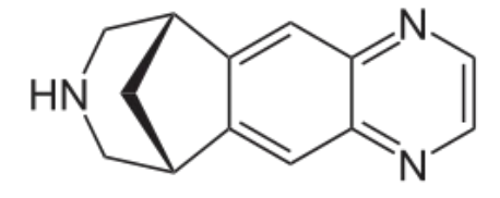
Varenicline
partial agonist at nAChR
Mecamylmine
Noncompetitive nAChR antagonist → Also an antihypertensive

a-Bungarotoxin
74 amino acid peptide
Competitive, irreversible antagonist of nAChR
Can be used to label (microscopy) and purify nAChR
M1,3,5
Gaq coupled receptors → Have excitatory effect on postsynaptic cell

M2,4
Gai coupled receptors → Have inhibitory effect on postsynaptic cell

Ga proteins

Acetylcholine Autonomic
Parasympathetic → “rest and digest”
Cardiac muscle expresses M2 muscarinic receptors
Inhibitory receptors → slows down heart
Smooth muscle and secretory organs express M3 muscarinic receptors
Increase smooth muscle contraction and secretion
Muscarine
Nonspecific agonist of mAChRs
Symptoms → Salivation, Sweating, Tearing, Diarrhea
Death by cardiac/respiratory failure
Antidote → atropine

Muscarinic Agonists
Muscarine
Arecoline (areca/betel nut) → (also nicotinic agonist)
Oxotremorine
Carbachol → (also nicotinic agonist)
Pilocarpine

Organophosphates
Acetylcholinesterase Inhibitors
Tabun & sarin are volatile (gas) nerve agents
Diazinon is a ____ insecticide

Carbamates
Reversible, covalent inhibitors of acetylcholinesterase (AChE)

acetylcholinesterase inhibitors in CNS
Uses include:
– Learning deficits associated w/neurodegeneration (Alzheimer’s)
– Cognitive impairment associated with schizophrenia
– Tacrine is no longer available owing to liver toxicity
acetylcholinesterase inhibitors in PNS
Myasthenia Gravis → loss of nAchRs at NMJ
Decreasing AchE activity helps compensate
Physostigmine
Acetylcholinesterase Inhibitor
Natural product from Calabar bean
Used medicinally, along with 2 semi-synthetic derivatives

Rivastigmine
Acetylcholinesterase Inhibitor
Used for CNS applications, such as treating dementia

Neostigmine
Acetylcholinesterase Inhibitor
Cannot cross into CNS, used for PNS applications

Nicotinic agonists
Act at neuromuscular junction to increase muscle tone/induce paralysis
Muscarinic agonists
Increase activity of parasympathetic nervous system
Secretory responses of the such as salivation and lacrimation (tearing).
Muscarinic antagonists
Inhibit the actions of the parasympathetic system → para-sympatholytic agents
SLUDGE
Symptoms of increased Ach activity in parasympathetic nervous system (mAChRs)
AChEI poisoning → possible nAChR symptoms plus:
• Salivation
• Lacrimation (tearing)
• Urination
• Diaphoresis (sweating)
• Gastrointestinal mobility (diarrhea)
• Emesis (vomiting)
Atropine
Nonspecific, competitive mAchR antagonist
Counteracts bradycardia
Counteracts muscarine and AchEI poisoning
Reduces salivation, diarrhea
Causes hallucinations at high doses

Benzatropine
Nonspecific competitive mAchR antagonist
Reduces parkinsonism, tremor

Glutamate
Major fast, excitatory NT of CNS
Non-essential amino acid
Does not cross blood brain barrier → Synthesized from precursors in brain
3 types of receptors
AMPA/Kainate
NMDA
Metabotropic
Glutamate Synapse

Glutamate Reuptake
Glutamine is metabolized into glutamate by glutaminase
Glutamate is removed from synaptic cleft by neurons and astrocytes
Astrocytes metabolize glutamate into glutamine, release glutamine, neurons take up glutamine and convert it back to glutamate

Glutamatergic signaling
Short term excitation → postsynaptic depolarization
Longer term signaling → synaptic plasticity
Excess glutamate → excitotoxicity

AMPAR
Ionotropic glutamate receptors
Mediate the majority of excitatory neurotransmission in the CNS
Tetramers of GluA1-4
Dimer of dimers → GluA1/2 or 2/3 most common
Homotetramers of GluA1 rare but exist
Ligand gated cation channels (usually Na+, sometimes Ca2+)
Ca2+ permeability depends on Q/R editing site of GluA2 subunit

GluA2 Q/R Editing
The mRNA GluA2 subunit of AMPARs is edited to replace a codon for glutamine (Q) to arginine (R)
This Q to R substitution makes AMPARs with GluA2 impermeable to calcium
This editing takes place in the vast majority of GluA2 mRNAs, meaning GluA2 containing AMPARs do not pass Ca2+

AMPAR Desensitization
AMPARs desensitize in presence of prolonged agonist

AMPAR Agonists
Research tools:
2-amino-3-(3-hydroxy-5-methylisoxazol-4-yl) propanoic acid (AMPA)
Quisqualate (also Group I mGluR agonist)
(S)-(-)-5-Fluorowillardiine

AMPAR Competitive Antagonists
Quinoxaline family
Usually abbreviated with “QX” at end
The most important of these is NBQX
Research tools

AMPAR Noncompetitive Antagonists
2,3-benzodiazepines
Anticonvulsants
Neuroprotective (reduces excitatory activity)
Tofisopram is available in Europe, not approved in US or Canada- CYP3A4 inhibition?

Voltage-dependent Blockers
Bind GluA2-lacking AMPARs
Or unedited GluA2 with Q residue
Block AMPAR-mediated Ca2+ current
Endogenous polyamine confers inward rectification

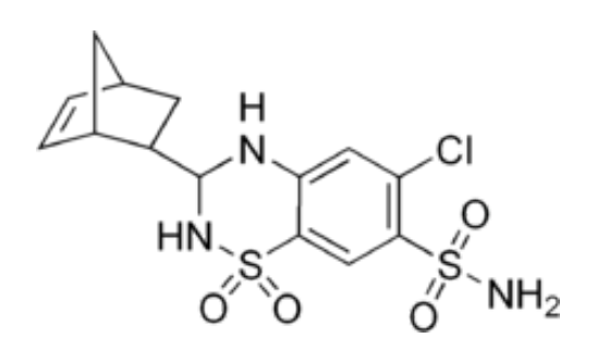
Cyclothiazide
AMPARs desensitize in presence of prolonged agonist
Desensitization can be reduced/blocked by _____
Positive allosteric modulator
Nootropics
Substances that improve cognition, concentration, memory
Ones we’ve seen before → cholinergic agents
Some AMPA modulators (ampakines) are ____ → Positive allosteric modulator
Ampakines
• Racetams (some, not all) → Aniracetam and Piracetam (other CNS effects)
• Benzylpiperidines: AMPAR-specific ampakines

NMDA Receptor
Tetrameric channels of GluN1-3 (old nomenclature: NR1-3)
GluN1 is obligatory subunitt → GluN1/2 most common
Similar topology to AMPARs
Ligand gated cation channels (Na+, Ca2+)
Also voltage gated (kind of)

Glutamate EPSCs

Glutamate/Glycine Co-agonists
Glutamate and glycine are NMDAR coagonists

Mg2+ NMDAR Block
Required for voltage dependent block of NMDARs at resting/hyperpolarized potentials → so pass current only when postsynaptic membrane is depolarized
NMDAR’s Asn residues in pore may confer Mg2+ block

NMDAR coincidence detectors
• Presynaptic glutamate release
• Postsynaptic depolarization

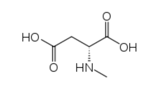
N-methyl-D-aspartic acid
Partial NMDAR agonist for the glutamate site
Cycloserine
Partial NMDAR agonist for the glycine site
May be useful adjunct therapy for schizophrenia
