Path Exam 2 Images
 Call Kai
 Learn
 Practice Test
 Spaced Repetition
 Match
 Flashcards
 Knowt Play
1/134

There's no tags or description
Looks like no tags are added yet.
135 Terms

Marasmus
Shrunken, wasted appearance with
loss of muscle and subcutaneous fat
mass
Concave abdomen with pronounced
ribs
Emaciated extremities
Head appears large relative to the
body
Thin, dry skin
Hair loss

Kwashiorkor
Edema with muscle atrophy (often
underappreciated) with normal body fat
Hepatomegaly (from fatty liver)
Distended abdomen
Rounded prominence of the cheeks
(“moon face”)
Thin, dry, peeling skin with
hyperkeratosis/hyperpigmentation

Russell’s sign from BN

Bitot’s spots - white plaques of keratinized
epithelial cells on the conjunctiva
Complication of Vitamin A deficiency

Rachitic rosary” –
overgrowth of
bone/cartilage at the
costochondral joints
and
Widening of wrists
Signs of Ricketts from vitamin D deficiency

Leg bowing from Rickets due to vitamin D deficiency

Osteoporosis seen on right. Result of vitamin D deficiency in adults.

Wernicke-Korsakoff Syndrome from vitamin B1 deificency
The thalamus / peri third ventricular region showing
bilateral multiple foci of petechial hemorrhages
(coronal section of the forebrain)

Fluid filled epidermal elevations from sun burn
Bullae if greater than 1 cm
Vesicles if less than 1 cm

Papules that form from folliculitis

Characteristic human dermatophyte infection
Patch: Flat lesion greater than 1 cm in diameter
Plaque: Broad flat papule greater than 1 cm in diameter

Cold sore vesicles due to HSV (herpes simplex virus)

Shingles taking form of multiple linear vesicles due to VZV (varicella zoster virus)

Papules with filliform architecture due to HPV
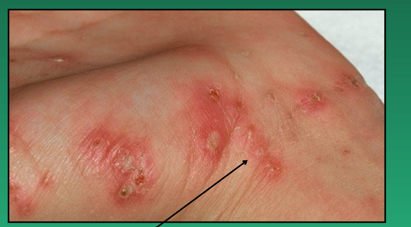
Scabies from mites burrowing into skin
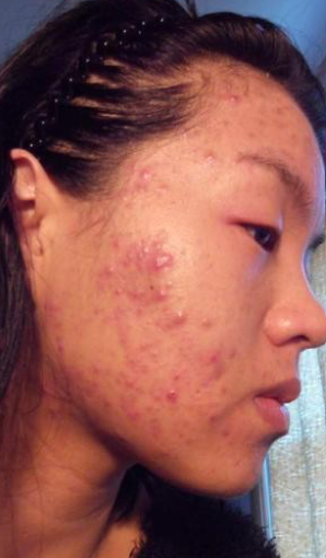
Inflammatory Acne papules and pustules from

Open comedones, AKA blackheads
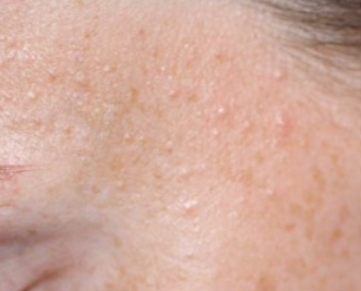
Closed comedones, AKA whiteheads

Eczema, allergic contact dermatitis

Psoriasis, erythematous patches and plaques with silvery scale, classically on extensor surfaces

Seborrheic Keratosis
Benign epidermal tumor
Presents as brown, waxy papules and plaques with furrowed surface

Basal cell carcinoma

Basal cell carc

Non-aggressive squamous cell carc

Aggressive squamous cell carcinoma

Squamous cell carc, hyperkeratotic plaque

Squam cell carc, ulcerated nodule

Solar lentigo

Lentigo simplex of childhood
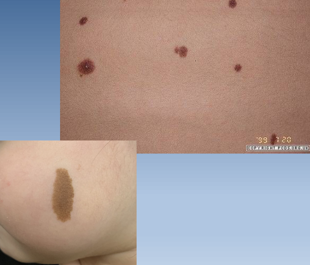
Melanocytic nevus (mole)

Melanoma
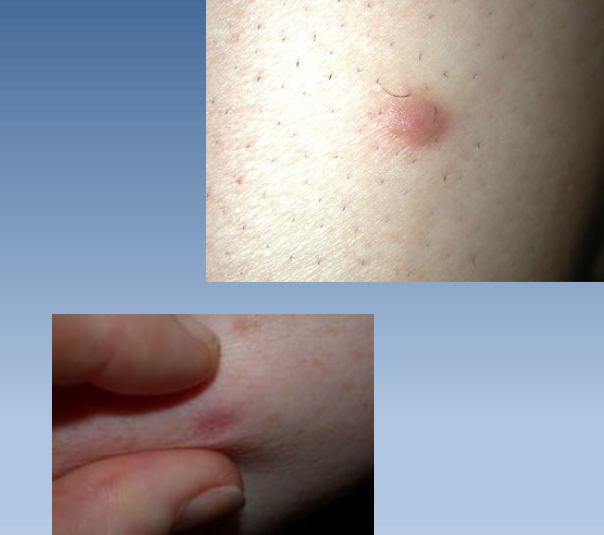
Dermatofibroma

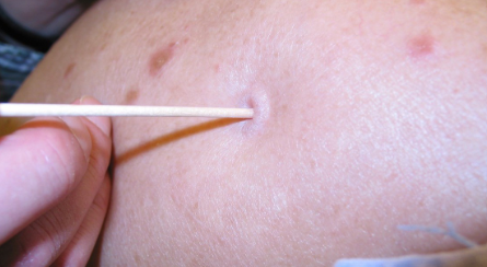
Hemangioma

Hemangioma

Bone with pus from osteomyelitis

Sinus tract formation from osteomyelitis

Aseptic bone necrosis

Paget’s disease bow legging seen on left

Irregularly shapen and thickened bone due to Paget’s disease

Skull thickening from paget’s disease

Paget’s disease caused skull and leg thickening with leg bowing

Comminuted fracture

Greenstick fracture

Compound fracture

Osteolysis

Osteosclerosis

Osteoid osteoma

Osteoid osteoma

Osteosarcoma
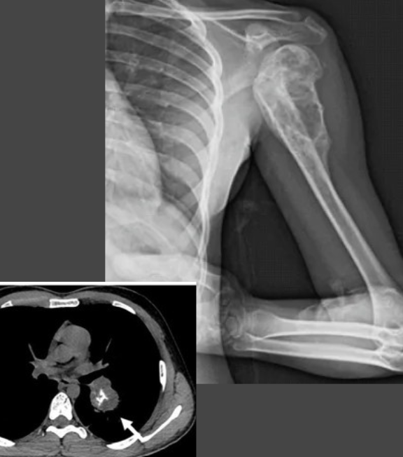
Osteosarcoma

Osteosarcoma

Osteosarcoma in metaphysis (middle arrow)

Chondrosarcoma
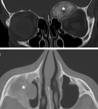
Chondrosarcoma

Chondrosarcoma

Ewings sarcoma

Ewings sarcoma in diaphysis

Upper line is synovium
THen meniscus
THen articular cart
Then ligaments

Degenerative joint disease seen on right

Joint mice from DJD
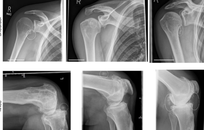
Loss of joint space from DJD

Bone cyst from DJD

Osteophyte formation from DJD
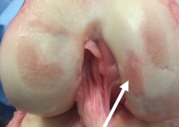
Thin art cart from DJD

Eburnation
Bone cyst

Osteophyte

Heberdens nodes from DJD

Bunion from DJD

Rheumatoid arthritis with rheumatoid nodules

RA Z deformity

RA swan neck deformity

RA swan neck deformity on left, boutonniere on right
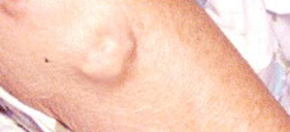
RA rheumatoid nodule

Scleritis from RA

INfectious arthritis

Bite mark from lyme arthritis

Gout

Uric crystals from gout

Tophus from chonic gout

Kidney from gout

Epiglottitis

Pneumonia

Regular lung

X ray of pneumonia, showing inflammaiton of airspaces that fills a lobe

Lobar pneumonia

Bronchopneumonia (show patchy)

Lung abscesses (from pneumonia

Empyema: pus formation on the pleura
Pleuritis: inflammation of the pleura

Ghon Complexes
On right is subpleural which is caused by primary infection
On left is central which is caused by lymph node drainage

Casseous necrosis and cavitation shown in Secondary TB

Milliary seen in Secondary TB (pale holes like millet seeds)

Thick bronchial mucus found in Asthma
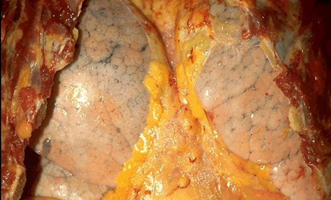
Hyperinflation of lungs from Asthma

Hyperinflated lung from Asthma

Honeycomb lung from chronic hypersensitivity pneumonitis

Chronic Bronchitis

Emphysema
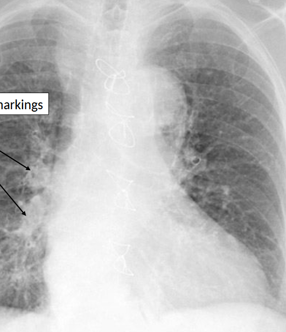
Increased bronchovascular markings from chronic bronchitis
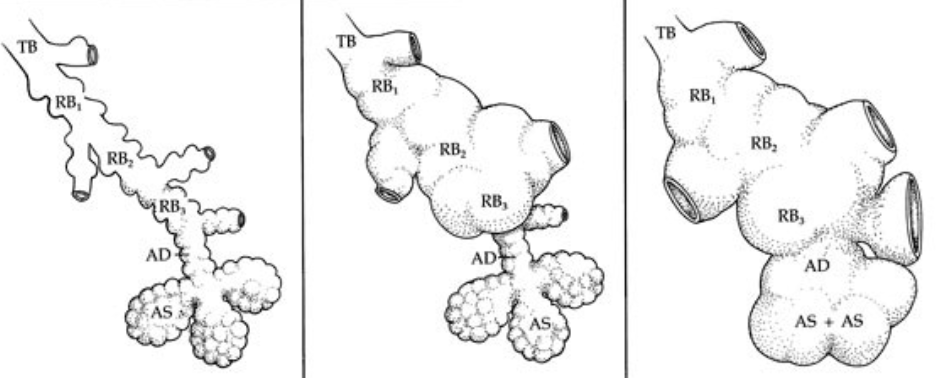
Left is normal
Middle is centriacinar emphysema
Right is panacinar emphysema