Humoral immune activation
 Call Kai
 Learn
Learn Practice Test
 Spaced Repetition
 Match
Match Flashcards
 Knowt Play
1/28

Earn XP
Description and Tags
B-cells activation, Germinal center and AID
Name | Mastery | Learn | Test | Matching | Spaced | Call with Kai | Chat |
|---|
No analytics yet
Send a link to your students to track their progress
29 Terms
What are the steps for B-cells activation? (T-dependent response)
Naive B cell circulates in blood and enters lymph node, checking the situation
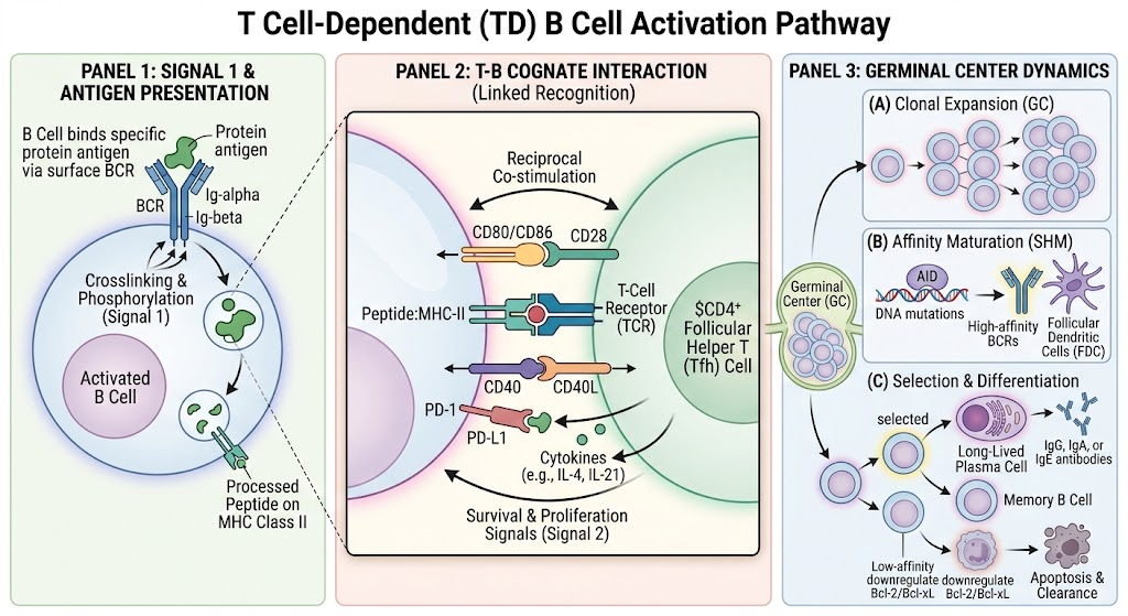
B cell finds and binds to specific antigen through B cell receptor (Signal One)
Antigen binding cross-links multiple B cell receptors together (when the antigens cross-links they move closer to each other to doe it)
Cross-linking activates signaling cascade internally through Ig alpha and Ig beta molecules (The phosphorylation of ITAMs-which lead to many pathways to make transcription factors (TF))
B cell internalizes the antigen and processes the antigen
B cell presents peptide on MHC II molecules on the surface
T follicular helper cell (TFH) recognizes the MHC-peptide complex with its T cell receptor
B cell and TFH bind to each other at the T-B border in the lymph node
10. B cell expresses CD40 and costimulatory molecules B7.1, B7.2 (CD80, CD86)
TFH expresses CD40 ligand and CD28
CD40-CD40 ligand binding provides Signal Two for B cell activation
CD80/CD86 binding to CD28 reinforces TFH activation
TFH produces cytokines: IL-2, IL-4, IL-5, and IL-21 (very important)
Cytokines bind to cytokine receptors on B cell, providing Signal Three
B cell receives all three signals and becomes fully activated
B cell undergoes clonal expansion, duplicating itself many times
B cell increases expression of costimulatory molecules B7.1 and B7.2 on surface
B cell increases expression of cytokine receptors
B cell expresses anti-apoptotic proteins to prevent cell death during proliferation
Upregulation of transcription factors for proliferation and B cell identity
B cell is now fully armed and ready to receive T cell help and stimulation
B cell is prepared for next steps: germinal center formation, AID function activation, differentiation into plasma cells or memory B cells
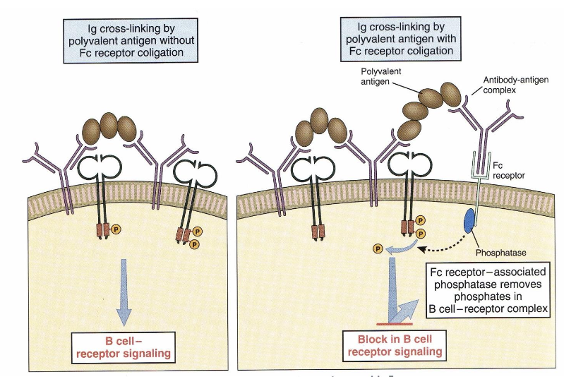
How can ITIMs (Immunoreceptor tryosine-based inghibitory motifs) inhibit signaling through BCR?
As an infection is cleared, secreted antibodies form antibody–antigen complexes with remaining pathogens.
The complex simultaneously binds to both the B-Cell Receptor (BCR) via the antigen and to an inhibitory Fc receptor via the antibody's tail region.
This binding brings the Fc receptor's intracellular ITIM motif near the BCR. The ITIM becomes phosphorylated and recruits intracellular phosphatases that strip phosphate groups from the BCR signaling complex.
This mechanism shuts down B-cell activation to prevent the overproduction of antibodies once sufficient levels are reached.
What are the parts for lymph node?

What other type of B-cells can u find in the body?
Follicular B cells (normal B-2 cells)
Marginal zone B cells: They stay at the marginal zone of the spleen.
B-1 B cells: They can be found in the peritoneum or omentum
Give me more information about follicular B-cells:
They are continuously produced
Mainly found in B cell follicles or in the lymph nodes
Are recirculating
Major producers of switched (IgG, IgA and IgE) antibodies, but also produce IgM antibodies
Are involved in germinal center (GC) and development of immunological memory
Interact with T cells during activation
What is T-independent B cell activation and how does it differ from T-dependent activation?
Primarily involves marginal zone B cells and B1 cells (not follicular/B2 cells)
Types of Antigens That Trigger TI Response:
Polysaccharides (sugars from bacterial cell walls)
Highly repetitive antigens with many copies of the same structure
Bacterial cell wall components with repeating sugar structures
Mechanism of Activation - Signal Generation:
Unlike T-dependent response, TI antigens have many repeating copies on their surface
These repetitive structures can cross-link many B cell receptors together at once (not just one or two like in normal activation)
This massive cross-linking creates a very strong signal that can activate the B cell without T cell help
The B cell can receive co-activation signals from:
Toll-like receptors (TLR) binding to bacterial components
Complement receptors binding to complement fragments attached to bacteria
These co-activation signals decrease the threshold for activation, allowing very fast activation
Why Pure Polysaccharides Cannot Use T-Dependent Pathway:
B cells can recognize polysaccharides (sugars), lipids, proteins, and mixtures
However, MHC II molecules only present peptides, not sugars
If the antigen is pure polysaccharide with no protein component, the B cell cannot present it on MHC II
Therefore, T cells cannot recognize it, and T cell help cannot be provided
This is why pure polysaccharide antigens must use the T-independent pathway
What are the out comes of TI B cell activation?
B cells can differentiate directly into plasma cells (antibody-producing cells) without going through germinal center
B cells can become memory B cells (similar to naive B cells but better)'
However, limited compared to T-dependent response:
Cannot form germinal centers
Cannot activate AID function (no somatic hypermutation or class switching)
Produce antibodies but only for a short period (about one week)
Results in a burst of antibodies that is relatively short-live
What are two ways antigens are transported from the periphery to the lymph node?
Antigen is transported via two main ways:
(1) Via lymphatics - antigens travel through lymphatic vessels, especially during edema or infection, reaching the lymph node outskirts where B cells are located.
(2) Via immune complexes and cells - small antigens pass through conduits (small roads in the lymphatic highway) directly to B cell areas. Larger antigens are delivered by subcapsular sinus macrophages. Antigens can also come as immune complexes or be transported by cells with complement receptors that pass them to subcapsular sinus macrophages, which then transfer them to follicular dendritic cells.

What is the function of follicular dendritic cells in the B cell follicle?
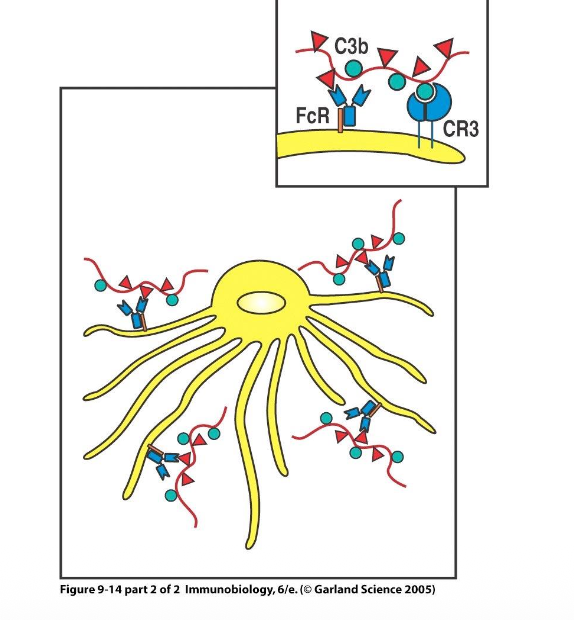
Follicular dendritic cells (FDCs) are special cells in the B cell follicle with long dendrites. They do not take up antigen themselves but act like double-sided tape, with antigen sticking to their surface. They have Fc receptors that bind antibody-coated antigens and complement receptors that bind complement-decorated pathogen pieces. These receptors ensure antigen stays available for B cells even when B cells are not immediately present. FDCs sit in the follicle displaying multiple different antigens on their surface, making antigen available for B cell activation and later in the germinal center.
How do activated T cells and B cells migrate to meet each other in the lymph node?
After activation, T cells upregulate CXCR5 and B cells upregulate CCR7. These chemokine receptors respond to chemokine gradients in the lymph node, causing T cells and B cells to move from their separate zones toward each other and meet at the border. This is necessary because T cells are activated by dendritic cells in one zone while B cells are activated in another zone; without migration, they would never have the chance to interact.

what is the primary role for IL-4?
Drives Class Switching: Directs B cells to switch antibody classes to IgE and IgG4 (human) or IgG1 (mouse).
Boosts Antigen Presentation: Upregulates MHC Class II and CD80/CD86 so the B cell can present antigen and receive helper signals more efficiently.
Promotes Survival: Upregulates anti-apoptotic proteins to prevent Germinal Center B cells from dying.
Shapes Cell Fate: Works together with IL-21 to guide whether the B cell becomes a Memory B cell or a Plasma cell.
What are all the costimulatory signals required for B cell activation and what do they do?
B cells receive multiple reinforcing signals:
(1) B7-1 and B7-2 (CD80, CD86) on B cells bind CD28 on T cells.
(2) ICOS ligand on B cells binds ICOS on T cells, reinforcing the Tfh program.
(3) CD40 ligand on T cells binds CD40 on B cells.
(4) Cytokines, particularly IL-21 (from the Tfh cell), which is the initiating cytokine for germinal center formation (prolifiration).
These signals ensure only properly activated B cells survive and expand. The adaptive immune system has multiple failsafe mechanisms at every step to ensure only good cells survive; if a cell was activated by mistake, it needs many reinforcing signals to survive.

What is a germinal center (GC) how does it form, what are its two zones, and what happens in each?
A germinal center is a microstructure within the lymph node formed by proliferating B cells after activation. It takes approximately 5-7 days to form.
(after activation some B cells make plasma cells (with IgM mostly and is it short lived and has unmutated antibodies) to fight the pathogen until the GC form and make more long lived plasma cells and memory cells)
The germinal center has two zones:
(1) Dark zone - B cells proliferate extensively and undergo somatic hypermutation (SHM). It appears dark under microscope because many cells cluster together.
(2) Light zone - B cells take up antigen from follicular dendritic cells, present it to Tfh cells, and undergo selection. It appears light because fewer cells are present as they are being checked for quality. B cells move between zones using adhesion molecules and chemokine receptors. To move from one zone to the other, they express different adhesion molecules and cytokine receptors.

What is AID and what two AID-dependent processes occur in the germinal center?
AID is an enzyme activated by CD40 ligand interaction. It mediates two crucial processes in the germinal center:
Somatic hypermutation (SHM) - introduces random mutations in the variable region of the antibody to increase affinity.
Class switch recombination (CSR) - changes the constant region of the antibody to different types (IgG, IgA, IgE) while keeping the variable region the same. Both processes are dependent on AID and are essential for generating improved antibody responses.
How does somatic hypermutation work and how does it lead to affinity maturation?
In the dark zone, AID introduces random mutations throughout the variable region of the antibody gene. Each B cell gets one or more mutations, creating a diverse population with different sequences. This is Darwinian evolution: mutations are random and unbiased with no direction. Then selection acts on this variation.
B cells with mutations that introduce stop codons or decrease affinity cannot bind antigen from FDCs and undergo apoptosis. B cells with slightly improved affinity pick up some antigen, present little to Tfh cells, and return to dark zone for more mutations. B cells with significantly improved affinity pick up lots of antigen, present lots to Tfh cells, receive survival signals, and differentiate into plasma cells or memory B cells.
The net result is affinity maturation - random mutation coupled with selection makes affinity increase over time. Without selection, affinity stays the same. Without mutation, affinity cannot increase.
What is the selection process in the light zone and what are the possible outcomes for a B cell?
In the light zone, B cells compete for limited antigen on FDCs. Possible outcomes:
B cell cannot express functional B cell receptor (e.g., stop codon) - cannot take antigen from FDCs - undergoes apoptosis.
B cell has lower affinity - cannot bind antigen well - cannot take it up from FDCs - undergoes apoptosis.
B cell has slightly improved affinity - picks up little antigen - presents little to Tfh cells - receives weak signal - returns to dark zone to try more mutations.
B cell has much better affinity - binds antigen much better - picks up lots of antigen from FDCs - presents lots to Tfh cells - receives strong survival signal - differentiates into plasma cell or memory B cell. This is a fight for limited antigen; higher affinity B cells are stronger and can pull out antigen, while weaker ones cannot compete.
How does affinity improve over time and what happens with repeated vaccinations or infections?
Affinity increases over time because random mutation combined with selection continuously eliminates worse cells and retains better ones. With repeated vaccinations or reinfections, naive B cells form germinal centers in the first response and improve affinity. Upon revaccination, memory B cells (which already have improved affinity from the first round) can form new germinal centers and start from a higher baseline affinity level. Then they can add more mutations on top of their already-improved affinity, achieving even better affinity. This process can repeat with each vaccination or infection, progressively improving affinity. Theoretically there is no limit to affinity improvement, only limited by signal strength at some point.
How long do germinal centers persist and what is the significance of long-lasting germinal centers?
Germinal center persistence varies: some are transient (2-3 weeks), while others induced by viral infections like influenza or mRNA vaccines can persist for 6-9 months. The signals leading to germinal center shutdown are unclear. One hypothesis for long-lasting germinal centers is that even after the pathogen is gone, continuing the process creates randomness and diversity that may generate memory B cells prepared for different pathogens or variants. Memory B cells generated from persistent germinal centers can be specific for variants never encountered. For example, vaccinated individuals who never got infected may develop memory B cells specific for Omicron variant just from random mutations in the germinal center, providing broad protection. This broadening of response is different from simple affinity maturation for the original antigen.
What is class switch recombination and why is it important to change the constant region?
Class switch recombination changes the constant region of the antibody while keeping the variable region identical. The constant region is very important for several functions. Different constant regions induce different effector functions. By changing to different constant regions (IgG, IgA, IgE), B cells can choose the better one for the specific pathogen they are fighting. This allows the same antibody specificity to be adapted for different types of immune responses and different pathogens.
What constant regions does a naive B cell express and how is this possible?
Naive B cells have both IgM and IgD on their surface as B cell receptors. This is possible through alternative splicing of the same VDJ segment in the DNA. In the bone marrow, the VDJ segment is rearranged. When the cell comes out in circulation in the lymph node, the DNA still contains all the constant region cassettes. Through alternative splicing, the cell can express either mu (M, IgM) or delta (D, IgD) from the same VDJ, allowing both antibody types on the surface simultaneously.

How are constant region genes organized in the B cell genome and how does class switch recombination work mechanistically?
Step 1: Cytokines open chromatin at the target constant region (e.g., epsilon for IgE), allowing transcription.
Step 2: AID enzyme creates DNA breaks by deaminating cytosine to uracil in both the IgM and target regions.
Step 3: DNA looping occurs, bringing the two switch regions together.
Step 4: The intervening DNA segment is deleted and excised, joining the VDJ region directly to the new constant region.
Result: Same variable region (same antigen specificity) but new constant region (new antibody class). This process is irreversible—once deleted, DNA cannot be recovered.

Why is it important that B cells are antigen-presenting cells and what is epitope mismatch?
B cells can grab non-protein parts of a germ (like a sugar coating) and pull the whole package inside, but they present a protein piece from that package to T cells. The T cell recognizes the protein, activates, and gives the B cell the helper signals needed to make strong, long-lasting antibodies against the sugar. This trick is used in conjugate vaccines—chemically linking a bacterial sugar to a protein ensures T cells can help B cells build durable immunity against non-protein targets.
How do cytokines determine which antibody class a B cell will produce during class switching? (examples)
Different cytokines produced by T helper cells and dendritic cells direct class switching to specific antibody types:
TGF-beta → IgA (especially for mucosal infections), IL-4 → IgE, and Interferon-gamma → IgG. This ensures the immune response matches the pathogen type and location. For example, mucosal infections trigger Th17 cells that produce IL-17 and TGF-beta, promoting IgA production for protection at mucosal surfaces.

What is AID, and how the mismatches are repaired? (Hypermutation)
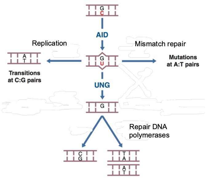
AID (Activation-Induced Deaminase) is an enzyme induced by CD40-CD40L interaction that deaminates cytosine to uracil in DNA.
This creates C-U mismatches that are repaired by three different pathways:
(1) Replication → C-G becomes T-A,
(2) UNG/APE pathway → C-G becomes G-C,
(3) Mismatch repair → C-G becomes A-T. Each repair pathway produces different mutations, allowing a single C-U mismatch to generate multiple possible mutations. In somatic hypermutation, this occurs sparsely throughout the variable region; in class switching, it occurs densely in switch regions and requires UNG activity to create double-strand breaks.
How the mismatches are repaired? (CSR)
It uses mismatch repair and APE/UNG repair pathways

How do B cells transition from producing membrane-bound antibodies to secreting soluble antibodies?
As B cells differentiate into plasma cells, RNA splicing removes the transmembrane anchor region from the antibody transcript, producing a secreted form instead of a membrane-bound form. This is not a DNA modification—it's a post-transcriptional change. This allows plasma cells to rapidly produce and secrete large quantities of antibodies without waiting for genetic recombination. All antibody classes except IgD can be expressed in both membrane-bound and soluble forms.

What are the key differences between memory B cells and plasma cells?
Memory B cells: Remain as B cells with improved B cell receptors (higher affinity from somatic hypermutation + class-switched antibodies like IgG or IgA), circulate in blood, long-lived (years), can divide, and rapidly generate new germinal centers upon re-exposure.
Plasma cells: Terminally differentiated antibody factories, secrete antibodies, migrate to bone marrow, cannot improve further, mostly non-dividing, and have variable lifespans (short-lived ~1 week for T-independent; long-lived months to years for T-dependent germinal center-derived cells).
Why do some plasma cells survive for years in the bone marrow, and what supports their survival?
Plasma cells from T-dependent germinal center responses migrate to the bone marrow where they can survive for months to years (some evidence suggests 60+ years). Survival is supported by:
(1) Stromal cells that produce CXCL12 chemokine,
(2) Adhesion molecules that anchor plasma cells in place, and
(3) Nutrient-rich microenvironmentPlasma cells are large cells with extensive cytoplasm packed with endoplasmic reticulum, requiring constant energy to continuously produce antibodies. This explains why childhood vaccinations (like measles) provide lifelong immunity—long-lived plasma cells continuously produce protective antibodies in circulation.
Why is the secondary immune response (upon re-infection or re-vaccination) faster, stronger, and different than the primary response?
Primary response: Naive B cells take 5-7 days to establish germinal centers and produce IgM antibodies initially.
Secondary response: Memory B cells rapidly initiate new germinal centers, producing higher-affinity antibodies that are already class-switched (IgG, IgA, etc.) rather than IgM. This is why vaccination works—the first dose primes memory cells, and subsequent doses or natural re-infection trigger a rapid, potent response. Additionally, memory T cells help activate both naive and memory B cells faster, creating multiple layers of protection.