Module 4 - Electrocardiogram
 Call Kai
 Learn
 Practice Test
 Spaced Repetition
 Match
 Flashcards
 Knowt Play
1/108

There's no tags or description
Looks like no tags are added yet.
109 Terms
precordial leads
chest leads V1-V6: view the heart’s electrical activity on a horizontal plane of the torso, defining anterior to posterior and left to right
normal arrythmias in terms of pattern
NSR, SB, ST, Aflutter, 3rd HB, Junctional, ventricular
regularly irregular arrythmias
aflutter, 2nd TI HB, bigemny, trigimeny
irregularly irregular arrythmias
SA, Afib, Atach, MAT, 2nd T2 HB
ECG big box rule
1 large square between R waves equals a heart rate of 300 bpm
2 large squares equal 150 bpm
3 large squares equal 100 bpm
4 large squares equal 75 bpm
5 large squares equal 60 bpm
6 large squares equal 50 bpm
8 large squares equal 37 bpm
limb leads
I, II, III, aVR, aVL, aVF: view the heart’s electrical activity on a vertical plane of the torso, defining superior to inferior and left to right
deflection on the ECG is positive when
electrical wavefront is travelling toward the positive electrode
leads 2,3,aVF view the heart from
inferior perspective on the vertical plane
leads 1 and aVL view the heart from
lateral perspective on the vertical plane
Leads V1 and V2 view the heart from
anterior septal perspective on the horizontal plane
Leads V3 and V4 view the heart from
anterior perspective on the horizontal plane
Leads V5 and V6 view the heart from
lateral perspective on the horizontal plane
what do the isoelectric lines represent in the ECG signal
periods in the cardiac cycle when there are no currents flowing towards the positive or negative electrodes of the ECG leads
one small box on ECG measurements and time
1mm x 1mm block = 40ms and 0.1 mV amplitude
one large box on ECG measurements and time
5mm x 5mm = 200ms and 0.5 mV amplitude
10mm high amplitude = ?mV
1
normal values for PR interval and QRS interval
120-200 ms
60-100 ms
heart rate equation
60000 / R-Rinterval in ms
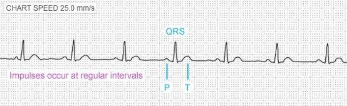
normal sinus rhythm
causes of arrhythmias
congential origins, heart diseases, drug or chemically induced
two basic underlying mechanisms of cardiac arrhythmias
abnormal automaticity and conduction issue
sinus node dysfunction types
sinus tachycardia, bradycardia, arrhythmia, and pause

sinus tachycardia

sinus bradycardia

sinus arrhythmia

sinus pause

sinus exit block
difference between sinus arrest and sinus exit block
in sinus arrest the basic rhythm doesn’t resume on time after the pause


premature atrial contraction
junctional arrhytmia types
premature junctional contractions, junctional rhythm, accelerated junctional rhythm, junctional tachycardia

premature junctional contraction

junctional rhythm
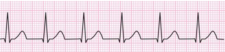
accelerated junctional rhythm

junctional tachycardia

AV block 1

second degree AV block Mobitz 1 Wenckebach

second degree AV block Mobitz 2

3rd degree AV block
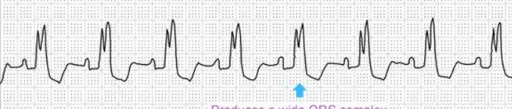
bundle branch block

right bbb

left bbb

atrial flutter (sawtooth)
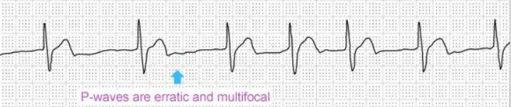
atrial fibrillation

atrial tachycardia

orthodromic narrow tachycardia

antidromic wide tachycardia

premature ventricular contraction
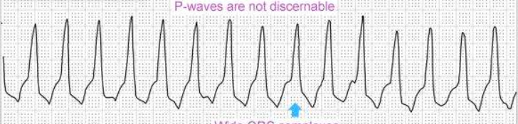
ventricular tachycardia

ventricular flutter

ventricular fibrillation
sinus tachycardia
SA node delivers impulses at a rate faster than 100bpm
sinus bradycardia
SA node fires at an abnormally slow rate, less than 60 bpm
sinus arrhythmia
discharges impulses irregularly thus irregular automaticity of the SA nose
sinus pause
SA node fails to fire but resumes to pacing after the escape beat occurs
sinus exit block
SA node impulse is blocked as it exits the SA node which prevents conduction into the atria
premature atrial contraction
due to the sudden discharge of an atrial ectopic focus. The premature beat can originate from any part of either atrium
extra P wave has an unusual shape
junctional arrythmias originate within
the AV node
premature junctional contraction
irritable automaticity focus in the AV junction suddenly fires a premature stimulus that is conducted to and depolarizes the ventricles. The P-wave may be hidden in the QRS, or just before or after the QRS. In addition, the QRS can be followed by a compensatory pause.
junctional rhythm
electrical impulses originate in the AV node and travel through the atrium and ventricle at about the same time. Junctional rates are usually slow — from 40 to 60 bpm — but regular
weak p wave
accelerated junctional rhythm
AV nodal rate accelerates to a rate faster than that of the sinus node and takes over the rhythm.
junctional tachycardia
automaticity of the AV junctional tissue is increased, sending out impulses between 100 and 200 per minute, most commonly at 140 per minute. The QRS complex is narrow, because as the AV junction fires, the impulse follows the normal conduction pathway into the ventricles to efficiently cause a ventricular contraction
heart block
failure to propagate electrical impulses appropriately
1st degree AV block
there is a slight delay in the conduction of a normal impulse from the atria to the ventricles. The ECG shows prolonged PR intervals, but a normal cycle of depolarization waves
causes of first degree AV block
• Myocardial infarction (MI)
• Coronary artery disease (CAD)
• Rheumatic fever
• Certain drugs
2nd degree AV block
by intermittent failure of atrial depolarizations to reach the ventricle
2nd degree AV block type 1 wenckebach
an intermittent delay in the conduction of normal sinus impulses from the atria to the ventricles. The PR interval becomes progressively longer until the QRS complex fails to appear. The sequence is then repeated
conduction ratio of 3:2
causes of mobitz 1 block
• Myocardial infarction (MI)
• Coronary artery disease (CAD)
• Rheumatic fever
• Certain drugs
second degree AV block mobtiz 2
to intermittent dropped beats preceded by constant P-R intervals.
The infranodal (bundle of His) tissue is most commonly the site of Mobitz II block. Note that advanced second-degree block refers to the block of two or more consecutive P-waves (that is 3:1 block).
how to differentiate mobitz 1 and 2
If the difference between these two P-R intervals is more than 20 milliseconds, then it is Mobitz I. If the difference is less than 20 milliseconds, then it is Mobitz II.
3rd degree AV block
there is no correlation between the P-waves and R-waves. The ventricles are beating at their own intrinsic rate (20 - 40 bpm)
bundle branch block
The blocked bundle branch delays full depolarization of the ventricle that it supplies.
Because the ventricles do not depolarize simultaneously, the QRS complex in the ECG will become wider, and two R waves will be visible.
right bundle branch block
the left ventricle will depolarize faster than the right ventricle. In the ECG, V1 shows a widened QRS with two R waves.
left bundle branch block
the right ventricle will depolarize faster than the left ventricle. In the ECG, V6 shows a widened QRS with two peaks.
atrial flutter
macro-reentry circuit in the atrium, originating in an atrial automaticity focus.
common type of atrial flutter occurs in the right atrium, with the macro-reentry circuit around the tricuspid valve annulus activated either in a counter-clockwise direction (typical common atrial flutter) or in a clockwise direction (typical reverse atrial flutter).
atrial fibrillation
supraventricular tachyarrhythmia with uncoordinated atrial electrical activation and consequently ineffective atrial contraction. AF is associated with the left atrium and in particular the pulmonary veins. The atrial rate may be 400 bpm or more.
atrial tachycardia
series of three or more consecutive atrial premature beats occurring at a rate greater than 100 beats per minute. Atrial tachycardia is usually paroxysmal, meaning it starts and ends abruptly. It begins suddenly with an irritable ectopic focus in the atrium discharging spontaneously, taking over the sinus node.
Atrioventricular reentrant tachycardia (AVRT)
supraventricular tachycardia (SVT), where the electrical conduction is going around a reentrant circuit involving an accessory pathway.
Wolff-Parkinson-White (WPW) syndrome
the presence of the accessory pathway is visible in normal sinus rhythm. Due to the premature depolarization (pre-excitation) of the ventricle, the ECG shows a delta wave, which is a slurred upstroke in the QRS complex that is associated with a short PR interval. (<120ms)
Atrioventricular nodal re-entrant tachycardia (AVNRT)
a paroxysmal SVT with a re-entrant circuit within or adjacent to the AV node
Premature ventricular contraction (PVC)
a broad, wide complex occurring earlier than a sinus beat would have been expected, and are followed by a full compensatory pause (when the distance between the beats before and after the PVC equals twice the normal cycle length)
Bigeminy
PVC every other beat
Trigeminy
PVC every third beat
Quadrigeminy
PVC every fourth beat
ventricular tachycardia
caused by a rapid discharge of ectopic foci from multiple sites in the ventricles. QRS complexes are wide and poorly shaped, and there are no related P-waves.
ventricular tachycardias are classified as being either
monomorphic or polymorphic
monomorphic ventricular tachycardia
electrical activity has a single point of origin
ventricular flutter
caused by reentry with a frequency of up to 300 bpm. The ECG shows a typical sinusoidal pattern.
ventricular fibrillation
chaotic, rapid firing of ventricular foci from all over the ventricle. Because the ventricle is quivering or twitching, there is no effective cardiac output.
when P-QRS relationship has a 1:1 and QRS complex of <160ms
suggests SVT
when P-QRS relationship has AV dissociation and QRS complex of >160ms or variable width
suggest VT

why for bundle branch block V1
so indicates left bundle branch block predominantly negative as the signal goes from RV to LV

why for bundle branch block V1
predominantly positve indicates right bundle branch block as blood is moving from LV to RV
sinuc tachycardia rate
100-180bpm
sinus bradycardia rate
40-60bpm
junctional rhtyhm rate
40-60bpm
accelerated junctional rhythm
60-100bpm
junctional tachycardia rate
>100bpm
first degree heart AV block rhythm
prolonged PQ interval
second degree mobitz 1 AV block rhythm and ratio
progressive prolongation of PR interval until dropped beat
3:2 p wave to QRS
second degree mobitz 2 AV heart block ratio
2:1 or 3:1 ratio for p wave to QRS