Treatments and Grafts
 Call Kai
 Learn
 Practice Test
 Spaced Repetition
 Match
 Flashcards
 Knowt Play
1/156

There's no tags or description
Looks like no tags are added yet.
Name | Mastery | Learn | Test | Matching | Spaced | Call with Kai | Chat |
|---|
No analytics yet
Send a link to your students to track their progress
157 Terms
Who is treatment indicated for in peripheral arterial disease?
People who have peripheral arterial disease (PAD) or are at high risk for developing PAD.
What determines the extent of intervention for PAD?
The extent/severity of the disease and the patient’s symptoms.
List the 2 main forms of medical therapy - Non-procedural (medical therapy = medication) for PAD.
Control risk factors
Pharmaceuticals
List the 4 risk-factor modifications used in medical therapy for PAD.
Weight control
Dietary changes: Low-cholesterol diet
Increased exercise
Smoking cessation
How can a low-cholesterol diet help patients with PAD?
It may enhance normal endothelial-cell metabolism
List the 2 benefits of increased exercise in PAD.
Promotes development of collateral circulation
Lowers blood pressure
Why is smoking cessation important in PAD?
Nicotine causes vasoconstriction and irritates the endothelium.
Medical Therapy, Pharmaceuticals:
What do Heparin and Lovenox do?
They are anticoagulants that prevent blood coagulation and thrombus extension.
Medical Therapy, Pharmaceuticals:
List the 3 situations in which Warfarin is listed for use.
After heparin or thrombolytic therapy
Arterial thrombosis
Graft failures
Remember: Warfarin prevents clot formation and extension, but does not dissolve existing clots*
List the 2 other types of medications listed for PAD on the anticoagulant slide / Medical Therapy, Pharmaceuticals; These are medictions that lower things…
Cholesterol-lowering medicine
Blood-sugar-lowering medicine
What does aspirin do in PAD medical therapy / Pharmaceuticals?
Aspirin is an antiplatelet that decreases platelet formation.
CHAT; Aspirin is helpful because it prevents platelets from sticking together and forming blood clots.
medical therapy / Pharmaceuticals
List the 2 effects of Trental.
Decreases blood viscosity
Allows RBCs to pass through stenotic lesions
Trental (pentoxifylline) helps blood flow more easily through narrowed arteries.
Decreases blood viscosity: Makes the blood less thick, allowing it to flow more easily.
Allows RBCs to pass through stenotic lesions: Makes red blood cells (RBCs) more flexible so they can squeeze through narrowed areas of an artery.
Example: If plaque buildup causes an artery to become narrow (stenosis), RBCs may have difficulty passing through. Trental helps RBCs bend and squeeze through the narrowed opening.
Remember: Trental does NOT remove plaque or widen the artery. It helps blood flow through the existing narrowing.
Trental helps by improving blood flow and oxygen delivery to the tissues.
medical therapy / Pharmaceuticals
What do antihypertensive drugs do to the vessel wall?
They decrease the force or pressure on the endothelial cells of the vessel wall.
List the 7 interventional or surgical treatments listed for vascular disease.
Pseudoaneurysm treatment
Angioplasty - Endovascular procedures
Stent/stent grafts - Endovascular procedures
Thrombectomy/embolectomy - Endovascular procedures
Atherectomy/endarterectomy - Endovascular procedures
Bypass graft – surgical procedure
Sympathectomy
A sympathectomy is a surgical procedure that cuts or blocks certain sympathetic nerves to help increase blood flow to an area.
Here's how it works:
The sympathetic nervous system causes vasoconstriction (narrowing of blood vessels).
Remember: Sympathectomy = blocks sympathetic nerve signals → decreases vasoconstriction → promotes vasodilation → improves blood flow.
It does not remove plaque or fix an arterial blockage.
What is a pseudoaneurysm?
A “false cavity” where blood leaks into the surrounding tissue and becomes walled off.
How does blood leak out in a pseudoaneurysm, and what connects it to the artery?
A tiny hole in the artery allows blood to leak out, and a neck connects the artery to the pseudoaneurysm cavity.
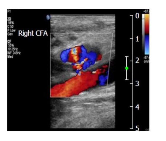
List the 2 examples of procedures or events that may cause a pseudoaneurysm.
Heart catheterization
Post-angioplasty
…. Post procedure / Trauma
List the 4 grayscale findings of a pseudoaneurysm.
( Has what appearance, Predominantly what echo apparence, what type of walls? and may be what?)
Irregular appearance
Predominantly anechoic
Irregular walls
May be bilobed (Bilobed means divided into two lobes or sections.)
List the 2 classic Doppler findings of a pseudoaneurysm. CD & SD
Color Doppler: Yin-yang sign
Spectral Doppler: To-and-fro signal (High velocity)

For Treatment How is ultrasound-guided compression performed on a pseudoaneurysm and what type of transducer is used?
Treatment - Compression
Pressure is exerted over the neck of the pseudoaneurysm with the transducer using a high-frequency linear transducer.
How long is pseudoaneurysm compression performed? What should you check for ? and should you repeat as needed?
Compress for 20–30 minutes, check for closure, and Yes, repeat as needed.
What is the goal of pseudoaneurysm compression?
To “seal” the hole in the artery.
What should be monitored during pseudoaneurysm compression? List 2
The native artery should be monitored and the pedal pulses palpated.
Yes, exactly! You're only trying to compress the neck of the pseudoaneurysm, NOT the native artery itself.
Compress the neck: Stops blood from entering the pseudoaneurysm, allowing it to clot and seal.
Monitor the native artery: Makes sure you're not accidentally blocking normal arterial blood flow.
Palpate pedal pulses: Confirms that blood is still reaching the foot.
Why is this important?
If you accidentally compress the native artery, you could reduce blood flow to the leg and cause ischemia (lack of adequate oxygenated blood to the tissues).
Remember: Compress the pseudoaneurysm neck to stop the leakage while maintaining blood flow through the native artery.
Why may sedation be needed during pseudoaneurysm compression?
Because the procedure may cause discomfort or pain.
List the 2 situations associated with a low success rate and high recurrence after compression.
Obese
Complex collections – multiple necks or collections
Pseudoaneurysm; Treatment - Thrombin Injection
What does thrombin do? - what is it and what does it convert?
Thrombin is a blood protein that converts fibrinogen to fibrin.
Fibrin: Strong, thread-like strands that form a mesh to hold a blood clot together.
Thrombin: Converts fibrinogen into fibrin, allowing a stable blood clot to form
What size needle and how much thrombin are used for pseudoaneurysm thrombin injection? & How fast is thrombin injected?
TB Syringe / 22-gauge needle - inserted into Pseudoaneurysm
½ mL of thrombin, slowly injected
How is a thrombin injection for a pseudoaneurysm performed? - what type of transducer and performed under what procedure and what guidance
It is performed using a high-frequency linear transducer, under sterile procedure and ultrasound guidance.
What happens to the pseudoaneurysm after thrombin is injected? - How quickly does it take to form?
Thrombus forms immediately within the pseudoaneurysm.
Here's why:
A pseudoaneurysm occurs when blood leaks through a hole in an artery and collects in surrounding tissue.
Thrombin is injected into the pseudoaneurysm, causing a blood clot (thrombus) to form.
The clot fills and seals off the pseudoaneurysm cavity, stopping blood from continuing to flow into it.
The native artery stays open so blood can continue flowing normally.
Think of it this way: We WANT the pseudoaneurysm cavity to clot because it's an abnormal collection of blood. We DON'T want the actual artery to clot because that would block circulation.
List the 3 benefits of thrombin injection
Safe
Effective and rapid
High success rate
What complication can occur with thrombin injection? & what are higher challenge to treat?
Arterial thrombus. Complex pseudoaneurysms are also more challenging to treat.

When may surgery be used to treat a pseudoaneurysm? & explain what they do?
When all other treatments have failed. The hole is then surgically closed.
List the 4 serious findings or complications associated with surgical treatment of a pseudoaneurysm.
Possibility of rupture
Limb ischemia, - Distal emboli, - Extensive soft-tissue damage
What does PTA stand for? & What is the purpose of PTA? (PCTA OR PTA) - what does it do to the vessel segments and this vessel segments containing what lesions
Percutaneous Transluminal Angioplasty.
Purpose: To dilate narrow vessel segments containing a focal atherosclerotic lesion.
List the 3 treatment approaches that may be used during Percutaneous Transluminal Angioplasty (PTA)
Balloon only
Stent only
Balloon and stent together
Technique: How is angiography used during angioplasty?
X-rays and dye are used to image the vessel.
What is done with the balloon-tipped catheter during angioplasty?
What does contrast highlights? Where is the catheter positioned ? what is then inflated?
Contrast highlights the lesion, the catheter is positioned within the stenosis, and the balloon is inflated

What does balloon inflation do to the plaque during angioplasty?
It pushes the plaque against the vessel wall.

What is the purpose of deploying a stent during angioplasty?
To keep the artery open

Angioplasty:
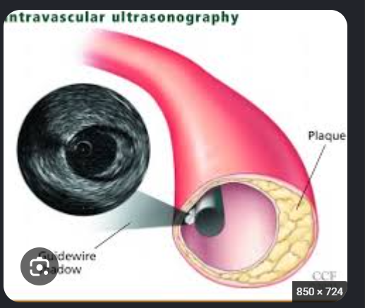
What is intravascular ultrasound, or IVUS? - Its what view inside the artery and uses the same what technology as everday use?
Ultrasound that provides a cross-sectional view from inside the artery, using the same basic ultrasound technology used normally.
Yes, exactly! IVUS is like putting a tiny camera inside the artery, but instead of using a camera, it uses ultrasound waves to create images.
How does IVUS work?
A small catheter with an ultrasound transducer at its tip is inserted into the artery.
The transducer sends out ultrasound waves from inside the artery.
These waves create a cross-sectional view of the artery, allowing doctors to see the lumen and vessel walls.
Doctors can identify plaque buildup, stenosis, and vessel size.
Why is IVUS helpful during angioplasty?
Before angioplasty: Helps determine how severe the narrowing is and measure the artery.
During stent placement: Helps doctors choose the appropriate stent size.
After stent placement: Helps confirm that the stent is properly expanded and positioned.
Remember: IVUS = Ultrasound performed from INSIDE the artery to provide a cross-sectional view of the vessel.
List the 3 benefits of Intravascular Ultrasound (IVUS)
Provides a more intimate look inside the What? Helps select the proper what for the Pt? Helps assure proper What placement ?
Provides a more intimate look inside the vessel
Helps select the proper treatment
Helps assure proper stent placement

Technique:
What does the Angioplasty do to the artery size?
What is angioplasty considered an alternative to?
Artery is dilated
A viable alternative to bypass-graft surgery
Artery is dilated: Angioplasty uses a balloon to widen a narrowed artery by pushing plaque against the artery walls. This allows more blood to flow through.
A patient has an artery narrowed by plaque buildup.
Option 1 – Angioplasty: The doctor inserts a balloon to widen the narrowed artery and improve blood flow.
Option 2 – Bypass graft surgery: The surgeon creates a new pathway for blood to travel around the blockage.
So what is your PowerPoint saying?
If angioplasty can successfully open the narrowed artery, the patient may not need bypass graft surgery at all!
However, if angioplasty doesn't work or the blockage is too severe, bypass surgery may be needed.
Remember: "Viable option" simply means a possible alternative treatment.
List the 2 most likely locations for angioplasty
Lower extremity — above and below the knee
Aortoiliac vessels
List the 5 locations evaluated during sonographic stent assessment. The Protocol
Sonographic stent assessment. GS/CD/SD
Pre-stent
Proximal margin
Mid-stent
Distal margin
Post-stent
List the 3 limitations of angioplasty
How does angioplasty target? Angioplasty works the best when the plaque is in what area and what size vessels? Angioplasty can be more difficult in what size arteries?
Specifically targeted
Focal lesions in large vessels
Small-diameter vessels
limitations of angioplasty, meaning when it works best and when it may not be effective.
1. Specifically targeted: Angioplasty treats a particular narrowed or blocked section of an artery, rather than treating the entire artery.
2. Focal lesions in large vessels: Angioplasty works well for short, localized areas of plaque buildup in larger arteries.
Focal = affects one specific area.
Large vessels = arteries with a larger diameter.
3. Small diameter vessels: Angioplasty can be more difficult in smaller arteries because there is less room for the balloon and catheter, and the artery may narrow again.
Remember: Angioplasty generally works best for short, localized stenoses in larger arteries. It can be more challenging in small arteries or when disease extends over a long section.
List the 2 Doppler findings considered Normal after AORTOILIAC Percutaneous Transluminal Angioplasty (PTA) - this is right after the post op
The CFA demonstrates a what waveform? Or the CFA may have a what waveform and Accleration time ?
CFA demonstrates a triphasic waveform
CFA may have a monophasic waveform with acceleration time <200 ms
Normal Triphasic Waveform
• Normal lower extremity arterial waveforms are triphasic, showing forward flow in systole, brief diastolic reversal, and late
diastolic forward flow.
AT <200 ms: Blood is still accelerating relatively quickly during systole, suggesting there isn't a severe upstream obstruction delaying blood flow.

In AORTOILIAC Percutaneous Transluminal Angioplasty (PTA) List the 2 abnormal Doppler criteria that indicate the aortoiliac vessels may need to be reevaluated.
List 3 things
Abnormal femoral pulses
Dampened monophasic waveform
(overtime)
AT >200 ms means prolonged acceleration time, suggesting a significant narrowing or blockage upstream (proximal to where you're measuring).
Why?
Normally, blood quickly reaches its peak velocity during systole.
If there's a significant stenosis upstream, blood flow is delayed.
This causes the Doppler waveform to have a slow, rounded systolic upstroke instead of a sharp peak.
In your PowerPoint (Aortoiliac PTA):
AT <200 ms: Considered acceptable after PTA.
AT >200 ms: Suggests abnormal blood flow, possibly due to residual or recurrent aortoiliac stenosis.
Remember for your flashcard:
Q: What does an acceleration time >200 ms in the CFA indicate?
A: Delayed systolic acceleration, suggesting significant proximal (aortoiliac) arterial disease.
Tardus-Parvus Pattern
• Tardus-parvus waveforms show dampened amplitude and prolonged systolic upstroke, indicating delayed flow from
upstream stenosis.
• Monophasic Waveform in Severe Disease
• Monophasic waveforms indicate critical arterial disease with loss of normal waveform complexity and severely altered
flow.
In AORTOILIAC Percutaneous Transluminal Angioplasty (PTA)
What change in ABI or TBI is considered abnormal between subsequent exams?
A decrease of 0.15 or greater
TBI = Toe-Brachial Index
It compares the systolic blood pressure in the toe to the systolic blood pressure in the arm to evaluate blood flow to the foot
Difference:
ABI: Compares ankle pressure to arm pressure.
TBI: Compares toe pressure to arm pressure.
A decrease in ABI or TBI of 0.15 or greater between exams may indicate worsening arterial blood flow after PTA.
What CFA Doppler finding may be considered abnormal after aortoiliac PTA? compared to the patients what?
A CFA Doppler waveform that is abnormal compared with the patient’s previous “normal.”
What PSV and velocity ratio indicate an abnormal finding after AORTOILIAC PTA?
PSV >300 cm/s
Velocity ratio >3.0
List the 2 consequences or Doppler criteria – abnormal associated with multilevel disease after aortoiliac PTA.
There is a Increased likelihood of what formation leading to limb what?
What typic of failure is common?
Increased likelihood of thrombus formation leading to limb ischemia
Angiographic failure is common
Multilevel disease means a patient has multiple areas of stenosis (narrowing) or blockage in their arteries, rather than just one.
Why does this increase thrombus formation and cause limb ischemia?
Multiple stenoses: Blood has difficulty flowing through several narrowed areas of the arteries.
Reduced blood flow: Blood flow becomes slower, especially downstream from the stenoses.
Thrombus formation: Sluggish blood flow increases the risk of blood clots forming.
Limb ischemia: The multiple narrowings or a blood clot can severely reduce blood flow, preventing enough oxygen from reaching the leg tissues.
List the 2 possible consequences when multilevel disease is not successfully treated
Failure to treat
Increase in chance for what loss & what placement/ treatment may be needed
Increased chance of limb loss
Bypass-graft placement may be needed
if severe arterial disease is left untreated, the patient has a higher risk of losing their limb and may need bypass graft surgery.
1. Increase in chance for limb loss: If the narrowed or blocked arteries aren't treated, blood flow may become so poor that tissues don't receive enough oxygen. This can lead to tissue death (necrosis) and possibly amputation.
2. Bypass graft placement: If angioplasty (PTA) fails or cannot adequately restore blood flow, the patient may need bypass surgery to create a new pathway around the blockage.
List the 3 reasons Arterial Percutaneous Transluminal Angioplasty (PTA) may need to be Repeated.
whats progressing? Abnormal what criteria? What about Clinical symptoms?
Progressing stenosis
Abnormal hemodynamic criteria
Clinical symptoms have returned
What velocity criteria suggest progressing stenosis after Percutaneous Transluminal Angioplasty (PTA) - what is the PSV and Velocity Ratio?
PSV >300 cm/s
Velocity ratio >3.0
What percentage of angiographic stenosis is considered normal? The goal is to establish normal what?
Less than 30%. The goal is to establish normal hemodynamics.
List the 3 common clinical signs of advanced arterial insufficiency. / chronic
Loss of hair growth over the dorsum of the toes and feet
Thickening of the toenails
Dependent rubor
List the 4 benefits or uses of stents/stent grafts.
Maintain what structure and what? of the vessel
Helps in limiting what lesions that reoccur
what is a stent used with - what type of device
very successful with what length disease segments
Maintain intraluminal structure and patency
Help limit restenosis
Used with balloon angioplasty
Very successful with short disease segments
List the 4 types of stents/stent grafts described
Flexible and malleable
Bare metal - They were used in peripheral vascular studies but are not used today.
Drug-eluting
Covered
What is the purpose of a drug-eluting stent?
It emits a drug to help prevent recurrence of stenosis
What is a covered stent listed as being used for? what repair
Endovascular aneurysm repair

List the 2 main purposes of stents/stent grafts
Treat what lesions? maintain the vessel what? Deployed post what angioplasty?
What repair? List the 2 most common where these are typical found ?
Treat stenotic lesions and maintain vessel patency after balloon angioplasty
Aneurysm repair, including AAA and popliteal artery aneurysms - the most common where a aneurysmal are

List the 3 basic steps of stent placement.
Perform an what? use a what angioplasty? stent is placed after what?
Perform an angiogram
Use balloon angioplasty
Place the stent after angioplasty

List the 2 limitations of stent/stent-graft placement. (think length and pt) & List 1 Complications***
Course and size of the vessel
Patient limitations
Complications ; Intimal hyperplasia
What can intimal hyperplasia cause in a stent? Leads to what happening
Leads to Restenosis
List the 3 mechanical stent complications mentioned- what can happen to the sent think movement
Migration
Twisting
Dislodgement
List the 5/7 additional complications of stents/stent grafts.
Restenosis/thrombosis
^Lower-extremity emboli
Dissection
Infection/hematoma
Pseudoaneurysm
What transducer and 3 imaging methods are used for stent surveillance? - like the mode and doppler
Use the highest-frequency transducer with:
B-mode
Color Doppler
Spectral Doppler
Which vascular protocols should be followed when evaluating a stent? List 3
Follow the appropriate protocol for:
Aortoiliac
Lower-extremity arterial (LEA)
Renal artery
List the 5 stent locations that should be sampled. Protocol
Pre-stent
Proximal margin
Mid-stent
Distal margin
Post-stent
What does a doubling versus tripling of velocity suggest in a stent?
Doubling of velocity = 50% stenosis
Tripling of velocity = 75% stenosis
List the 3 complications of AAA stent grafts.
Endoleak, AAA, Popliteal artery
Stent migration, twisting, or dislodgement
Infection/hematoma
Aortic Graft/Stent Abnormal
List the 5 types of Aortic endoleak descriptions
What is still filling aneurysm sac?
Endoleaks are characterized by the source of the vessel of what into the aneurysmal sac.
Type 1 where is the leak at? List 2 attachmente site and what occluder
Type 2 Sac fills via an what branch? what is this leak called? does it spontneosly resolve list 2 artery we see this
Type 3 within the what material? this is do to the what failure and list 3 types of failures
Type 4 is what graft - intentional.. Small What in the graft’s fabric allow what pressure what to pass through, rather than what around the graft edges like in Type
what list 2 endoleaks.
Type 5 what is it called the sac continus to what - idopathic
pressure is still transmitted in the what sac
Aortic endoleaks
Blood is still filling aneurysm sac
Endoleaks are characterized by the source of the vessel of inflow into the aneurysmal sac.
5 types
• Type 1: leak is at the end of graft
• Proximal attachment site, distal attachment site, or
iliac occluder
• Type 2: sac fills via an aortic branch
• Retroleak
• Spontaneously resolve
• Lumbar or IMA
Aortic endoleaks
5 types
Type 3: within the graft material
Mechanical failure
Graft defect, hole or separation
Type 4: porous graft-intentional
Small pores in the graft’s fabric allow low-pressure blood to pass
through, rather than leaking around the graft edges like in Type
1 or 3 endoleaks. (Society for Vascular Surgery)
Type 5: Endotension
Sac continues to expand – idiopathic
Pressure is still transmitted in the aneurysmal sac.
What is the PREFERRED follow-up method for AAA stents? - what type of imaging
CTA.
What type of stent is specifically listed for duplex-sonography follow-up? List 2 the last one is a maybe
A popliteal-artery endoluminal stent. The slide also questions the use of duplex for AAA stents.
A popliteal artery endoluminal stent is a small metal mesh tube placed inside the popliteal artery (artery behind the knee) to help keep it open or treat an aneurysm.
Let's break it down:
Popliteal artery: Artery located behind the knee.
Endoluminal: Means inside the blood vessel.
Stent: A small tube that helps keep an artery open.
Why is it used?
Stenosis: If plaque narrows the popliteal artery, a stent can help keep it open and improve blood flow.
Popliteal aneurysm: If the artery develops a weakened, bulging wall, a covered stent graft can be placed inside it so blood flows through the graft instead of into the aneurysm sac.

What transducer and imaging modes are used for sonographic evaluation of an AAA stent graft?
Use the highest-frequency transducer with:
B-mode
Color Doppler
Spectral Doppler
List the 3 sonographic steps for evaluating an endoluminal aneurysm repair.
Cine ___of the ____within the ____using ____Doppler
______Doppler sampling within the ______aneurysm
Measure the ____dimension of the vessel including the ________
Cine clip of the stent within the aneurysm using color Doppler
Spectral Doppler sampling within the thrombosed aneurysm
Measure the AP dimension of the vessel including the aneurysm
Yes! After EVAR, we WANT blood to clot inside the aneurysm sac, but NOT inside the stent graft.
Here's why:
Before EVAR: Blood flows into the aneurysm, putting pressure on its weakened walls and increasing the risk of rupture.
After EVAR: The stent graft redirects blood flow away from the aneurysm sac.
Blood outside the graft: The blood trapped in the aneurysm sac becomes stagnant and forms a clot.
Why this is good: Clotting helps prevent continued blood flow into the sac, which can allow the sac to shrink over time.
What's the difference?
Clot inside aneurysm sac (outside graft) = GOOD, because we want the sac excluded from blood flow.
Clot inside stent graft = BAD, because it could block blood flow to the legs.
Remember: After EVAR, we want blood to flow through the stent graft, NOT through the aneurysm sac.
One important point: Clotting alone doesn't guarantee the aneurysm is safe. The sac must also be sealed off from blood flow and pressure, which is why we monitor for endoleaks.

List the 3 questions to ask when using color Doppler to assess for an endoleak.
Is there what in the aneurysmal sac?
What about the size of the aneurysmal sac?
Is there what flow present in the aneurysmal sac?
Is there blood in the aneurysmal sac?
Has the aneurysmal sac decreased in size?
Is blood flow present within the sac?
What is the purpose of a thrombectomy or embolectomy?
To remove what from an affected arterial segment? This is considered a surgical what?
To remove emboli from an affected arterial segment. It is considered a surgical emergency.
List the 3 basic steps / Technique of a thrombectomy/embolectomy.
Uses what as an Interventional procedure?
The catheter tip is placed beyond where
What is inflated and catheter is withdrawn removing the what?
Use angiography as an interventional procedure
The catheter tip beyond the thromboembolism
Inflate the balloon and withdraw the catheter to remove the thromboembolus
Angiography: Uses X-rays and contrast dye to visualize arteries and identify blockages.
Interventional procedure: A minimally invasive treatment performed using catheters inserted into blood vessels.
Example:
A patient has a narrowed artery due to plaque buildup.
Angiography is performed to locate the narrowing.
The doctor uses angioplasty (a balloon) to widen the artery.
A stent may be placed to help keep the artery open.
Remember: Angiography = visualizes the blockage. Angioplasty = treats the blockage.
Angiography alone is a diagnostic procedure, but it can also be used to guide an intervention.
The balloon helps because it pushes against the blood clot so the doctor can pull the clot out of the artery.
Think of it like this:
The artery is blocked by a blood clot, preventing blood from reaching the leg.
The doctor passes a deflated balloon past the clot.
The balloon is inflated behind the clot, making it large enough to catch the clot when pulled backward.
The doctor pulls the balloon back, dragging the clot out of the artery.
Now that the clot is removed, blood can flow through the artery again, delivering oxygen to the leg.
Why not just pull the catheter out without inflating it?
Because the catheter is too small to catch the clot. Inflating the balloon allows it to pull the clot along with it.

What is the purpose of an atherectomy?
Removal of obstructing lesions.
How is plaque removed during an atherectomy?
A what device at the catheter tip does what to the plaque.
A rotational device at the end of the catheter cuts or pulverizes the plaque.
Pulverizes means crushes or breaks something into very tiny pieces.
In vascular procedures, such as atherectomy, it means a device breaks up plaque inside an artery into tiny particles.

What happens to the material removed during an atherectomy?
collected in a what compartment in the catheter.
The excised material is collected in a storage compartment in the catheter.
Excised material means tissue or other material that has been surgically cut out and removed from the body.
In your vascular PowerPoint:
Excised = Surgically cut out or removed.
Material = What was removed, such as plaque or a blood clot.
List the 2 limitations of atherectomy.
what generation from the devices and what particles?*
Heat generation from the devices
Microembolization of particles****
These are two potential complications of atherectomy, which is a procedure that removes plaque from inside an artery.
1. Heat generation from devices: The device used to remove plaque can generate heat from friction or mechanical energy. This heat can potentially damage the arterial wall.
2. Microembolization of particles: When the device breaks up or removes plaque, tiny pieces of plaque can break off and travel downstream through the bloodstream.
These particles may block smaller arteries.
This can decrease blood flow and cause ischemia (lack of adequate oxygen to tissues).
Remember:
Heat generation → Can damage the artery.
Microembolization → Plaque particles travel downstream and may block smaller vessels.
The goal of atherectomy is to remove plaque and improve blood flow, but these are risks associated with the procedure.
What advantage of atherectomy? Reduced risk of what list 2
Reduced risk of peripheral emboli and intimal dissection.
What is an endarterectomy?
Surgical removal of plaque, including the intima, media, and plaque.
List the 2 additional surgical steps that may occur during an endarterectomy.
A temporary what is placed and what is placed over the incision?
A temporary bypass may be placed.
A patch is placed over the incision.
- Temporary bypass (shunt): Keeps blood flowing while the surgeon removes plaque from the artery, preventing tissues from losing oxygen.
- Patch: Covers the incision after plaque removal and helps keep the artery wide open, preventing narrowing.
Remember: Shunt = Maintains blood flow during surgery. Patch = Prevents narrowing after surgery.

How can an endarterectomy patch affect ultrasound? It may limit what?
It may limit visualization.
List the 3 complications or limitations of an endarterectomy. What is the most common?
Myointimal hyperplasia ****(most common?)
Intimal flap at the distal end
Restenosis
During an endarterectomy, the surgeon removes plaque from inside the artery. Sometimes, part of the inner lining can become loose, creating an intimal flap.
Why is this a problem?
The flap can stick out into the artery and:
Disrupt normal blood flow.
Cause narrowing or blockage.
Increase the risk of thrombus (blood clot) formation.
What is the purpose of a bypass graft? & this is done such as when what 2 things was unsuccessful
To bypass an occluded or narrowed diseased arterial segment, such as when angioplasty or stenting was unsuccessful
What is required for a bypass graft to have a high success rate?
The patient must have patent distal runoff vessels, meaning patent arteries beyond the obstruction.
: The arteries beyond the blockage must be open so blood can continue flowing to the leg after the obstruction is removed.
Remember: Removing a blockage only helps if blood has somewhere to flow afterward!
When a patient has multilevel disease, which lesions are usually bypassed first?
Proximal lesions.
This means that when a patient has multiple arterial blockages, surgeons usually treat the blockage closest to the heart first.
Let's break it down:
Proximal lesion: A narrowing or blockage closer to the heart.
Multilevel disease: Multiple blockages at different locations in the arteries.
Bypassed first: The surgeon creates a new pathway around the proximal blockage before treating blockages farther down.
Why treat the proximal blockage first?
Blood must travel from the heart down toward the foot.
If there's a blockage higher up, it reduces blood flow to everything below it.
Bypassing the proximal blockage first improves blood flow to the arteries farther down the leg.
The surgeon can then determine whether the remaining blockages need treatment.
List the 2 main types of bypass-graft material. (think of different procedures)
Synthetic material
Native veins
What synthetic graft material is listed in the PowerPoint? List 2
Polytetrafluoroethylene (PTFE), or Gore-Tex.
List the 2 preferred native-vein graft methods.
Reversed saphenous vein
In-situ vein
List the 3 native-vein options considered last-resort alternatives.
Spliced veins
Arm veins
Small saphenous vein
Spliced veins are two or more pieces of veins that are surgically connected together to create one longer vein for bypass graft surgery.
Why is this done?
A patient needs a bypass graft to go around a blocked artery.
The surgeon may not have one vein that's long enough or suitable for the entire bypass.
They connect multiple vein segments together to create a longer graft.
The graft creates a new pathway for blood to flow around the blockage.
Example: If the great saphenous vein isn't long enough, the surgeon may connect it to another suitable vein segment to complete the bypass.
Remember: Spliced veins = multiple vein segments sewn together to create one longer bypass graft.
List the 4 complications of bypass grafts
Pseudoaneurysm
Hematoma
Arterial thrombosis
Perforating veins that were not ligated
What can happen if perforating veins are not ligated during bypass-graft surgery?
An arteriovenous fistula can form.
An arteriovenous fistula (AVF) forms when there is an abnormal connection between an artery and a vein, allowing blood to flow directly from the artery into the vein.
How does this happen during bypass surgery?
The surgeon uses the great saphenous vein as a bypass graft.
The vein has smaller branches (perforating veins) that connect to the venous system.
If those branches are not ligated (tied off), they remain open.
When arterial blood starts flowing through the bypass graft, some blood can travel through those branches into the veins, creating an AV fistula.
But WHY does blood enter those veins?
Because arterial pressure is much higher than venous pressure.
Blood naturally flows from an area of higher pressure to an area of lower pressure.
So blood flows from:
High-pressure bypass graft → Open vein branch → Low-pressure venous system
Why is this a problem?
Blood can take this shortcut into the veins instead of continuing down the bypass graft toward the foot.
This may reduce blood flow reaching the foot.
Think of the bypass graft as a highway for blood.
The whole purpose of the bypass graft is to carry blood down to the leg and foot.
Now imagine this:
The surgeon uses the great saphenous vein as a bypass graft to carry blood down the leg.
But this vein has smaller veins branching off it, like side roads coming off a highway.
If those smaller veins are left open, some blood can escape into those veins instead of continuing down the graft toward the foot.
This means less blood may reach the foot, which is a problem because the patient already has poor circulation.
So why do we tie them off?
Because we want blood to stay inside the bypass graft and travel toward the foot, rather than take another pathway into the venous system.
Think of a water hose: Imagine you're trying to get water to the end of a hose, but there are extra openings along the sides. Some water escapes through those openings instead of reaching the end.
Tying off those openings keeps the water moving toward the end of the hose.
That's exactly why surgeons ligate those vein branches.
The one thing I want you to remember:
We close off the smaller connecting veins so arterial blood doesn't escape from the bypass graft into the venous system.
What does “ligate” mean
To tie up or close off an artery
When are Gore-Tex grafts considered reliable?
When the distal anastomosis is above the knee.
Above the knee: Arteries are generally larger and have better blood flow, making it less likely for the graft to develop a blood clot.
Below the knee: Arteries are smaller and often have poorer blood flow, increasing the risk of clot formation and graft failure.
Example: A femoral-to-above-knee popliteal bypass using Gore-Tex is generally more successful than a femoral-to-below-knee popliteal bypass using Gore-Tex.
Remember: Gore-Tex grafts are more reliable above the knee because larger arteries and better blood flow help keep the graft open (patent).
How are the patency rates of Gore-Tex grafts below the knee?
Low
Means bypass grafts placed below the knee have a lower chance of staying open over time.
Patency: The graft remains open and allows blood to flow through it.
Low patency rate: The graft is more likely to become narrowed or blocked.
Why?
Arteries below the knee are generally smaller.
Blood flow through the graft may be reduced.
Slower blood flow increases the risk of thrombus (blood clot) formation.
This increases the chance of graft failure.
Remember: Gore-Tex grafts have lower patency rates below the knee because smaller arteries and poorer blood flow increase the risk of clotting and graft blockage.