CR1 KCP1 Basic Tissues
 Call Kai
 Learn
 Practice Test
 Spaced Repetition
 Match
 Flashcards
 Knowt Play
1/45

There's no tags or description
Looks like no tags are added yet.
46 Terms
What is the formal histological definition of a tissue?
A group of cells with similar structure that function together as a unit.
List the four basic tissue types and state the primary role of each.
1. Epithelial (covers body surfaces, lines cavities, forms glands); 2. Connective (underlies and supports other tissues); 3. Muscle (responsible for movement); 4. Nervous (receives, transmits, and integrates information).
7 main functions of epithelial tissue?
Protection, absorption, sensory reception, ion transport, secretion, filtration, and formation of slippery surfaces for movement.
Describe the cell dimensions for Squamous, Cuboidal, and Columnar epithelial cells.
Squamous: wider than tall. Cuboidal: as tall as wide. Columnar: taller than wide.
What defines a pseudostratified epithelium?
A single layer of cells that appears stratified because the cell nuclei are at different levels.
What is Endothelium and where is it located?
Simple squamous epithelium lining the inside of blood vessels, lymphatic vessels, and heart chambers.
What is Mesothelium and where is it located?
Simple squamous epithelium lining closed body cavities (pleural, pericardial, peritoneal cavities).

Primary functions of Simple Squamous Epithelium?
Allows material passage via diffusion and filtration, and secretes lubricating substances.
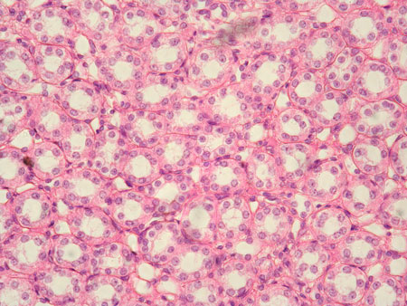
Primary locations of Simple Cuboidal Epithelium?
Kidney tubules, and ducts/secretory portions of small glands.
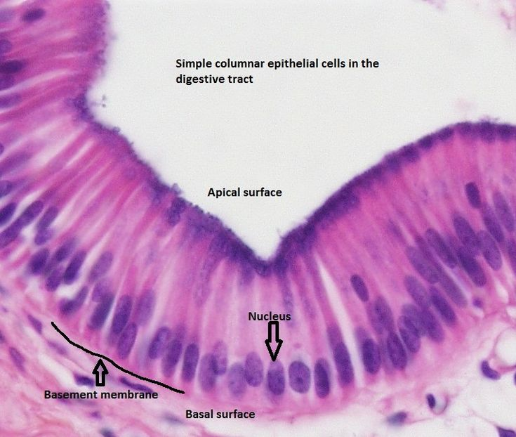
Where is Ciliated vs. Non-ciliated Simple Columnar Epithelium found?
Ciliated: Bronchi, uterine tubes, uterus. Non-ciliated: Digestive tract, bladder.

What is the primary location and function of Pseudostratified Columnar Epithelium?
Ciliated type lines the trachea and upper respiratory tract; secretes mucus and ciliated action moves mucus.

Contrast the locations of Keratinized vs. Non-Keratinized Stratified Squamous Epithelium.
Keratinized: Skin (epidermis). Non-keratinized: Lining of esophagus, mouth, and vagina.

Primary function of Stratified Squamous Epithelium?
Protects underlying tissues against abrasion.

Name three locations where Stratified Cuboidal Epithelium is found.
Sweat glands, salivary glands, and mammary glands.

Where is Stratified Columnar Epithelium located in the body?
Male urethra and the ducts of some glands.
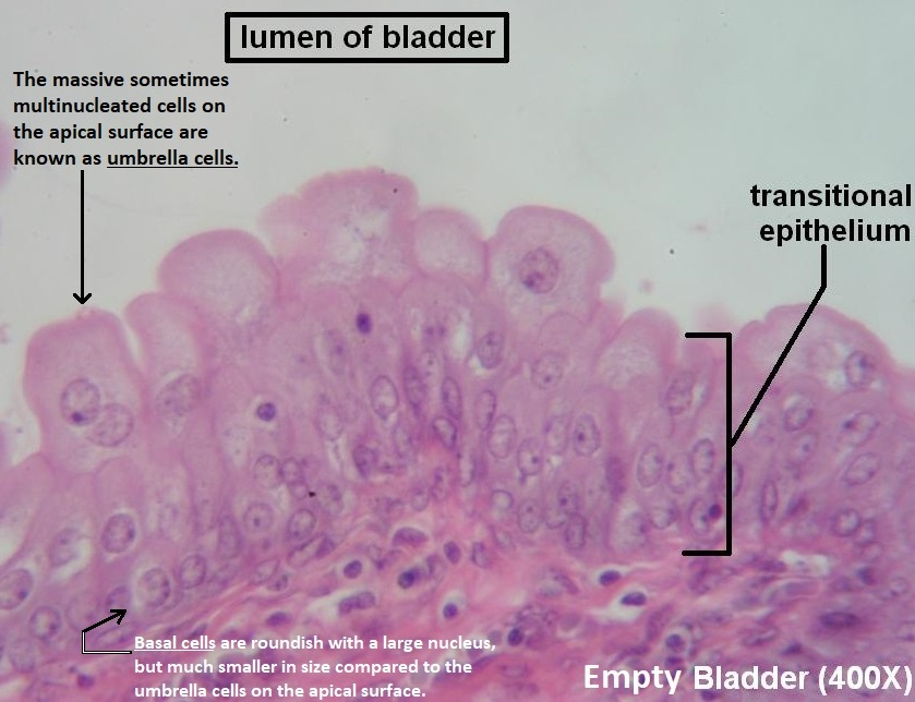
What is the special functional property of Transitional Epithelium and where is it found?
Allows urinary organs to expand and stretch; lines the bladder, ureters, and urethra.
What is the basic distinction between Exocrine and Endocrine glands?
Exocrine glands discharge secretions via a duct onto body surfaces or into cavities. Endocrine glands are ductless and secrete hormones directly into the blood.
Differentiate between Serous, Mucous, and Mixed glandular secretions.
Serous = Watery discharge. Mucous = Slimy, chemically inert secretion. Mixed = Combination of watery and mucous discharge.
Explain Merocrine secretion and give an example.
Secretory product is released without cell damage (cells remain intact). Example: Submandibular / salivary glands.
Explain Apocrine secretion and give an example.
The apical portion of the cell is pinched off/detached along with the secretion. Example: Mammary gland.
Explain Holocrine secretion and give an example.
The entire cell disintegrates and is discharged with the secretion. Example: Sebaceous gland.

Define Tubular, Acinar, and Tubulo-alveolar secretory units.
Tubular = Elongated tube-shaped lumen. Acinar/Alveolar = Small grape-like or sac-like group of cells. Tubulo-alveolar = Contains both shapes.

Differentiate between Simple, Branched, and Compound exocrine glands based on duct arrangement.
Simple = Unbranched duct. Branched = Multiple secretory units empty into a single unbranched duct. Compound = Highly branched duct system draining into larger ducts.
What embryonic germ layer does connective tissue originate from?
Mesenchyme (mesenchymal origin).
What are the two primary structural components of Connective Tissue?
Cells and Extracellular Matrix (ECM).
What two sub-components make up the Extracellular Matrix (ECM)?
Ground substance and Fibers.
What three major class molecules compose the Ground Substance?
Proteoglycans, Multiadhesive glycoproteins, and Glycosaminoglycans (GAGs).
List all 8 cell types present in connective tissue proper.
Mesenchymal cells, Fibroblasts/Fibrocytes, Macrophages, Leucocytes (WBCs), Plasma cells, Mast cells, Adipocytes (fat cells), and Chromatophores (pigment cells).
What structural protein forms Collagen fibers, and how are they arranged?
Collagen protein; unbranched fibrils arranged in wavy, parallel bundles.
Functional property and location requirement of Collagen fibers?
Provides great tensile strength combined with flexibility; found wherever great strength is required.
What protein forms Elastic fibers, and how much can they stretch?
Elastin protein; can stretch up to 1.5x their original length.
Structural appearance of Elastic fibers?
Fine, thin fibers that branch and anastomose forming an irregular network.
What protein forms Reticular fibers, and where are they particularly abundant?
Reticulin protein; abundant in hemopoietic tissues and soft tissue organ frameworks.
Structural description of Reticular fibers?
Extremely fine, delicate short fibers that branch and anastomose into a net-like meshwork.
What are the three types of Loose Connective Tissue?
Areolar tissue, Adipose tissue, and Reticular tissue.
Where is Areolar tissue located in the body?
Underneath epithelial tissue of skin, in visceral organ walls (stomach, trachea), and in blood vessel walls.
What is Adipose tissue structurally composed of?
Modified areolar tissue whose matrix contains a large number of adipose cells (adipocytes) alongside fibrocytes and macrophages.
What is the primary function of Reticular connective tissue?
Forms a supportive framework (stroma) for cells in lymph nodes, glands, and hemopoietic organs.

Differentiate between Dense Regular and Dense Irregular Connective Tissue.
Dense Regular: Collagen fibers run in parallel rows between bundles (tendons, ligaments). Dense Irregular: Collagen fibers arranged randomly in different patterns (skin dermis).
List the three specialized connective tissues.
Cartilage, Bone, and Blood.
What are the three histological types of Cartilage?
Hyaline cartilage, Fibrocartilage, and Elastic cartilage.

Detailed histology and properties of Skeletal Muscle?
Striated, tubular, multi-nucleated fibers; voluntary control; usually attached to the skeleton.

Detailed histology and properties of Smooth Muscle?
Non-striated, spindle-shaped, uni-nucleated fibers; involuntary control; lines walls of internal hollow organs.

Detailed histology and properties of Cardiac Muscle?
Striated, branched, uni-nucleated fibers; involuntary control; found only in the heart wall.
Name the structural hierarchy of skeletal muscle from outermost wrapping to innermost filament.
Epimysium (covers muscle) → Perimysium (covers fascicle) → Endomysium (covers muscle fiber/cell) → Myofibrils → Thick & Thin filaments.
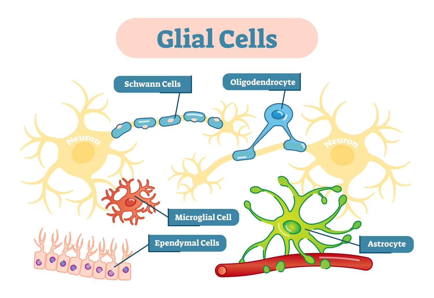
Name the 4 glial cells and state their precise location and functions.
Astrocytes: Supportive cells in the CNS.
Oligodendrocytes: Myelinate axons in the CNS.
Schwann cells: Myelinate axons in the PNS.
Microglia: Macrophages of the CNS.