lecture 8 regulation of the cell cycle and apoptosis
 Call Kai
 Learn
 Practice Test
 Spaced Repetition
 Match
 Flashcards
 Knowt Play
1/26

There's no tags or description
Looks like no tags are added yet.
27 Terms
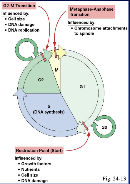
what are the phases of the cell cycle in order, and which make up interphase?
G1 → S → G2 → M. interphase includes G1 (cell growth), S (DNA replication), and G2 (preparation for division).

during M phase, what happens in mitosis, and what happens in cytokinesis?
during mitosis, chromosomes condense, line up, and separate their sister chromatids into two daughter nuclei. during cytokinesis, the cytoplasm divides, forming two daughter cells.

progression through cell-cycle checkpoints requires cyclin-dependent kinases (Cdks). what three types of targets do Cdks phosphorylate?
proteins that carry out the events of the current cell-cycle stage.
transcription factors that increase production of proteins needed for those events.
transcription factors that increase production of the next cyclin, helping the cell progress to the next stage.
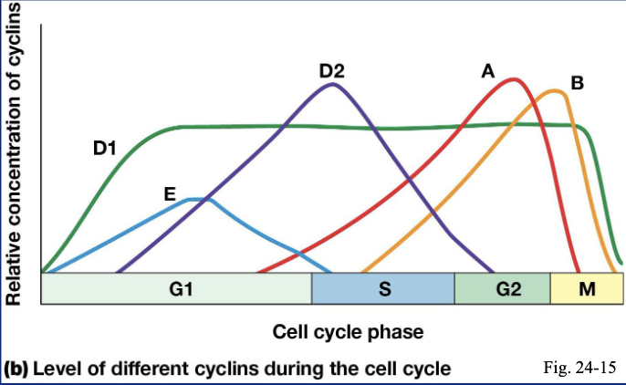
which cyclin–Cdk pair controls each stage of the cell cycle?
G1 → cyclin D + Cdk4 or Cdk6
G1/S transition → cyclin E + Cdk2
S phase → cyclin A + Cdk2 or Cdk1
M phase → cyclin B + Cdk1

what two things does cyclin binding do for a Cdk?
cyclin binding activates the Cdk and gives it substrate specificity, helping determine which proteins it phosphorylates.
what three mechanisms regulate cyclin-dependent kinases (Cdks)?
cyclin binding is required for Cdk activation.
phosphorylation can activate or inhibit a Cdk, depending on which site is phosphorylated.
Cdk inhibitory proteins (CKIs) bind Cdks or cyclin–Cdk complexes and inhibit their activity.
how does growth factor signaling drive a cell through G1 toward the restriction point?
growth factor signaling activates MAP kinase.
MAP kinase phosphorylates transcription factors that increase transcription of immediate early response genes.
these genes encode transcription factors such as Myc, Fos, and Jun, which increase production of cyclin D and other proteins needed to progress toward the restriction point.

once cyclin D is produced during G1, how does it lead to cyclin E production?
cyclin D activates Cdk4 and Cdk6, which phosphorylate the retinoblastoma protein (Rb). phosphorylated Rb releases E2F, a transcription factor that increases expression of cyclin E, the G1/S cyclin.

once cyclin E is produced, how does the cell pass the restriction point and move toward S phase?
cyclin E activates Cdk2. active Cdk2 phosphorylates proteins that help the cell pass the restriction point and progress toward S phase.

during late G1, how does cyclin A–Cdk build up while staying inactive?
cyclin E–Cdk2 activity causes cyclin A levels to rise. Cdk inhibitory proteins (CKIs) bind and inhibit the cyclin A–Cdk complexes as they accumulate.

once inactive cyclin A–Cdk has built up, what triggers the abrupt start of S phase?
very late in G1, cyclin E–Cdk2 activity rises enough to phosphorylate the CKIs, causing them to release cyclin A–Cdk. the accumulated complexes become active in a surge, triggering the abrupt onset of DNA synthesis.

during G2, how does cyclin B–Cdk1 build up while staying inactive?
cyclin A–Cdk activity causes cyclin B levels to rise. cyclin B binds Cdk1, but an inhibitory phosphate on Cdk1 keeps the complexes inactive as they accumulate.

once inactive cyclin B–Cdk1 has built up, what triggers the abrupt start of M phase?
late in G2, cyclin A–Cdk phosphorylates and activates a phosphatase. the phosphatase removes the inhibitory phosphate from Cdk1, activating cyclin B–Cdk1. active cyclin B–Cdk1 then activates more of that same phosphatase, creating positive feedback and a surge in Cdk1 activity that triggers entry into M phase.

at the metaphase-to-anaphase transition, what must happen before sister chromatids can separate, and which checkpoint ensures this?
the spindle assembly checkpoint delays anaphase until all kinetochores are properly attached to the mitotic spindle. once this condition is met, sister chromatids separate, marking the start of anaphase.
before anaphase, what holds sister chromatids together, and what keeps them from separating too soon?
cohesins hold sister chromatids together. separase can cleave cohesin, but securin binds separase and keeps it inactive.

once all kinetochores are properly attached to the mitotic spindle, how does the anaphase-promoting complex (APC/C) trigger sister chromatid separation?
the spindle assembly checkpoint allows APC/C to become active. APC/C is a ubiquitin ligase that tags securin for degradation by the proteasome. this frees separase to cleave cohesin, allowing sister chromatids to separate.

why does an unattached kinetochore keep the anaphase-promoting complex (APC/C) from triggering anaphase?
unattached kinetochores keep Mad-dependent checkpoint signaling active. this sequesters Cdc20, the protein APC/C needs to recognize and bind its substrates.

once all kinetochores attach to the mitotic spindle, what happens to Mad signaling and Cdc20?
Mad signaling drops quickly, and Cdc20 is released. Cdc20 can then activate APC/C, allowing it to tag securin for degradation and trigger anaphase.

after APC/C triggers sister chromatid separation, how does it help the cell exit mitosis?
APC/C tags cyclin B and other mitotic proteins for degradation by the proteasome. as cyclin B is degraded, Cdk1 activity drops, allowing the cell to exit mitosis.

what controls the three major transitions in the cell cycle?
G1 restriction point: growth factors promote cyclin–Cdk activity, leading to Rb phosphorylation and E2F release. DNA damage can halt progression through p21, which inhibits cyclin–Cdk activity.
G2 → M: activation of cyclin B–Cdk1 drives entry into mitosis.
metaphase → anaphase: once all kinetochores are properly attached to the spindle, APC/C triggers sister chromatid separation. it also targets cyclin B for degradation, helping the cell exit mitosis.
how does DNA damage activate p53?
DNA damage activates ATM or ATR, which activate checkpoint kinases. these kinases phosphorylate p53, stabilizing it so it can increase expression of p21 and PUMA.

once p53 is activated, how do p21 and PUMA lead to different outcomes?
p21 inhibits cyclin–Cdk complexes, keeping Rb from being phosphorylated. this arrests the cell cycle and gives the cell time to repair its DNA.
PUMA inhibits anti-apoptotic proteins such as Bcl-2, promoting apoptosis (programmed cell death).

what are the five hallmarks of apoptosis (programmed cell death)?
orderly breakdown of organelles and the cytoskeleton
DNA fragmentation
membrane blebbing (the membrane bulges outward)
formation of membrane-bound apoptotic bodies
typically no inflammation, because apoptotic bodies are cleared without spilling their contents

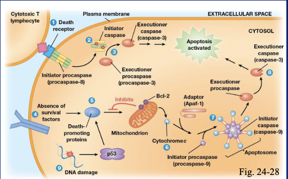
when is apoptosis a normal process, and what signals activate the extrinsic pathway?
apoptosis is a normal part of development and metamorphosis. the extrinsic pathway starts with signals from outside the cell, such as tumor necrosis factor (TNF) produced by immune cells or Fas ligand on the surface of cytotoxic T cells.
what triggers the intrinsic pathway of apoptosis?
the intrinsic pathway starts when the cell responds to stress such as DNA damage, loss of contact with the extracellular matrix (ECM), or insufficient nutrients or survival signals such as hormones.
how do the extrinsic and intrinsic apoptosis pathways activate initiator caspases?
extrinsic: extracellular signals bind death receptors, which trigger initiator caspase activation.
intrinsic: mitochondria release cytochrome c, which binds an adaptor protein that helps activate initiator caspases.
once initiator caspases are active, how do they produce the hallmarks of apoptosis?
initiator caspases activate executioner caspases. these proteases cleave many cellular proteins, producing changes such as cytoskeleton breakdown, membrane blebbing, and apoptotic body formation.