Module 7 - Linear Measurements in PLAX and PSAX
 Call Kai
 Learn
 Practice Test
 Spaced Repetition
 Match
 Flashcards
 Knowt Play
1/7

There's no tags or description
Looks like no tags are added yet.
8 Terms
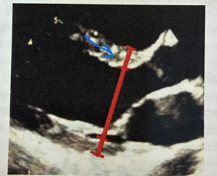
What does the blue arrow measure?
Identify the normal ranges for both males and females for this anatomy.
The IVS is being measured
IVS
M - .6 – 1.0 cm
F - .6 - .9 cm

What does the shaded green segment measure?
Identify the normal ranges for both males and females for this anatomy.
What caliper placement is used in this measurement?
LVIDd
LVIDd
M - 4.2 – 5.8 cm
F – 3.8 - 5.2 cm
Inner edge of LV to leading edge of LV at end-diastole

What does the shaded green segment measure?
Identify the normal ranges for both males and females for this anatomy.
PWT
PWT
M - .6 – 1.0 cm
F - .6 - .9 cm

What does the shaded green segment measure?
Identify the normal ranges for this anatomy in both males and females.
What caliper placement is used in this measurement?
LVIDs
LVIDs
M – 2.5 – 4.0 cm
F – 2.2 – 3.5 cm
Inner edge to leading edge of end-systole

What does the yellow line measure?
Identify the normal ranges for this anatomy in both males and females.
What caliper placement is used in this measurement?
Left atrium at end systole
LA
M – 3.0 – 4.0 cm
F – 2.7 – 3.8 cm
Measurement is leading-edge to leading edge

What do the blue,pink, and green lines measure?
What caliper placement is used in this measurement?
Blue: Sinus of Valsava (SOV)
Pink: Sinotubular Junction (STJ)
Green: Ascending Aorta
Measurement is made from leading-edge to leading edge

What do the red and yellow lines measure?
Red- Proximal RVOT
Yellow- Distal RVOT
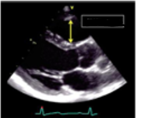
What is being measured in this image?
The proximal RVOT.