Ch. 32 Orthopedic Injuries
 Call Kai
 Learn
Learn Practice Test
 Spaced Repetition
 Match
Match Flashcards
 Knowt Play
1/100

There's no tags or description
Looks like no tags are added yet.
Name | Mastery | Learn | Test | Matching | Spaced | Call with Kai | Chat |
|---|
No analytics yet
Send a link to your students to track their progress
101 Terms
fascia
fibrous tissue
how man bones are in the body
206
skeletal muscle
also called striated muscle or voluntary muscle
attaches to bones + usually crosses at least one joint
makes up the largest portion of the body’s muscle mass
primary functions: movement + posture
cardiac muscle
cardiovascular system
smooth muscle
also called involuntary muscle
found in the walls of most tubular structures of the body
contracts + relaxes to control the movement of the contents within the structures
bone marrow produces:
red blood cells
white blood cells
platelets
joint
forms wherever two bones come into contact
ligaments
band of fibrous tissue that connect bones to bones
support and strengthen a joint
articular cartilage
pearly white substance
covers ends of bones
allow ends of bones to glide easily
joints are lubricated by _____ fluid
synovial (joint)
zone of injury
the area of potentially damaged soft tissue, adjacent nerves, and blood vessels surrounding an injury to a bone or a joint
force may be applied to the limb in the following ways
direct blows
indirect forces
twisting forces
high-energy injuries
osteoporosis
a disease that weakens bones, making them brittle and more prone to fracture
displaced fracture
deformity or distortion of the limb
often can be associated with crepitus
check for differences in the injured limb vs. the uninjured limb
nondisplaced (hairline) fracture
a simple crack of the bone
may be difficult to distinguish from a sprain or simple contusion
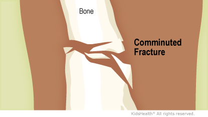
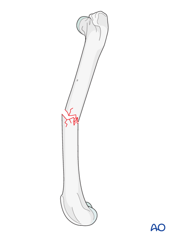
comminuted fracture
a fracture in which the bone is broken into more than two fragments
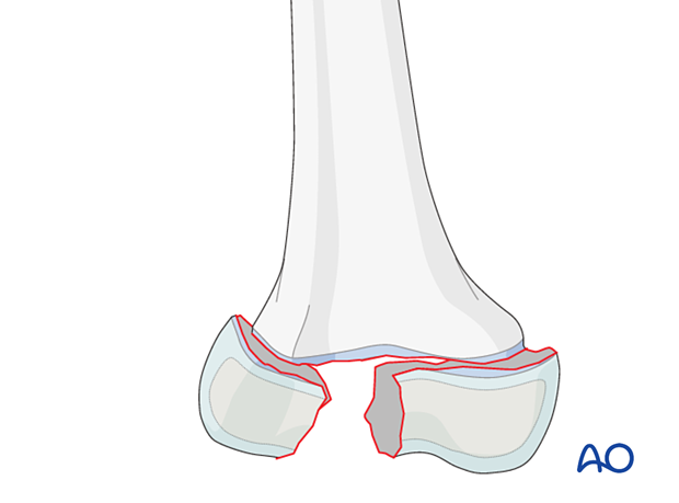
epiphyseal fracture
a fracture that occurs in a growth section of a child’s bone and may result in growth abnormalities
greenstick
an incomplete fracture that passes only partway through the shaft of a bone but may still cause substantial angulation
occurs in children
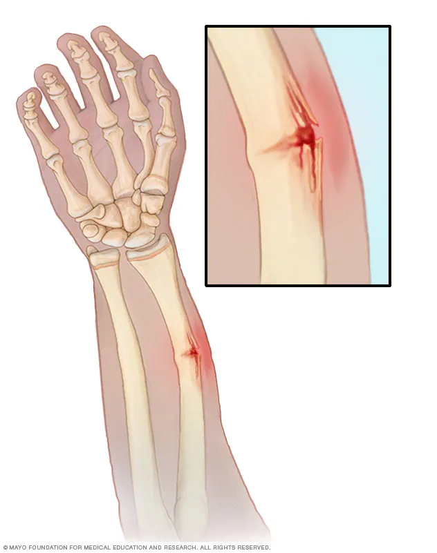
incomplete fracture
a fracture that does not run completely through the bone
a nondisplaced partial fracture
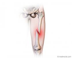
oblique fractures
a fracture in which the bone is broken at an angle across the bone
usually the result of a sharp, angled blow to the bone
pathologic fracture
a fracture of weakened or diseased bone
seen in pt.s with osteoporosis, infection, or cancer
produced by minimal force
spiral fracture
a fracture caused by a twisting or spinning force
causes a long, spiral-shaped break in the bone
sometimes the result of abuse in young children
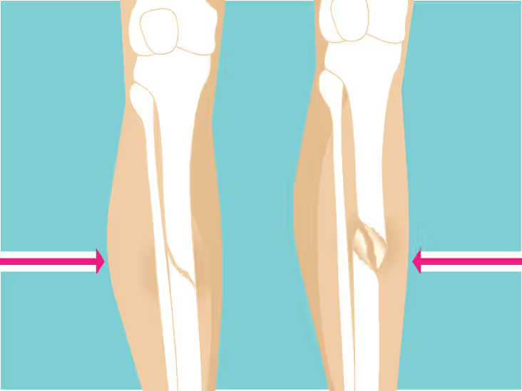
transverse
a fracture that occurs straight across the bone
usually the result of a direct and relatively high-energy blow
suspect fractures if pt. presents with:
deformity
tenderness
guarding
swelling
bruising
crepitus
false motion
exposed fragments
pain
locked joint
point tenderness
pain elicited by palpation with a finger at the site of an injury
crepitus
a grating or grinding sensation
can be felt and sometimes heard when fractured bone ends rub together
NEVER push the ends of a ______ ______ back into place
protruding bone
OPQRST
O = Onset
P = Provocation
Q = Quality
R = Region/radiation
S = Severity
T = Time
dislocation
disruption of a joint in which the bone ends are no longer in contact
supporting ligaments are often torn
signs + symptoms of a dislocated joint
marked deformity
swelling
pain that worsens with movement
tenderness on palpation
loss of normal joint motion
numbness or impaired circulation
sprain - partial dislocation
occurs when a joint is twisted or stretched beyond its normal range of motion
supporting capsule and ligaments are stretched or torn - results in injury to the ligaments, articular capsule, synovial membrane, and tendons
can range from mild - severe
some pt.s might hear a “snap”
do not involve deformity
signs + symptoms of a sprain
guarding
swelling and ecchymosis
pain prevents pt. from moving limb
instability of the joint
strain (mulled muscle)
a stretching or tearing of the muscle and/or tendons
occurs because of an abnormal contraction or from excessive stretching
may range from from minute separation to complete rupture
no ligament or joint damage typically occurs
no deformity is present
pt. may report a “snap” when muscle tears
signs + symptoms of a strain
pain
report increased sharp pain with passive movements
swelling
bruising
severe weakness of the muscle
tenderness
preventing contamination following an open fracture:
brush away any obvious debris on the skin surrounding the fracture
do NOT try and get debris directly out of the fracture site
minor injuries
minor sprains
fractures or dislocations of digits
moderate injuries
open fractures of digits
nondisplaced long bone fractures
nondisplaced pelvic fractures
major sprains of a major joint
serious injuries
displaced long bone fractures
multiple hand and foot fractures
open long bone fractures
displaced pelvic fractures
dislocations of major joints
multiple digit amputations
laceration of major nerves or blood vessels
severe, life-threatening injuries
multiple closed fractures
limb amputations
bilateral femur fractures
critical injuries
multiple open fractures of the limbs
suspected pelvic fractures with hemodynamic instability
6 Ps of musculoskeletal assessment
pain
paralysis
paresthesia (numbness or tingling)
pulselessness
pallor
pressure
what should you do before and after applying a splint
assess the pt.s circulation, motor function and sensation
document whether they are absent or present
caring for musculoskeletal injuries
cover open wounds with dry, sterile dressings + apply pressure. Apply tourniquet if bleeding can’t be controlled
asses pulse, motor, and sensory function
apply a splint + elevate extremity approximately 6 in
reassess pulse, motor, and sensory function
apply cold packs if there is swelling - do NOT place directly on skin
position pt. for transport
secure injured area
always splint extremities prior to moving pt. unless they are in _____ ______
immediate danger
splinting helps prevent:
further damage
laceration of the skin from broken bones
restriction of distal blood flow
excessive bleeding of the tissues
increased pain from movement
if you encounter resistance to limb alignment, _________
splint the limb in its deformed position
rigid (nonfarmable) splints
applied to the sides, front, and/or back of an injured extremity
requires 2 people
applying a rigid splint
provide gentle support + in-line traction for the limb
asses distal pulse, motor, and sensory function
place splint alongside or under the limb
pad between the limb and splint as needed
secure the splint to the limb with bindings
asses + record distal pulse, motor, and sensory function
most dislocations should be splinted __ _____
as found
formable splints
structural aluminum malleable splints (SAM) + vacuum splints
can be molded and remolded as needed
becomes rigid when molded into “T” shape
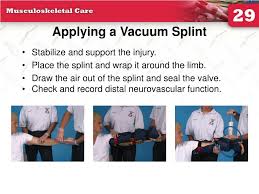
applying a vacuum splint
*still asses distal pulse, motor, and sensory function before applying splint
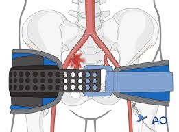
pelvic binders
used to splint the bony pelvis to reduce hemorrhage
hazards of improper splinting
compression of nerves, tissues, and blood vessels
delay in transport of pt. with a life-threatening injury
reduction of distal circulation
aggravation of the injury
injury to tissue, nerves, blood vessels, or muscles as a result of excessive movement of the bone or joint
injuries to the scapula:
almost always the result of a forceful direct blow to the back
could possibly injure thoracic cage, lungs, and heart
carefully asses these pt.s for difficulty breathing
provide supplemental O2
abrasions, contusions, and swelling are also common
can be splinted with a sling
Acromioclavicular (AC) joint separations
common in sports
distal end of the clavicle will often stick out
pt. will report pain
can be splinted with a sling
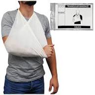
sling
sling must apply gentle upward support to the olecranon process of the ulna
knot of sling should be tied to one side of the neck
swathe should be used to bind the arm to the chest wall
leave fingers exposed
asses distal pulse, motor, and sensory function before + after applying sling
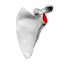
glenoid fossa
the part of the scapula that joins with the humeral head to form the glenohumeral joint
shoulder dislocations
humeral head most commonly dislocates anteriorly
pt. will guard shoulder and often hold it in a fixed position away from chest wall
shoulder will appear squared off or flattened
humeral head will protrude anteriorly underneath the pectoralis major in the anterior chest wall
axillary nerve may be compressed - causes a numb patch on the outer aspect of the shoulder
document this finding
numbness in the hand may occur
splinting shoulder dislocations
splint in position most comfortable for pt.
consider placing pillows/blankets between arm and chest wall
then, apply a sling + swathe
splinting a fractured humerus
apply traction to realign the fracture fragments
support fracture site with one hand and use the other to hold the two humeral condyles just above the elbow
pull gently in line with the normal axis of the limb
splint with a sling and swathe supplemented by a padded board + splint on lateral aspect of arm
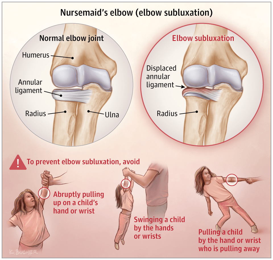
nursemaid’s elbow
fracture of the olecranon process of the ulna
can result from direct pressure or indirect forces
often associated with lacerations + abrasions
pt. will be unable to extend elbow
fractures of the radial head
generally occurs as a result of a fall on an outstretched arm or direct blow
attempts to rotate elbow will cause discomfort
care of elbow injuries
asses distal neurovascular functions periodically
splint elbow in position of strong pulses and good cap refill
2 padded boards, one applied to each side of the limb secured with roller bandages is usually best
bones above + below elbow should also be immobilized
SAM splints can also be molded to fit the arm
cold, pale hand or weak/absent pulse - further care should be dictated by physician
pulseless + significantly deformed - apply gentle manual traction in line with the long axis of the limb
if no pulse returns, splint arm in most comfortable position
nightstick fracture
an isolated fracture to the ulna shaft
colles fractures
fractures of the distal radius
stabilizing fractures of the forearm or wrist
padded boards, air vacuums, or pillow splints can be used
if possible, elevate the injured limb above the heart to help with swelling
splinting the hand and wrist
support the injured limb + move the hand into position of function
place a soft roller bandage in the palm
apply a padded board splint on the palmar side with fingers exposed
secure the splint with a roller bandage
pelvis fractures
several liters of blood may drain into pelvic space and retroperitoneal space
can result from direct compression or indirect forces
immediately treat for shock
most reliable sign is simple tenderness and instability on palpation
assess pelvic tenderness by:
place the palms of your hands over the lateral aspect of each iliac crest, apply firm + gentle inward pressure on pelvic ring
place palm over the anterior aspect of each iliac crest, and apply firm downward pressure
use palm of hand to firmly but gently palpate the pubic symphysis
dislocations of the hip
takes significant force
most dislocations are posterior
commonly occurs when knees hit dashboard in MVCs
injury to the sciatic nerve often occurs
pt. will have severe pain, tenderness, and will resist any attempt to move joint
posterior dislocation - pt. typically lie with knee drawn up and thigh rotated inward over the other leg
anterior dislocation - pt.’s leg is extended straight out, externally rotated away from midline
sciatic nerve
largest peripheral nerve in the body
controls the activity of muscles in the posterior thigh, below the knee, and sensation in the leg and foot
how to check for injury:
carefully assess sensation + motor function in the lower extremity
splinting dislocated hips
splint in position of dislocation
support injured limb with pillows and rolled blankets - especially under the knee
secure entire limb to backboard with long straps
fractures of the proximal femur
characteristic deformity - leg externally rotated, injured leg is usually shorter than the other leg
gentle rolling will cause pain but no further damage
pain might be referred to knee
splinting proximal femur fractures
assess pulse, motor, and sensory function
age + severity will dictate method of splinting
geriatrics with isolated hip fractures do NOT require a traction splint
femoral shaft fractures
500 - 1,000 mL of blood loss is possible
handle these pt.s with extreme care
periodically assess distal neurovascular function
remove clothing from affected limb
splinting a femoral shaft fracture
apply gentle longitudinal traction
gradually turn leg from the deformed position
apply traction splint
goals of in-line traction
stabilize the fracture fragments to prevent excessive movement
align the limb sufficiently to allow it to be placed in a splint
avoid potential neurovascular compromise
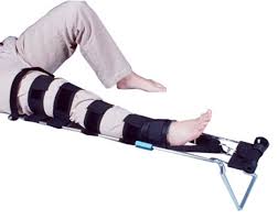
applying a traction splint
expose injured limb + check pulse, motor, and sensory function
place splint beside uninjured limb to find correct length
support the injured limb as partner fastens the ankle hitch above the foot
continue to support the limb as your partner applies gentle in-line traction to the ankle/foot
slide the splint in position under the injured limb
pad the groin + fasten the ischial strap
connect the loops of the ankle hitch to the end of the splint. Then, tighten ratchet to the point that the splint holds adequate traction
secure + check the support straps
asses pulse, motor, and sensory functions
secure pt. to backboard
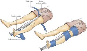
applying a sager traction splint
after exposing the area, asses distal pulse, motor, and sensory function.
position splint against the medial aspect of the injured leg, with perineal cushion resting against the ischial tuberosity
adjust thigh straps so that it lies anteriorly when secured
secure thigh strap
apply padded ankle harness just above the ankle
attach harness to the crossbar
extend the splint’s inner shaft to apply traction of abt 10% of body weight (up to 15 lbs)
secure splint with elasticized cravat bandages
secure pt. to a backboard with a figure 8 configuration
use the pedal pinion strap to secure the feet together
reassess distal pulse, motor, and sensory function
injuries to the knee ligaments
occur when abnormal bending or twisting occurs
pt. will report pain in joint and be unable to move it
swelling, ecchymosis, point tenderness, and a joint effusion are common
joint effusion
excessive fluid in the joint
splinting knee ligament injuries
several different splints can be used
splint should extend from hip joint to the foot
splint in bent position if there is too much pain when trying to straighten leg
knee dislocations
may threaten the limb
may be associated with vascular occlusion
ligaments may be damaged or torn
severe pain + inability to move joint
posterior knee dislocations - occur from extreme hyperextension; high risk of injury to the popliteal artery
medial dislocations - direct blow to the lateral part of the leg; high risk of injury to the medial ligaments
vascular occlusion
a blockage in a blood vessel (artery or vein) that cuts off or severely reduces blood flow, depriving tissues of vital oxygen and nutrients, leading to potential damage
splinting knee dislocations
splint knee in position if was found if there are strong distal pulses
do not attempt to move if these distal pulses are good
straight leg - apply rigid splints to both sides of the limb
bent knee - use parallel padded boards secured at hip and ankle joints
fractures about the knee
adequate distal pulse + no significant deformity → splint with knee straight
adequate pulse + significant deformity → splint in position of deformity
pulse is absent below injury → contact medical control
NEVER use a traction splint of you suspect a fractured knee
dislocation of the patella
splint knee in position found
stabilize + apply padded board splints to the medial and lateral aspects of the joint
patella may go back into place spontaneously
use a long padded leg splint
report to hospital staff
injuries to the tibia and fibula
should be stabilized with a padded rigid long leg splint or an air splint
after splinted, affected leg should be secured to the opposite leg
apply gentle traction
ankle injuries
hard to tell the difference between a sprain and a fracture
dislocations of the ankle is commonly associated with fractures of one or both malleoli
most frequent MOI = twisting
managing ankle injuries
dress open wounds
asses distal neurovascular function
correct any gross deformity by applying gentle longitudinal traction to the heel
before releasing traction, apply a splint - can use a rigid, air, or pillow splint (must extend up the leg to the level of the knee joint)
foot injuries
heels - injury occurs from falls or jumps
force may be transmitted up the legs to the spine
treat for spinal injuries
dislocated feet - take off shoe and assess distal neurovascular functions
stabilize extremity but leave toes exposed
significant swelling
never allow foot/leg to dangle off of the stretcher
treatment for sprains + strains
treat as if they are fractures
RICES
protect from weight-bearing activity
compartment syndrome
limb-threatening condition
localized tissue swelling within a compartment
ischemia results and anaerobic metabolism compensates for the loss of O2
often develops between 6-12 hours after injury
early signs + symptoms
pain that is out of proportion to injury, pain on passive stretch of muscles within the compartment, altered sensation, pallor, decreased power
how to treat suspected compartment syndrome
splint affected limb - keeping at level of heart
provide immediate transport
reassess neurovascular status frequently
amputations
control bleeding
partial amputations - stabilize part with bulky compression dressings and splint
complete amputation - wrap clean part in sterile dressing and place in a bag. Place bag on top of ice
atrophy
the wasting away, shrinking, or decrease in size and strength of a body part, organ, or tissue
causes: disease, disuse, malnutrition, nerve damage, or hormonal changes
effects: weakness + reduced function
skeletal muscle tissue is directly attached to bones by _____
tendons
characteristics of proximal humeral fractures
significant swelling, but no significant deformity of the upper arm
neurovascular compromise uncommon
if present, any or all of the brachial plexus may be affected
concurrent soft-tissue injuries possible
possible rotator cuff injury
characteristics of a midshaft humeral fracture
gross angulation of the arm
marled instability + crepitus of fracture fragments
possible neurovascular compromise
possible entrapment of the radial nerve
pt. cannot extend or dorsiflex the wrist/fingers and may report numbness on the dorsum of the hand; classic wrist drop