radiology
 Call Kai
 Learn
Learn Practice Test
 Spaced Repetition
 Match
Match Flashcards
 Knowt Play
1/367

There's no tags or description
Looks like no tags are added yet.
Name | Mastery | Learn | Test | Matching | Spaced | Call with Kai | Chat |
|---|
No analytics yet
Send a link to your students to track their progress
368 Terms
no
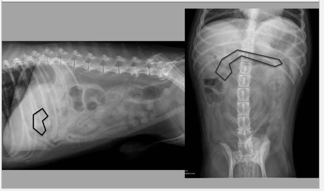
should neonatal, immature (up to 6 months of age), athletic, and cachexia animals have excellent, good, decreased, or no peritoneal/retroperitoneal detail
excellent
should mild/moderate obese animals have excellent, good, decreased, or no peritoneal/retroperitoneal detail
decreased
does young animal with brown fat and small amount of fluid, decreased intra-abdominal fat, abdominal fluid, large masses, carcinomatosis/sarcomatosis, steatitis, and peritonitis/retroperitonitis cause increased or decreased peritoneal/retroperitoneal detail
increased
does gas in peritoneal/retroperitoneal space cause increased or decreased peritoneal/retroperitoneal detail
good
should animals with normal body condition have excellent, good, decreased, or no peritoneal/retroperitoneal detail
decreased
should animals with extreme obesity have excellent, good, decreased, or no peritoneal/retroperitoneal detail
pneumoperitoneam
what cause of increased peritoneal detail is caused by hollow viscous rupture, penetrating wound, iatrogenic (recent surgical intervention), abscess rupture (gas forming bacteria)/perforated emphysematous/cystitis/cholecystitis
mediastium
pneumoretroperitoneum is generally the result of an extension of a pneumo what
bate’s body
term for nodular fat necrosis in cats abdomen
vessel
mineralization of what is usually associated with endocrinopathies and chronic uremia in abdomen
inflammation
dystrophic mineralization of any structure is secondary to prior what in abdomen
adrenal glands
what organ in aged cats is often mineralized in abdomen
liver, spleen, kidney, adrenal gland, ovary, stomach
6 abdominal organs of lateral craniodorsal mass effect (L, S, K, A, O, S)
liver, gallbladder, stomach, pancreas
4 abdominal organs of lateral cranioventral mass effect (L, G, S, P)
spleen, mesenteric lymph node, digestive tract, pancreas, uterus, ovaries, testes
7 abdominal organs of lateral central mass effect (S, M, D, P, U, O, T)
medial iliac lymph node, colon, rectum, ureters
4 abdominal organs of lateral caudodorsal mass effect (M, C, R, U)
uterus, prostate, urinary bladder, testes
4 abdominal organs of lateral caudoventral mass effect (U, P, U, T)
liver, stomach, spleen head, adrenal gland, kidney, ovary
6 abdominal organs of VD left cranial mass effect (L, S, S, A, K, O)
liver, gallbladder, stomach, duodenum, pancreas, kidney, adrenal, ovary
8 abdominal organs of VD right cranial mass effect (L, G, S, D, P, K, A, O)
spleen, mesenteric lymph node, digestive tract, pancreas, uterus, ovaries, testes
7 abdominal organs of VD central mass effect (S, M, D, P, U, O, T)
medial iliac lymph node, colon, rectum, uterus, ureters, prostate, bladder, testes
8 abdominal organs of VD caudal mass effect (M, C, R, U, U, P, B, T)
liver
what organ is in cranial abdomen between diaphragm and stomach with sharp well defined and smooth margins
parallel
liver and gastric access should be what in lateral
spine
liver gastric axis is parallel to what in VD
metabolic hepatopathy, toxicity, viral hepatitis, infiltrative neoplasia, acute inflammatory disorder, hepatic congestion
6 causes of generalized hepatomegaly (MH, T, VH, IN, AID, HC)
hepatic lipidosis, steroid hepatopathy, diabetes mellitus
3 causes of metabolic hepatopathy which causes hepatomegaly (HL, SH, DM)
nodular hyperplasia, neoplasia, cyst, abscess, hematoma, hepatic lobe torsion
6 causes of focal hepatomegaly (NH, N, C, A, H, HLT)
cyst, hematoma, abscess, neoplasia, nodular hyperplasia, granuloma, extra abdominal, extramedullary hematopoiesis
8 causes of masses that cause hepatomegaly (C, H, A, N, NH, G, EA, EH)
generalized
rad findings of what kind of hepatomegaly include rounded caudoventral margin, caudal shift gastric axis, margins extend beyond costal arches, caudal displacement of cranial abdominal organs
focal
rad findings of what kind of hepatomegaly include rounded margin, creates abdominal mass effect, masses can be pedunculated and caudal to stomach
microhepatia
rad findings of hepatomegaly or microhepatia include caudoventral margin not seen, cranial shift of gastric axis, cranial displacement of organs of abdomen, calculi, renomegaly with PSS, rule out PPDH
chronic hepatitis, hepatic cirrhosis, congenital portosystemic shunt
2 differentials for microhepatia include what with what, and what (CH, HC, CPSS)
hepatolith
mineral hepatic opacity located in intrahepatic biliary ducts
choledocolith
mineral hepatic opacity located in common bile duct
cholecystolith
mineral hepatic opacity located in gall bladder
spleen
what abdominal organ has a fixed head in left dorsocranial abdomen and a mobile tail with sharp well defined margins
breed, drug induced, infiltrative neoplasia, extramedullary hematopoiesis, splenic congestion, splenic torsion, systemic infectious disease
7 causes of generalized splenomegaly (B, DI, IN, EH, SC, ST, SID)
nodular hyperplasia, neoplasia, hematoma, abscess
4 causes of focal splenomegaly (NH, N, H, A)
no
does this rad have excellent, good, decreased, or no peritoneal/retroperitoneal detail
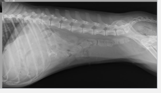
good
does this rad have excellent, good, decreased, or no peritoneal/retroperitoneal detail

excellent
does this rad have excellent, good, decreased, or no peritoneal/retroperitoneal detail

decreased
does this rad have excellent, good, decreased, or no peritoneal/retroperitoneal detail

loss of retroperitoneal detail, mass effect
what 2 things does this rad have

severe peritoneal effusion
this loss of peritoneal/retroperitoneal detail is caused by what

increased, penumoperitoneum
is there increased or decreased peritoneal detail, and what is it caused by

pneumoretroperitoneum
what is this a radiograph of

generalized hepatomegaly
what is this a radiograph of
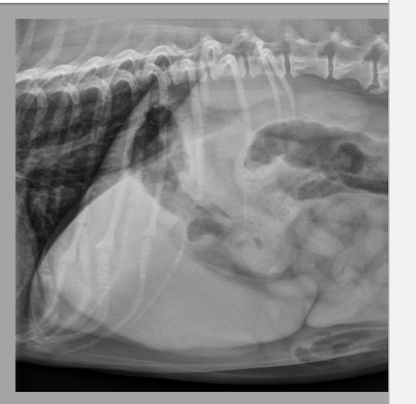
focal hepatomegaly
what is this a radiograph of

microhepatia
what is this a radiograph of

generalized splenomegaly
what is this a radiograph of
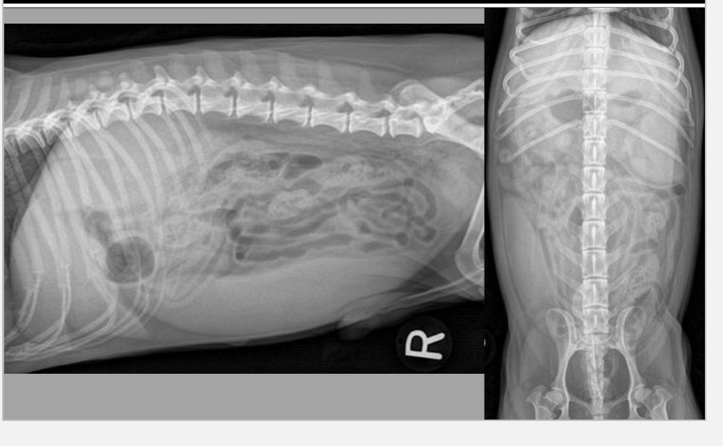
generalized splenomegaly
what is this a radiograph of

focal splenomegaly
what is this a radiograph of

focal splenomegaly
what is this a radiograph of
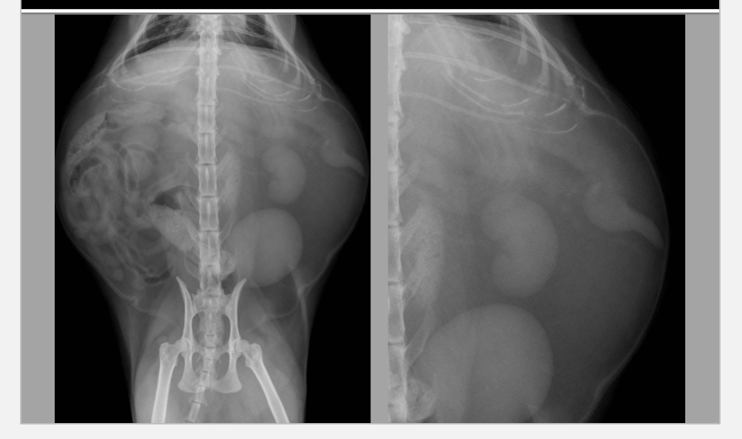
generalized hepatosplenomegaly
what is this a radiograph of

splenic torsion
what is this a radiograph of

overconditioned
8 YO FN beagle with a history of intermittent vomiting and diarrhea: is the patient underconditioned, normal condition, or overconditioned

good
8 YO FN beagle with a history of intermittent vomiting and diarrhea: is the peritoneal and retroperitoneal detail good or bad

liver, moderate, generalized, large
8 YO FN beagle with a history of intermittent vomiting and diarrhea: what organ is affected, is it mild, moderate, or severe, is it generalized or focal, and is it large or small

caudoventral, last ribs, caudodorsal, mass effect
8 YO FN beagle with a history of intermittent vomiting and diarrhea: the liver is moderately diffusely enlarged with rounded what margins, extending further beyond what, causing what deviation of the gastric axis, which is causing what

spleen, generalized, large
8 YO FN beagle with a history of intermittent vomiting and diarrhea: what organ besides the liver is affected, is it generalized or focal, and is it large or small

moderate generalized hepatomegaly, equivocal generalized splenomegaly
8 YO FN beagle with a history of intermittent vomiting and diarrhea: 2 conclusions of this case

vacuolar/metabolic hepatopathy, hepatitis, infiltrative neoplasia, hepatic congestion
8 YO FN beagle with a history of intermittent vomiting and diarrhea: 4 differentials for moderate generalized hepatomegaly (V/M H, H, I N, H C)

vascular congestion, sedation, extramedullary hematopoiesis, lymphoid hyperplasia
8 YO FN beagle with a history of intermittent vomiting and diarrhea: differentials for equivocal generalized splenomegaly are V C secondary to S, E H, L H
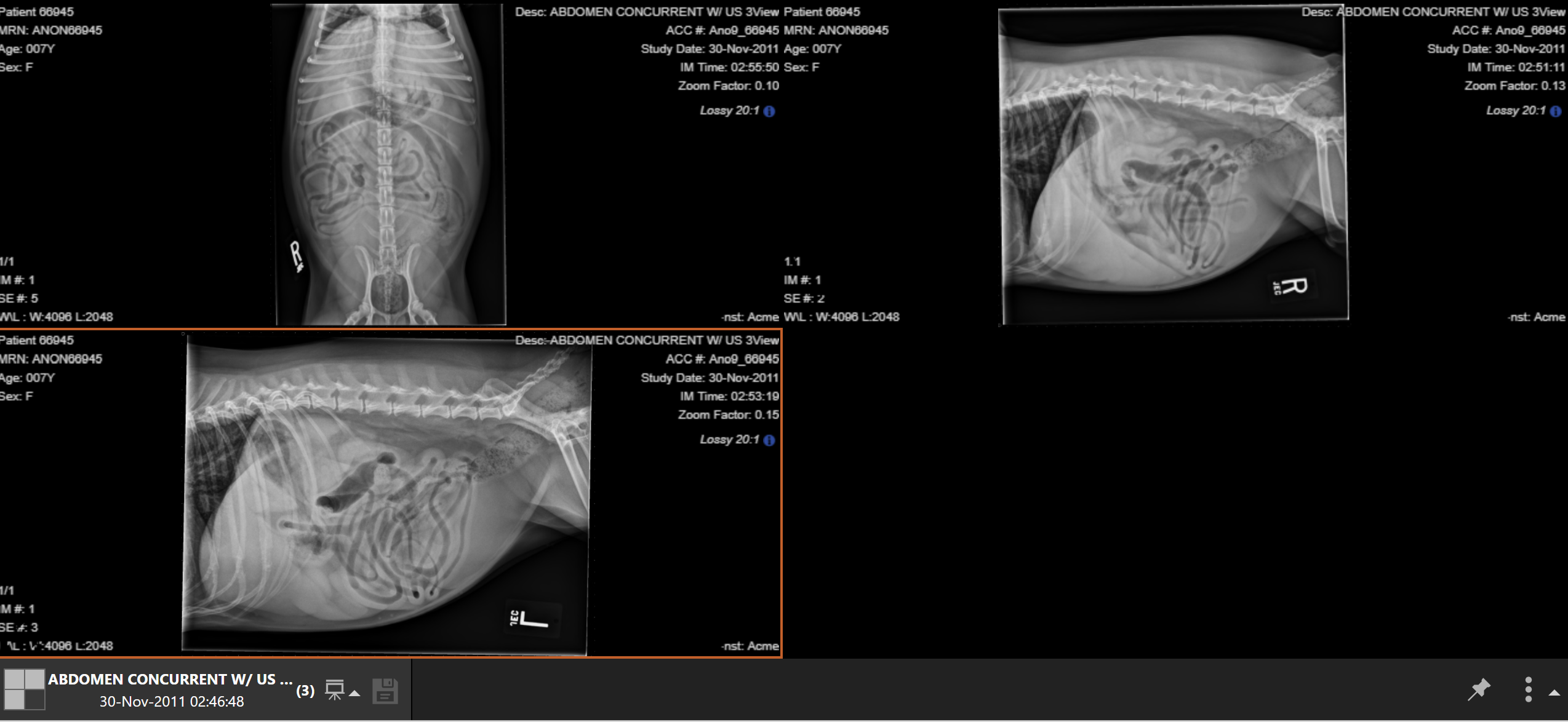
overconditioned
16 YO FN labrador decreased appetite: is this patient underconditioned, normal condition, or overconditioned

spondylosis deformans, multifocal
16 YO FN labrador decreased appetite: what is seen on the thoracic, lumbar, and lumbosacral vertebrae, and is it focal or multifocal

fat opaque masses
16 YO FN labrador decreased appetite: what is seen along the dorsal and ventral body wall and proximal thighs

left caudal lung lobe
16 YO FN labrador decreased appetite: there is a round, soft tissue opaque nodule located where (aside from body wall)

good
16 YO FN labrador decreased appetite: is the peritoneal and retroperitoneal detail good or bad

cranioventral
16 YO FN labrador decreased appetite: there is a mass located in what abdomen division

liver, pedunculated mass, yes, stomach, transverse colon, small intestines
16 YO FN labrador decreased appetite: what organ is affected, and it is affected by what, and is there a mass effect, if yes what 3 organs is it displacing

pedunculated hepatic mass, pulmonary nodule, vertebral degenerative changes, multifocal body wall and proximal thigh fatty masses
16 YO FN labrador decreased appetite: 4 conclusions of the case (P D M, P N, V D C, M B W P T F M)
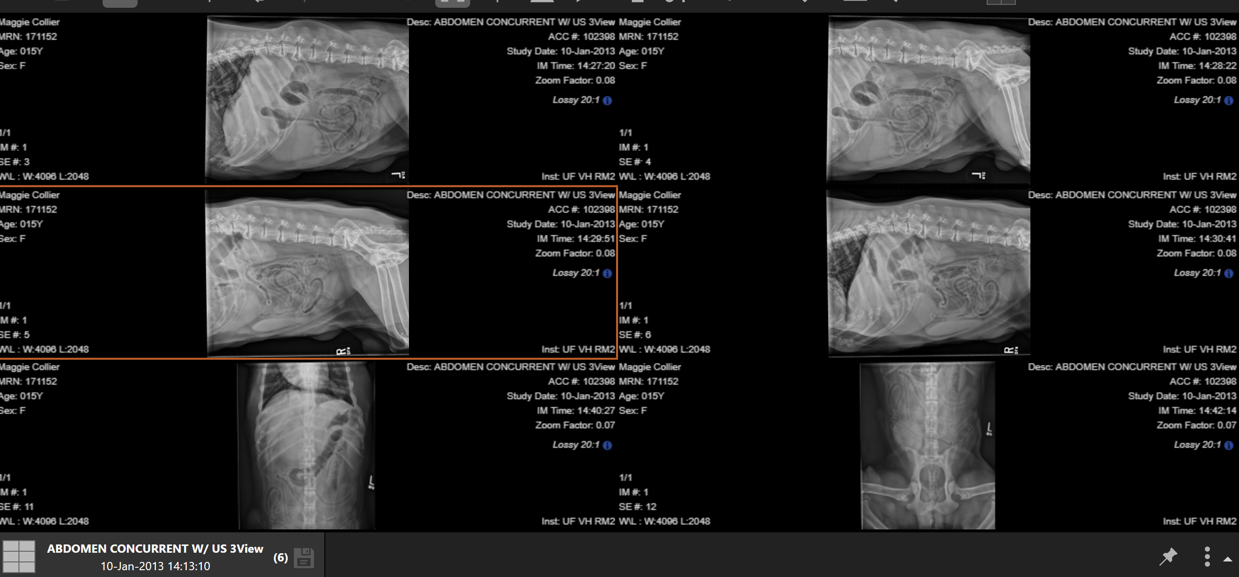
malignant neoplasia, hepatocellular carcinoma
16 YO FN labrador decreased appetite: primary differential for pedunculated hepatic mass is what, such as what (M N, H C)
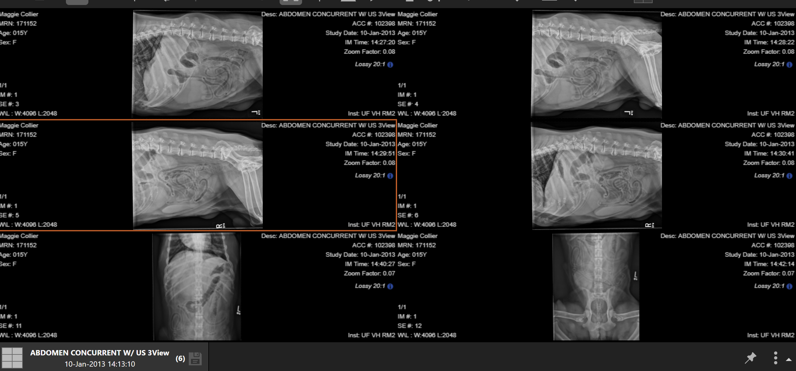
metastatic
16 YO FN labrador decreased appetite: pulmonary nodule is concerning for what disease

underconditioned
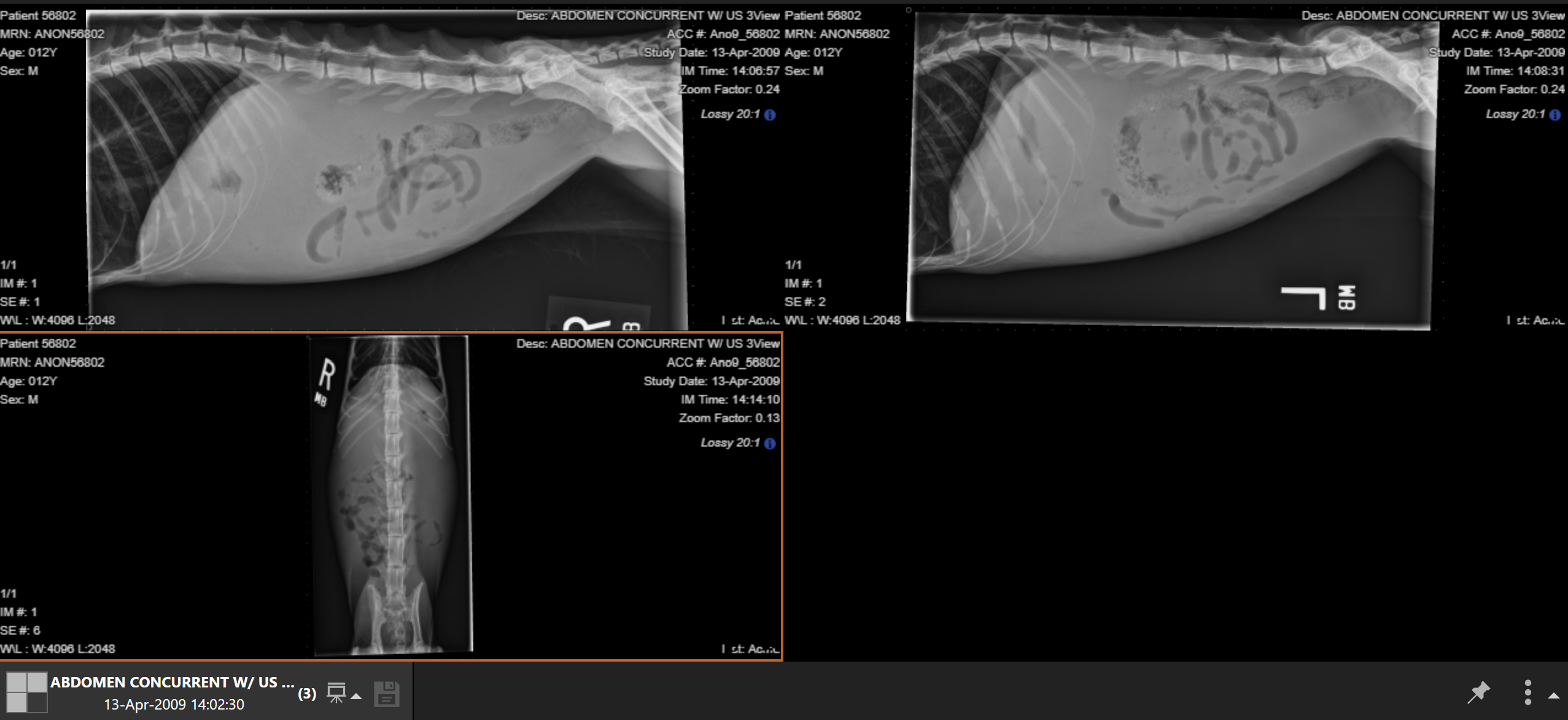
12 YO MN DSH with 1 month history of weight loss: is this patient overconditioned, normal condition, or underconditioned

distended
12 YO MN DSH with 1 month history of weight loss: is the patient’s abdomen normal or distended

6 lumbar vertebrae
12 YO MN DSH with 1 month history of weight loss: what is present in the vertebral column
lost
12 YO MN DSH with 1 month history of weight loss: is there good peritoneal/retroperitoneal detail or is it lost

no
12 YO MN DSH with 1 month history of weight loss: is there a mass effect

complete loss of peritoneal detail, abdominal distention, peritoneal effusion, fat, no, cachexia, lumbosacral transitional vertebra, L7
12 YO MN DSH with 1 month history of weight loss: conclusion for this case is C L P D with associated A D compatible with P E, retroperitoneal loss of detail may be associated with lack of what, can retroperitoneal effusion be ruled out, C, L T V with suspected sacrilization of what vertebrae

overconditioned
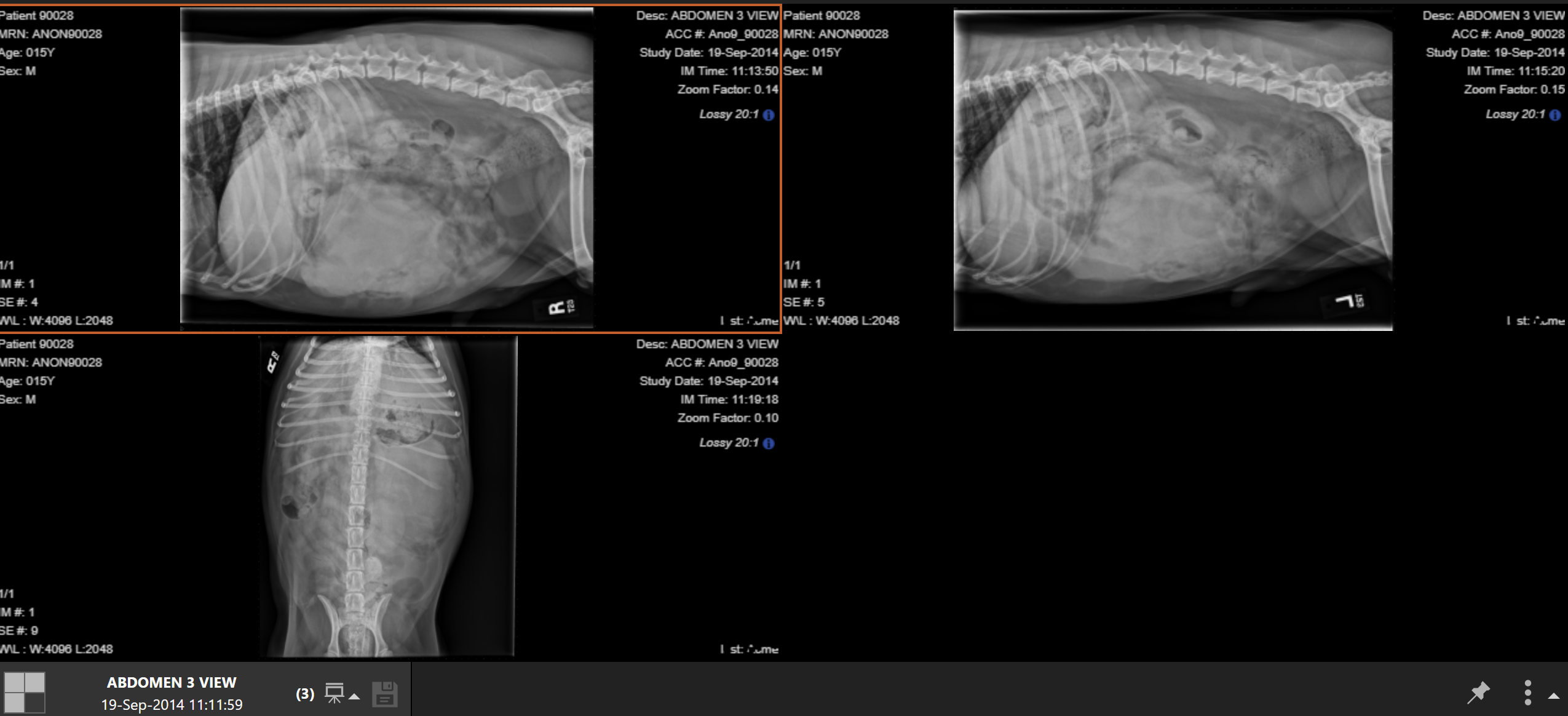
15 YO MN beagle abdominal pain and acute collapse: is the patient overconditioned, normal condition, or underconditioned
good
15 YO MN beagle abdominal pain and acute collapse: is the retroperitoneal detail good or is it lost

increase, decreased, ventral, left
15 YO MN beagle abdominal pain and acute collapse: there is an increased or decreased what opacity with multiple wispy striations and associated increased or decreased peritoneal detail noted along the dorsal or ventral part and left or right side of the abdomen

mass, yes, colon, small intestine, left kidney
15 YO MN beagle abdominal pain and acute collapse: within the mid ventral and left side of the abdomen there is what, and is there a mass effect, if yes what is displaced

yes
15 YO MN beagle abdominal pain and acute collapse: is the stomach in a normal position

normal
15 YO MN beagle abdominal pain and acute collapse: are the small intestines within normal limits for size or are they too big or too small

left, central, ventral peritoneal mass, spleen, malignant neoplastic, hemangiosarcoma, peritoneal effusion, hemorrhage, undigested food, no
15 YO MN beagle abdominal pain and acute collapse: conclusion for this case is L C V P M likely originating from what organ, can be compatible with a M N process such as a what, P E likely compatible with what, gastric contents are likely compatible with what, can foreign material be ruled out

overconditioned
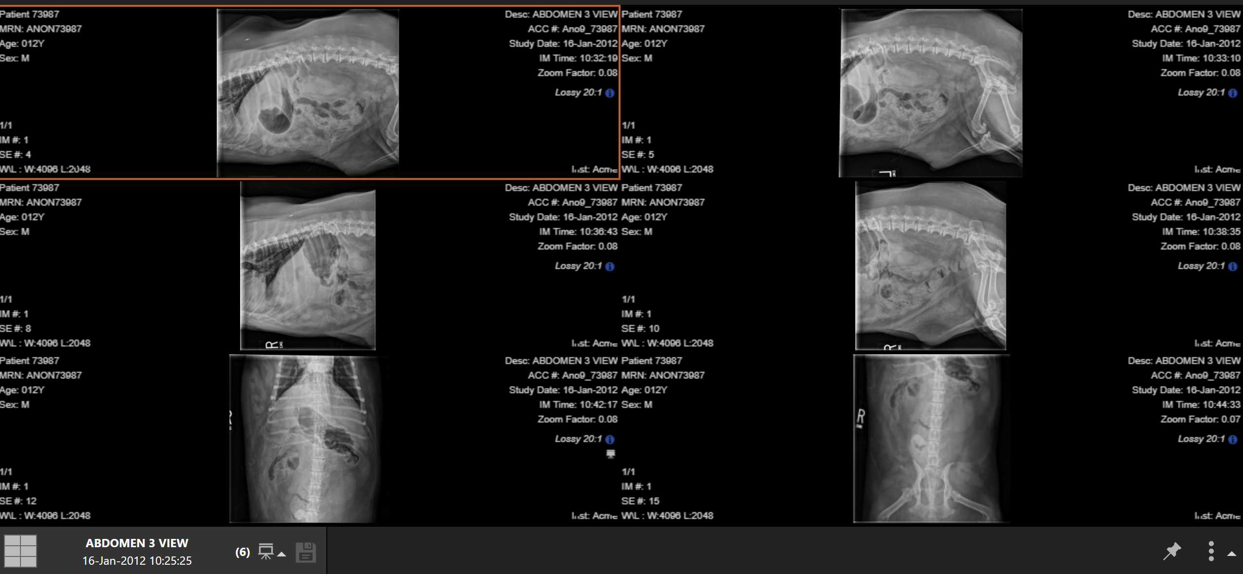
12 YO MN welsh corgi hit by car: is this patient overconditioned, normal condition, or underconditioned
normal
12 YO MN welsh corgi hit by car: does the visible thorax appear normal

multifocal spondylosis deformans
12 YO MN welsh corgi hit by car: there is M S D involving multiple thoracic and lumbar as well as lumbosacral vertebrae
are there fractures present in the ribs, and if so which ribs are affected
12 YO MN welsh corgi hit by car: yes, 10, 11, 12, 13

small intestine
12 YO MN welsh corgi hit by car: along the right caudal thoracic and right cranial abdominal wall, there is an increase in thickness containing multiple tubular thin soft tissue opaque structures resembling what

osseous fragments
12 YO MN welsh corgi hit by car: laterally to the iliac wings there are 2 sharply marginated what seen cranioventrally to the pelvis

heterogenous, increase, soft tissue
12 YO MN welsh corgi hit by car: between the caudoventral abdominal wall and penis, is there a homogenous or heterogenous increase or decrease in what opacity
decrease
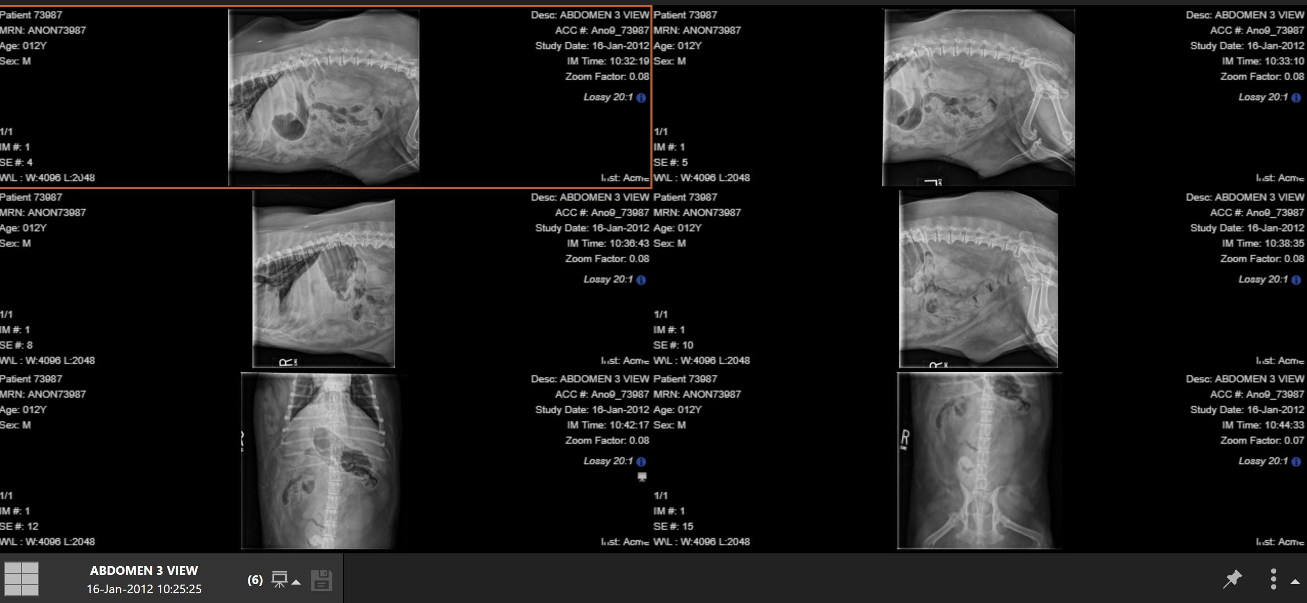
12 YO MN welsh corgi hit by car: is there an increase or decrease in retroperitoneal detail

yes, colon
12 YO MN welsh corgi hit by car: is there a mass effect, and if yes what is it displacing

traumatic right cranial abdominal wall rupture, herniation, small intestines, right thoracic, abdominal wall, bilateral prepubic tendon avulsion, prepubic inguinal hernias, retroperitoneal effusion, blood, urine, excretory urogram, small periotneal effusion, steatitis, artifactual, 10-13 rib fracture, vertebral degenerative changes
12 YO MN welsh corgi hit by car: conclusion for this case is T R C A W R with what of what within R T and A W, B P T A with associated P I H, retroperitoneal decreased detail and associated mass effect are suggestive of R E such as what and what, an E U may help distinguish between the two, equivocal decreased peritoneal detail differentials are S P E, S, and A, left sided ___-___ R F, V D C

no
is the pancreas normally seen in radiographs
left
what pancreas lobe is caudal to stomach and is the larger lobe in cats
right
what pancreas lobe is dorsomedial to descending duodenum and is the larger lobe in dogs
pancrea
what organ is this