Hematopoiesis: erythrocytes
 Call Kai
 Learn
Learn Practice Test
 Spaced Repetition
 Match
Match Flashcards
 Knowt Play
1/9

There's no tags or description
Looks like no tags are added yet.
Name | Mastery | Learn | Test | Matching | Spaced | Call with Kai | Chat |
|---|
No analytics yet
Send a link to your students to track their progress
10 Terms

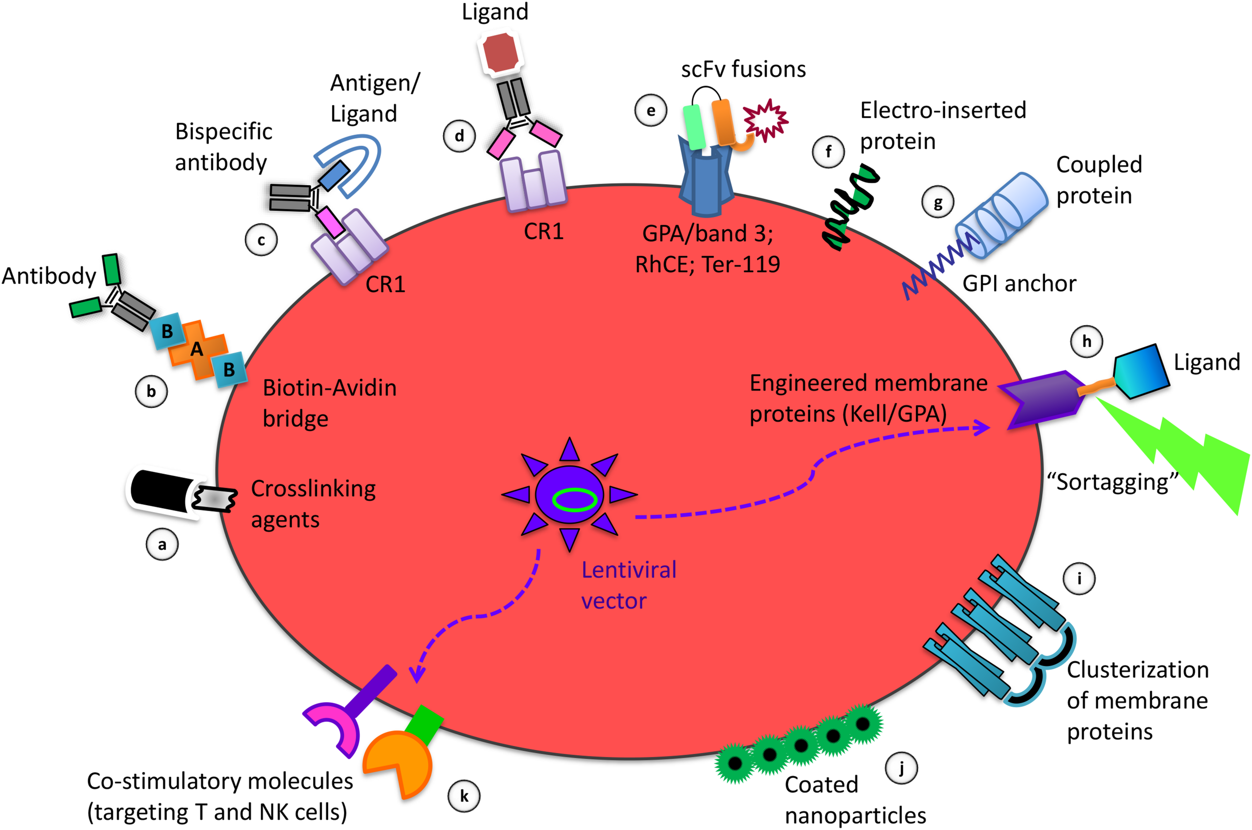
RBC: Spectrin & Glycophorin
Spectrin:
Peripheral membrane protein
Intertwined alpha and beta chains→ forms flexible tetramers
Forms microfilament skeletal structure
Connects RBC skeleton network together, stabilizes junction between spectrin and actin
Glycophorin
Integral membrane/ transmembrane protein
Negative charge (sialic acid)→ RBCs repel each other
Interacts with outside antigens and transports them inside
Cytoskeleton allows cell support, stability, flexibility, deformability
Anchors inner spectrin skeleton to outer lipid membrane
Glygophorin A (GPA) most common, but there’s A-E variants


RBC: Actin and Ankyrin
Actin
Junctional complex between spectrin tetramers
Links integral membrane proteins (like band 3) via proteins like ankyrin and protein 4.1
Ankyrin
Links integral membrane proteins to spectrin-actin cell cytoskeleton intracellularly
Cluster and positions ion channels, ion exchangers, transporters, cell-adhesion molecules to specialized membrane domains
Have 33-AA repeats


RBC: Band 3, Band 4.1
Band 3
Ion exchange: Trades chlorine for bicarbonate (HCO₃⁻) across membrane→ CO2 transport
Structural supprot: connects outer lipid payer to internal skeleton tia ankyrin
Band 4.1
Links inner skeleton proteins to outer membrane proteins (ex: GPC and band 3)
General structural and flexible support

RBC ions
Maintained by Na+/K+ -ATPase (sodium potassium pump)
Transports 3 Na+ outside, and 2 K+ inside
Failure= Na+ enters, K+ leaves cell→ Lysis
Na+ higher on outside, K+ higher on inside
High extracellular K+ could also be caused by hemolysis after centrifugation
Hemolytic anemia: Lysis of RBCs

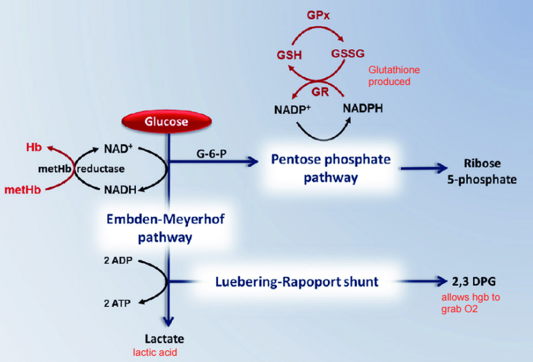
RBC metabolism- ATP
No nucleus= no oxidative respiration to metabolize glucose and obtain ATP
RBCs use glycolysis via Embden-Meyerhof pathway (EMP) (mnemonic: MeyeRBC)
Glucose→ Absorbed through plasma membrane→ metabolizes to lactic acid (instead of pyruvic acid)→ 2 ATP per glucose
Goes through aerobic phosphorylation and anaerobic glycolysis


RBC metabolism- oxygen
Via Luebering-Rapport shunt (mnemonic: Wrapper like wrapping up oxygen)
Produces 2,3-DPG or BPG (2,3 DIphosphoglycerate)
Presence= Closed salt bridge→ Prevents Hgb to grab O2 (tense state)
Absence= open salt bridge→ Delivery of O2 to tissues. Offloads 25%


Oxygen dissociation curve
Sigmoid curve
Bohr effect: Hgb’s O2 binding affinity inversely related to acidity and concentration of carbon dioxide
In lungs (pressure is 100 mm Hg), Hgb fully saturated: has all O2 it can hold.
At 40 mm, Hgb saturated 50% but willing to give up 50% of it’s O2
normal blood: pH 7.4. 75% Hb has O2, 25% can be released to tissues
Right shift: lower pH (7.2), Increased 2,3 DPG, higher body temperature, more CO2→ more Oxygen offloaded


RBC metabolism- glutathione
Via Pentose Phosphate Pathway (PPP)