A&P - Lab Practical Unit 2
 Call Kai
 Learn
Learn Practice Test
 Spaced Repetition
 Match
Match Flashcards
 Knowt Play
1/76

There's no tags or description
Looks like no tags are added yet.
Name | Mastery | Learn | Test | Matching | Spaced | Call with Kai | Chat |
|---|
No analytics yet
Send a link to your students to track their progress
77 Terms
6 Classifications of Bones
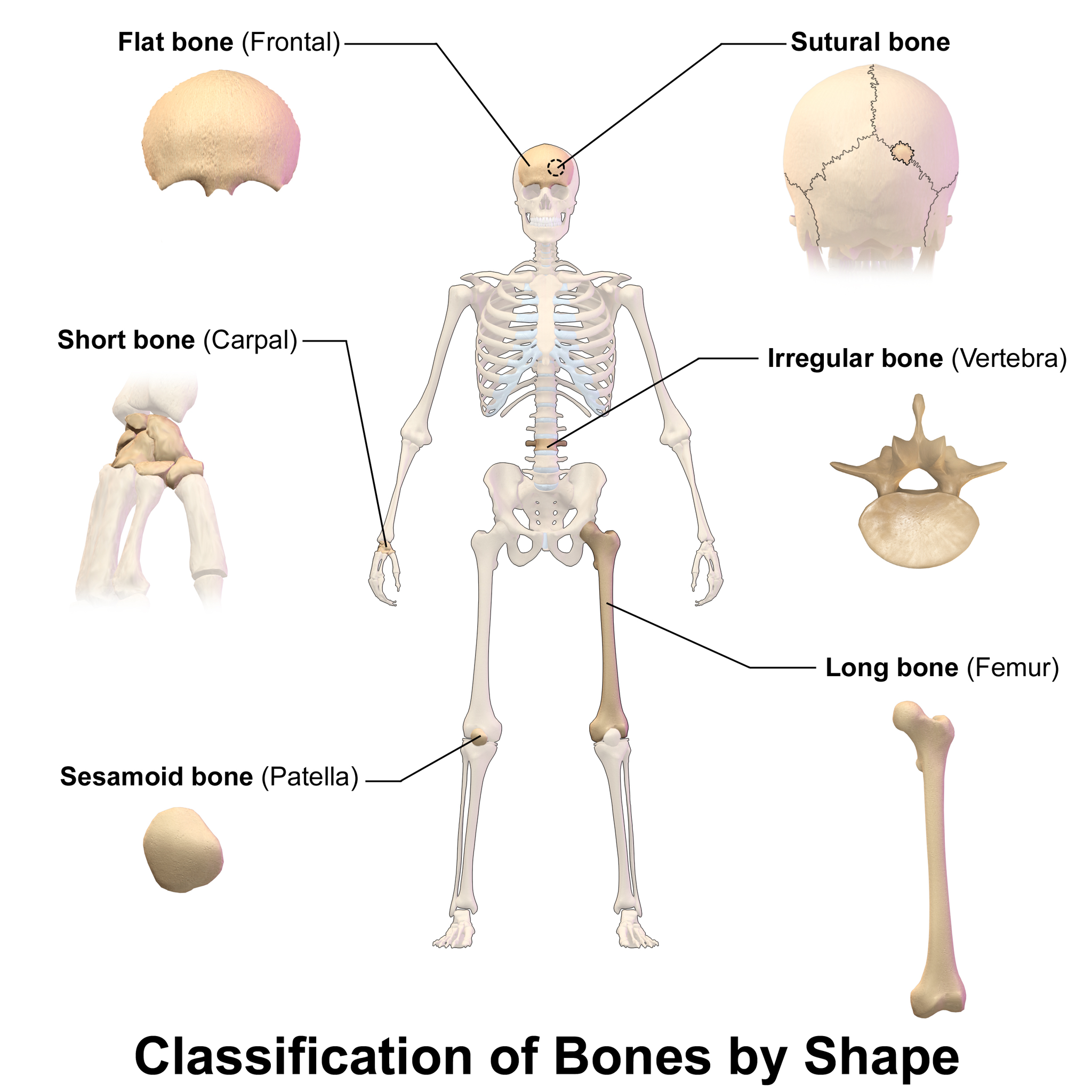
Long
Short
Flat
Irregular
Sesamoid
Sutural (Wormian)
Epiphysis
The end region of a long bone

Diaphysis (shaft)
The shaft/main region of a long bone

Metaphysis
The region of a long bone between the epiphysis and diaphysis

Medullary cavity
The central cavity of a long bone, containing bone marrow

Cortex (cortical bone)
The dense outer region of bone, i.e. compact bone
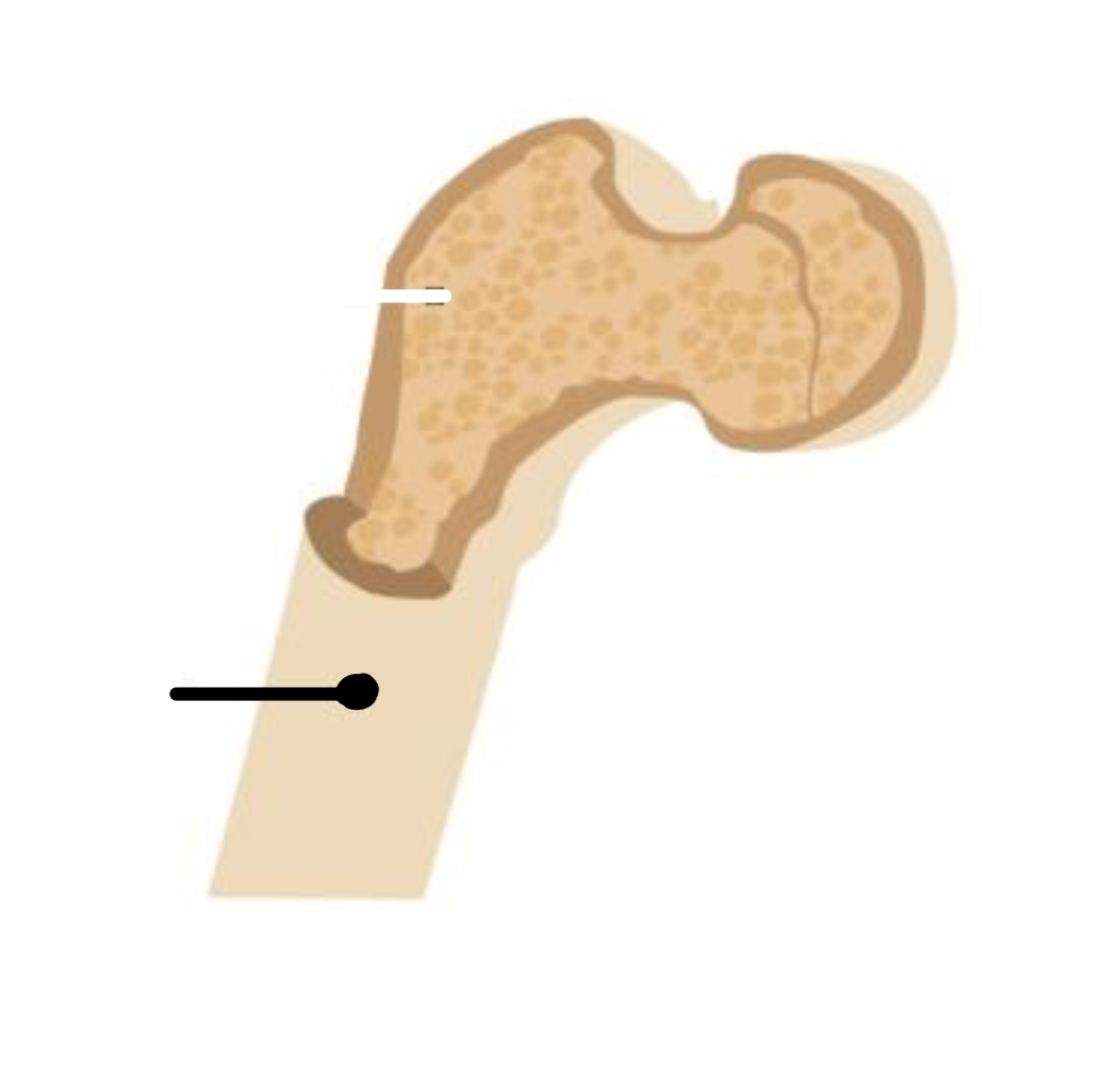
Endosteum
The membrane lining the medullary cavity of bone

Periosteum
The membrane covering the outer surface of bone

Red bone marrow
Found in the epiphysis/spongy bone; site of blood cell formation


Yellow bone marrow
Found in the medullary cavity; functions mainly as fat/energy storage
Fossa (fossae)
A shallow depression on a bone
example: olecranon fossa of the humerus
Foramen (foramina)
An opening in a bone, usually a passageway for nerves or vessels
example: foramen magnum of the skull
Meatus, canal
A tunnel through bone
example: external auditory meatus of the temporal bone
Sinus, antrum
An enclosed cavity in a bone
example: frontal sinus
Processes (bone markings)
Projections extending from a bone's surface
Condyle
A rounded process, usually found near articulations
example: femoral condyle
Epicondyle
A projection above a condyle
example: epicondyle of humerus
Head (bone marking)
A rounded process at the end of a bone
example: head of femur
Crest (bone marking)
A narrow ridge
example: iliac crest
Spine (bone marking)
A slender, pointed process
example: scapular spine
Trochanter, tuberosity, tubercle
Variously-shaped bumps/processes near articulations or muscle attachments; example: greater trochanter of femur
Trabeculae
The structural struts/plates of spongy (cancellous) bone
Haversian (central) canal
The central canal of an osteon that allows passage of blood vessels
Perforating (Volkmann's) canals
Canals that run perpendicular to the central canal, connecting blood vessels between osteons
Sutures (skull)
Immovable joints connecting the bones of the skull
types include: coronal, sagittal, lambdoid, and squamous
Coronal suture
The suture between the frontal bone and the two parietal bones

Sagittal suture
The suture between the two parietal bones

Lambdoid suture
The suture between the parietal bones and the occipital bone

Squamous suture (skull)
The suture between the temporal bone and the parietal bone

Wormian (sutural) bones
Extra, irregular isolated bone pieces that occur within a suture of the cranium in addition to the usual centers of ossification

Frontal Bone features
Supraorbital margin, supraorbital notch or foramen, and frontal sinus
Parietal Bones
2 bones forming the sides and roof of the cranium

Temporal Bones — features
Mandibular fossa, external auditory meatus, internal auditory meatus, zygomatic process, styloid process, mastoid process, carotid canal
Sphenoid Bone — features
Greater and lesser wings, sella turcica, hypophyseal fossa, optic canals (foramina), foramen ovale
Occipital Bone — features
External occipital protuberance, foramen magnum, occipital condyles, jugular foramen, hypoglossal canal
8 Cranium (braincase) bones
Frontal
2 Parietal
2 Temporal
Sphenoid
Occipital
Ethmoid
Maxilla — features
Palatine process (hard palate), zygomatic process, maxillary process, alveolar process, infraorbital foramen
Mandible — features
Body, ramus, angle, mandibular condyle (condylar process), coronoid process, alveolar process, mandibular foramen, mental foramen
Facial bones (named, unpaired/other)
Maxilla, Mandible, Palatine bones, Zygomatic bones, Lacrimal bones, Nasal bones, Vomer; also forms the Nasal septum
22 Facial bones
The bones of the face, as distinguished from the 8 cranium/braincase bones
Vertebral Column composition
26 bones total in an adult; 24 individual vertebrae grouped into cervical, thoracic, lumbar, and sacral types, plus the sacrum and coccyx (fused caudal vertebrae)
Sacrum and coccyx
The caudal end of the vertebral column; consist of variable numbers of vertebrae that fuse together during development
3 main types of vertebrae
Cervical, Thoracic, and Lumbar

General vertebra features
Body (centrum), transverse process (lateral spine), spinous process (dorsal spine), vertebral canal (spinal canal), intervertebral foramen, intervertebral disc
Cervical vertebrae (7) — unique features
Transverse foramina, bifid spinous process, superior articular facets, inferior articular facets
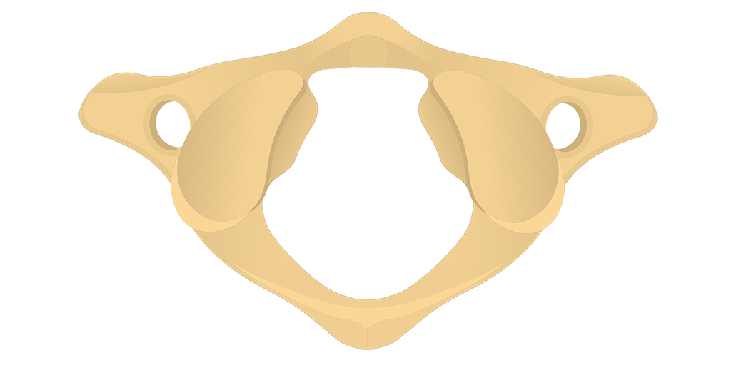
C1 (Atlas)
The first cervical vertebra; articulates with the skull
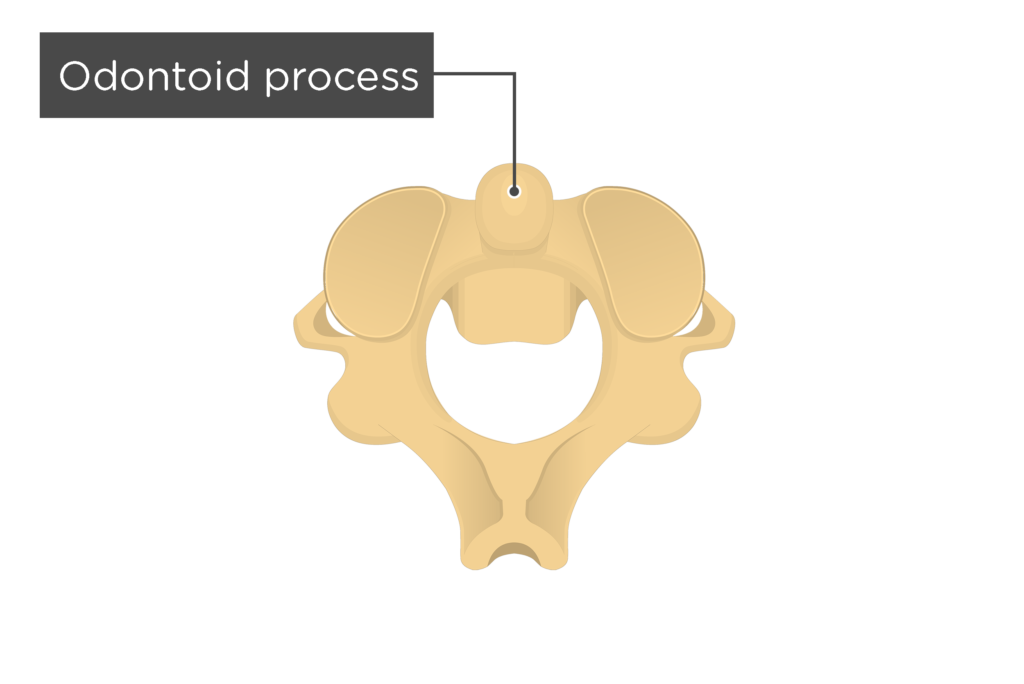
C2 (Axis)
The second cervical vertebra; has the odontoid process (dens)
Thoracic vertebrae (12) — unique feature
Rib facets: transverse costal facets on the transverse processes, and demifacets on the vertebral bodies

Lumbar vertebrae (5) — unique feature
Lack transverse foramina and rib facets

Sacrum (5 fused) — features
Base, apex, sacral canal, sacral foramina, transverse lines, sacral curve, sacral hiatus, sacral cornu, sacral promontory
Coccyx
Several fused vertebrae forming the tailbone, at the caudal end of the vertebral column

Thorax (rib cage) — function
Protects the organs of the chest cavity; provides attachment for muscles involved in respiration and upper limb movements; includes the sternum and ribs
Sternum — features
Manubrium, body, xiphoid process, jugular (suprasternal) notch, costal notches
Ribs — classification
12 pairs total
7 pairs of true ribs
have costal cartilages connecting them to the sternum)
5 pairs of false ribs
first 3 pairs, vertebrochondral ribs, merge with cartilage of the 7th rib
last 2 pairs are floating ribs that attach to thoracic vertebrae and do not connect to the sternum
Rib — features
Head, neck, tubercle, body, sternal end, costal cartilage
Pelvic Girdle (os coxa)
Formed by fusion of the ilium, ischium, and pubis into a single hip bone
Ilium — features
Acetabulum, iliac crest, greater sciatic notch, iliac spines (anterior superior & inferior; posterior superior & inferior)
Ischium — features
Ischial tuberosity, ischial spine, lesser sciatic notch, obturator foramen
Pubis — feature
Pubic symphysis
Femur — features
Head, neck, shaft, greater trochanter, lesser trochanter, linea aspera, medial and lateral condyles, intercondylar notch or fossa
Tibia — features
Head (medial and lateral condyles), tibial tuberosity, anterior crest or margin, medial malleolus
Fibula — features
Head, lateral malleolus
Tarsals
Calcaneus, talus, navicular, cuboid, and the cuneiforms (medial, intermediate, and lateral)
Foot bones (besides tarsals)
Metatarsals and Phalanges
Knee Joint — bones
Distal end of femur, proximal end of tibia, proximal end of fibula, and the patella
Knee Joint — tendons/ligaments
Quadriceps tendon, patellar ligament, tibial collateral ligament, fibular collateral ligament, anterior cruciate ligament, posterior cruciate ligament, and the medial & lateral menisci
Pectoral Girdle
The articulation of the upper arm and the trunk; includes the scapula and the clavicle
Scapula — features
Glenoid fossa (cavity), scapular spine, inferior angle, acromion process, coracoid process, vertebral (medial) border, axillary (lateral) border, supraspinous fossa, infraspinous fossa
Clavicle — features
Acromial end and sternal end
Shoulder Joint — bones
Scapula, clavicle, and head of humerus
Shoulder Joint — ligaments
Acromioclavicular, coracoclavicular, glenohumeral, coracohumeral, coracoacromial
Humerus — features
Head, greater tubercle, lesser tubercle, intertubercular (bicipital) groove, deltoid tuberosity, capitulum (lateral condyle), trochlea (medial condyle), olecranon fossa, coronoid fossa, medial and lateral epicondyles
Radius — features
Head, neck, radial tuberosity, styloid process, ulnar notch
Ulna — features
Semilunar (trochlear) notch, olecranon process, coronoid process, radial notch, styloid process
Carpals — Distal Row
Trapezium, Trapezoid, Capitate, Hamate
Carpals — Proximal Row
Navicular (scaphoid), Lunate, Triquetrum, Pisiform
Hand bones (besides carpals)
Metacarpals and Phalanges