NUR305: Fluid, Electrolyte, and Acid-Base Homeostasis
 Call Kai
 Learn
Learn Practice Test
 Spaced Repetition
 Match
Match Flashcards
 Knowt Play
1/115

There's no tags or description
Looks like no tags are added yet.
Name | Mastery | Learn | Test | Matching | Spaced | Call with Kai | Chat |
|---|
No analytics yet
Send a link to your students to track their progress
116 Terms
Methods of maintaining homeostasis
Fluids are dispersed into various compartments in the body and move among the compartments, Electrolytes and fluids work together, and Acid-base balance.
Acid-base imbalance leads to:
electrolyte imbalance
60-40-20 Rule
Total Body Weight is 60%, Intracellular Fluid is 40 %, and Extracellular Fluid is 20%
Fluid Compartments in the body
moves based on flow concentration gradient, moves until concentration is same on both sides
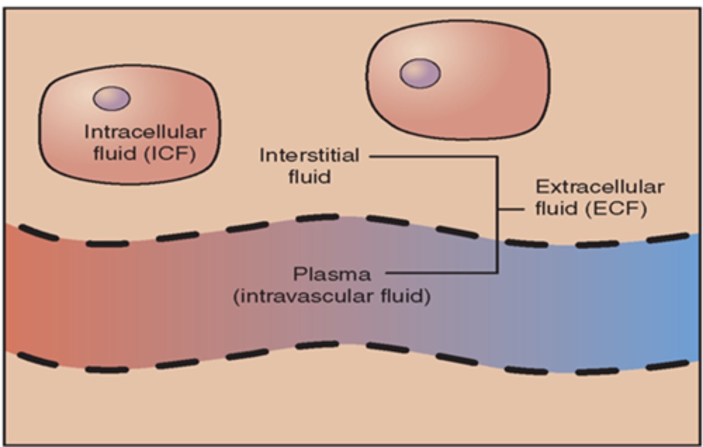
interstitial space
space between cells (edema forms here)
intravascular
inside blood vessels
Intracellular fluid
where most of the fluid is located, Fluid inside the cell, 2/3 of the body's water, and Balance maintained primarily through the cell membrane's permeability.
Extracellular fluid
includes fluid outside of the cell, 1/3 of total body fluid (ex: interstitial fluid, intravascular fluid, cerebrospinal fluid, transcellular fluid)
Intracellular electrolytes
potassium, magnesium, phosphates, sulfates, amino acids
Extracellular electrolytes
sodium, chloride, and bicarbonate, glucose
Transcellular fluids
peritoneal, pleural, pericardial cavities, joint spaces, lymph system , eyes, GI tract, and cerebrospinal fluid.
Osmosis
movement of water across membrane from an area of low solute concentration to an area of high solute concentration.
Oncotic pressure
(exerted by solutes in the cell) pulls fluid into the cell.
Osmotic pressure
specifically the movement of water
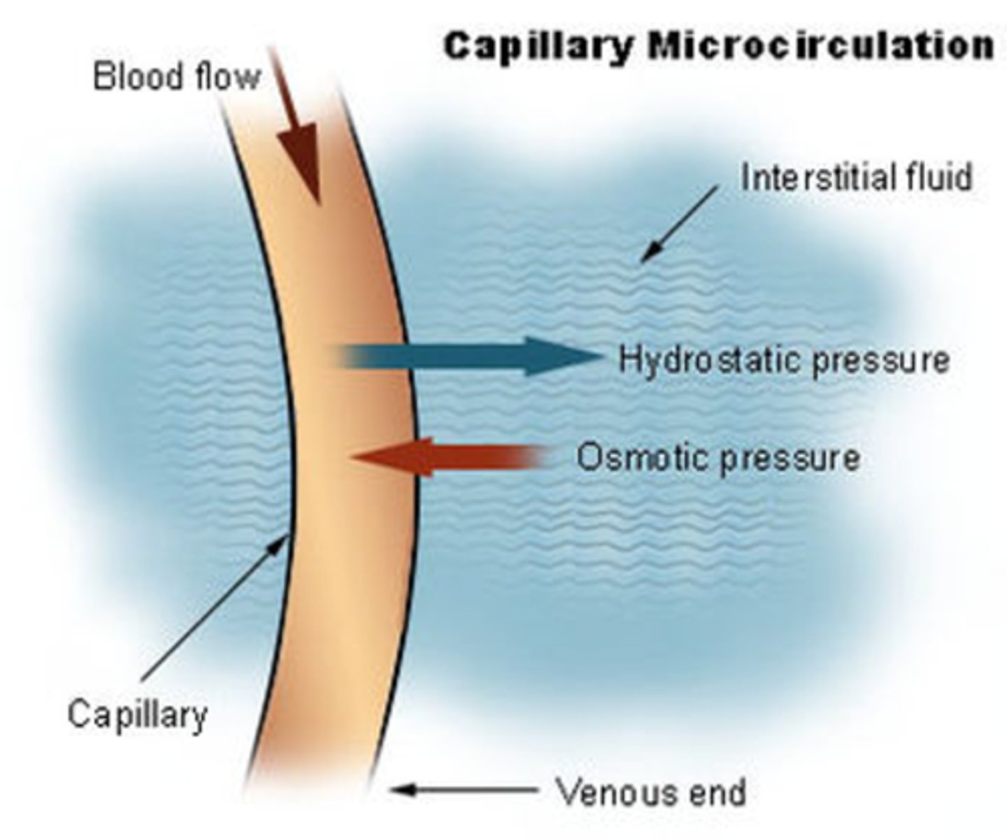
Hydrostatic pressure
the pushing force of fluid in the blood vessels (from heart generating it)
Capillary oncotic pressure
pulls fluid back into the capillaries, fluid is drawn by solutes (proteins, other substance).
Albumin
produced by liver, plasma protein that plays a crucial role in maintaining blood volume and pressure
Osmosality of blood stream
normal range: 275-295 milliosmoles (mOsm), the concentration of particles dissolved in your body fluid
Hypotonic
below 275 mOSm
Hypertonic
above 295 mOsm
Isotonic NaCl
0.9% NaCl (same tonicity as plasma, less will leave the blood stream)
Hypertonic NaCl
3% or above 0.9% NaCl (fluid will shift from cellular and interstitial spaces to the intravascular space)
Hypotonic NaCl
0.45% NaCl or below 0.9% (would hydrate the cells and interstitial spaces)
Dehydration causes
hypotension, tachycardia, dizziness, fatigue
Fluid are added to the body by
ingestion of foods and liquids
Fluids are lost from the body by
urine and feces
Fluids are also lost by insensible (immeasurable) methods
perspiration (sweating) and the respiratory tract (coughing, fast breathing)
Control of fluid balance is maintained by:
the hypothalamus, posterior pituitary gland, adrenal glands, kidneys, and the heart
Hypothalamus
thirst regulation, the thirst control center
posterior pituitary gland
anti-diuretic hormone (ADH), keeps fluid from all going into the urine
adrenal gland
aldosterone
kidneys
renin-angiotensin-aldosterone (R-A-A) mechanism (powerful vasoconstrictors)
the heart
atrial natriuretic peptide (ANP), counteracts the aldosterone and RAA mechanism
SIADH
too much ADH, causing too much fluid in the blood
diabetes insipidus
to little ADH, causing too little fluid in blood
Aldosterone
A hormone released to conserve water when needed. Acts to cause sodium and water reabsorption in the renal tubules
Atrial Natriuretic Peptide
A hormone released when the atria of the myocardium is overstretched. indicates increased fluid volume. (opposite of aldosterone and RAA) Stimulates renal vasodilatation to increase urine output.
Osmoreceptor cells
in the hypothalamus, sense changes in fluid volume status. Decreased fluid volume (increased osmolarity) triggers our thirst mechanism.
Hypovolemia
decreased blood volume, major physiologic stimulus for thirst
ADH (antidiuretic hormone)
Released by the posterior pituitary. Helps to regulate fluid volume by controlling water losses. Promotes reabsorption of water from renal tubules into the blood.
Fluid Intake
Thirst regulates fluid intake, Intake is about 2200 to 2700 ml/day (24 hours)
Hormonal regulation of bodily fluids
Antidiuretic hormone, Renin-angiotension-aldosterone mechanism, and Atrial natriuretic peptide
Fluid Output
Fluid is lost through kidneys, skin, lungs, and GI tract, Insensible loss: skin, lungs, and perspiration
Fluid Excess/Overload problems
Edema, Hypervolemia, Water intoxication
Edema
excess fluid in the interstitial space, Can be localized in one area such as in the feet, and Can be generalized throughout the body (anasarca).
Hypervolemia
excess fluid in the intravascular compartment; aka. fluid volume excess.
Water intoxication
fluid excess in the intracellular space; can lead to rupture of the cells
Causes of Edema
Loss of proteins in the plasma (some cause: liver disease, burns, poor nutrition, blood loss, Hemorrhage), Leaky capillaries (vasodilated), and fluid increase (change in osmolarity), or too much sodium and water kept by kidneys. (could be too much ADH)
Edema Formation
An alteration in capillary hemodynamics that favors the movement of fluid from the vascular space into the interstitial area.
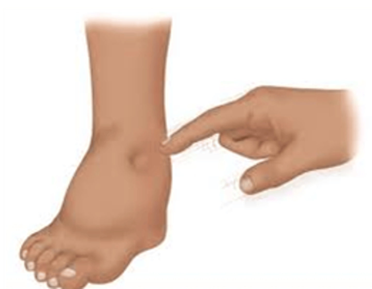
Peripheral edema
skin dents with pressure (pitting)
Periorbital edem
swelling around the eyes

Cerebral edema
fluid in the brain
Symptoms of more severe edema
Dyspnea (feel like they can’t breathe), Tachypnea (more than 20 respirations per minute), Bounding pulse (very strong pulse), and Tachycardia: (higher than 100 beats per minute)
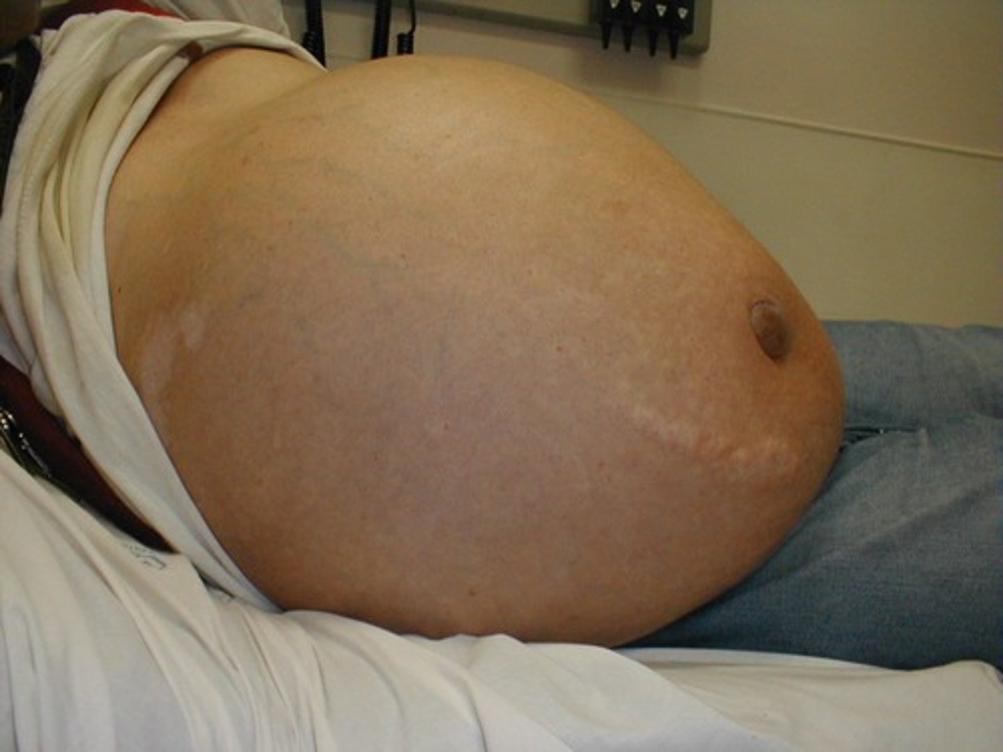
Ascites (third spacing)
accumulation of fluid in the peritoneal cavity
Jugular vein distention
A visual bulging of the jugular veins in the neck that can be caused by fluid overload
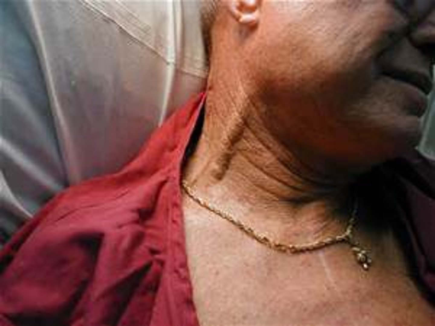
Clinical Manifestations of Fluid Excess
Jugular vein distension, Hypertension, Polyuria (lots of urine), Rapid weight gain (one of the best methods to assess fluid status), and Crackles (fluid in the airways of the lungs)
Diagnosing Fluid Excess
Urine specific gravity below 1.010 (low: solution diluted, high: solution concentration high), Urine & blood osmolality, Complete Blood Count (CBC)
Treating fluid excess (starting with least invasive)
Administering medications (diuretics), Restricting sodium and fluids, and Sit the patient up in bed, so they can breathe (high Fowler’s), Giving hypertonic fluids (last resort, Shifts the excess fluid to the intravascular space so that it can be excreted)
Fluid Deficit
Dehydration, Occurs when the total body fluids are insufficient to meet the body’s needs, Hypovolemia, As fluid volumes decrease, levels of blood cells and electrolytes will increase due to the hemoconcentration.
Causes of fluid deficit
Inadequate fluid intake: Poor oral intake or Inadequate IV fluid replacement, Excessive fluid losses: (GI losses, Excessive diaphoresis, Hemorrhage, Diabetes mellitus: high glucose levels + high H2O loss and Diabetes Insipidus: not enough ADH)
Clinical Manifestations of fluid deficit
thirst, hypotension, tachycardia, weak thready pulse, dry mucous membrane, wrinkled tongue, decreased skin turgor, oliguria (scanty urine, small amounts), weight loss, altered state of consciousness, anuria (no urine), dry skin
Diagnosing fluid deficit
history and physical, measure accurate intake and output, daily weight (very effective), lab work (CBC: blood cells high, blood specific gravity: high, BUN: high)
Treating fluid deficit
identify underlying cause, treat the cause, replace fluid (oral for mild loss, IV for greater loss: isotonic or hypotonic)
Electrolytes
electrically charged minerals that are found in Blood, Urine, Other body fluids (sodium, potassium, chloride, calcium, magnesium, phosphorus)
Ion Exchange in the Kidneys
the body is trying to balance the positive (cations) and negative (anion) ions
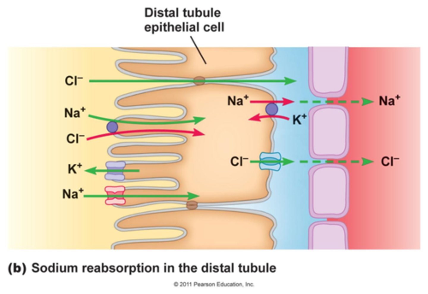
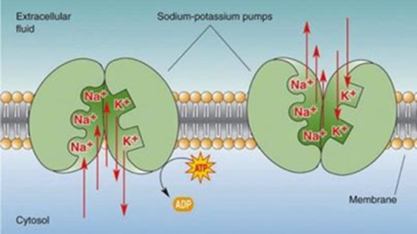
Sodium Potassium Pump
across cell membranes, generates
action potential, transport substances
Sodium
most significant cation (135-145 mEq/L), most prevalent electrolytes of extracellular fluid, likes chloride, regulated by kidneys and aldosterone
Primary function of sodium
Control serum osmolality and water balance, Help maintain acid-base balance when combined with HCO3 (bicarbonate), and Facilitates muscle and nerve impulses via the sodium-potassium pump.
Sodium Intake and Loss
Obtained primarily via dietary intake, Losses: The kidneys, Vomiting & diarrhea, Nasogastric tube suctioning (NGT), Extensive burns, and in excessive sweating (heat exhaustion and stroke)
Hypernatremia
sodium greater than 145 mEq/L, water shifts from the intracellular and interstitial spaces to the intravascular compartment.
causes of hypernatremia
Excessive Sodium: Excessive ingestion, Hypertonic IV administration (usually high salt), and Cushing’s syndrome: cortisol & aldosterone excess. Deficient Water: Decreased ingestion, Inability to drink water, or Too much output: Vomiting/diarrhea, Diuretic use, Diabetes insipidus
Clinical manifestations of hypernatremia
increased temperature, warm flushed skin, increased thirst
Hypernatremia affect on Body systems
Neurological: Irritability and agitation, Headache, Seizures, Lethargy, and Coma. Cardiac: Tachycardia, and a Weak, thready pulse. Decreased urine output (with DI would have high urine output)
Diagnosing Hypernatremia
history and physical, Lab (blood and urine), CT/MRI (looking for cause)
Cannot correct hypernatremia too quickly:
Brain can become accustomed to high sodium and as the sodium level drops, water moves into the cerebral cells causing cerebral edema. (Seizure precautions and Neurologic checks)
Hyponatremia
sodium less than 135 mEq/L, water shifts into the brain cells, causing cerebral edema. Nerve conduction also becomes impaired as sodium levels fall. Also can result from increased water gains (dilutional hyponatremia).
Causes of Hyponatremia
Deficient Sodium: diuretic, GI loss, excessive sweating, insufficient aldosterone levels, adrenal insufficiency (low cortisol/Addison's disease), diet. Excessive Water: Hypotonic IV, hyperglycemia, excessive water ingestion, renal failure, SIADH (too much ADH), heart failure (cannot remove fluid)
Clinical Manifestations of hyponatremia
poor skin turgor, edema, Neurological: anorexia, headache, lethargy, seizures, confusion, coma. Muscular: muscle weakness, diminished deep tendon reflexes
Treating hyponatremia
treat underlying cause, (Excessive water, oral intake may be limited, Efficient sodium, oral intake may be increased) must be corrected slowly (Could overload the heart if replace sodium too quickly. Seizure precautions, neurological checks)
Chloride (Cl-)
95-105 mEq/L, major extracellular anion, abundant in cerebrospinal fluid where it binds to sodium (water balance), Absorbed in the colon, Kidneys are primarily responsible for chloride excretion, Diet is the main source of chloride
Chloride Function
Assist in fluid distribution by attaching to sodium or water, Found in gastric secretions, pancreatic juices, and bile, and Unites with H+ in the stomach and forms HCL (acid-base balance)
Hyperchloremia
greater than 105 mEq/L, Increased chloride intake: Hypernatremia, Hypertonic IV solution, Metabolic acidosis. Decreased chloride excretion: Hyperparathyroidism, Hyperaldosteronism, Renal failure
Diagnosing and Treating Hyperchloremia
Diagnosing: Obtain history, Perform physical assessment, Lab work: Urinalysis and Arterial blood gases (looking for metabolic acidosis). Treating: Correct the underlying cause, May give diuretics, and May give bicarbonate.
Hypochloremia
less than 95 mEq/L, caused by Decreased chloride intake: Hyponatremia or Water intoxication. Increased chloride excretion: Diuretics, Vomiting or Other gastrointestinal losses (loses sodium, therefore chloride)
Potassium
Normal range 3.5 -5.0 mEq/L, primary intracellular ions, play crucial role in electrical conduction, acid-base balance, and metabolism. Regulated by: Sodium-potassium pump and Kidneys. (Diet is the primary source, Can affect muscle and nerve cells, and cardiac rate, rhythm, and contractility)
Hyperkalemia
greater than 5.0 mEq/L, unusual, may be medical emergency, can be caused by: Impaired secretion of potassium, Increased intake of potassium (oral supplements, salt substitutes, rapid intravenous administration), and A release of potassium out of the cell.
built-in mechanisms that try to prevent hyperkalemia
Removing potassium from the blood after a meal, Vomiting, and Excretion by the kidneys.
electrolytes lost through NG suctioning:
potassium, sodium, and chloride
Hyperkalemia by Deficient Excretion
Renal failure, Addison’s disease – decreased levels of aldosterone decreases potassium excretion, Medications, an other Rare genetic disorder
NEVER give potassium IV
undiluted (MUST be diluted) can cause pain, necrosis, etc.
Hyperkalemia by release of potassium from cells
caused by acidosis (Increase in serum hydrogen, H+, levels going into the cell cause potassium to shift out of the cells), Blood Transfusions: Cause blood cell lysis which releases the intracellular potassium, and Burns or other injuries: causing major cellular damage can also cause cell lysis and thus the releasing of intracellular potassium.
Clinical Manifestation of Hyperkalemia
Cardiovascular: bradycardia, dysrhythmias, EKG changes – peaked T-wave, & cardiac arrest. Neuromuscular: muscle weakness, paresthesia, flaccid paralysis & respiratory depression (diaphragm muscle). Gastrointestinal: abdominal cramping, nausea, & diarrhea.
Treating Hyperkalemia
May give sodium bicarbonate to treat acidosis, (Need to treat the acidosis first, potassium will shift back into the cell and you can obtain a true potassium level). Calcium gluconate may be given to minimize dysrhythmias. May also give insulin to facilitate cellular exchange of H+ and K+. Increase excretion: dialysis, medications (kayexalate , diuretics), IV fluids
Hypokalemia
less than 3.5 mEq/L, usually results from excessive loss, inadequate intake, or increased potassium cellular uptake. Gastrointestinal loss: vomiting, diarrhea, nasogastric suction, laxatives. Urinary loss: diuretics (increase urination)
Causes of Hypokalemia
endocrine disorders (Cushing's syndrome: decreases sodium excretion which therefore will increase potassium excretion), certain medications, deficient intake: malnutrition, extreme dieting, alcoholism. Increased potassium cellular intake: Alkalosis and Insulin excess
Low potassium can be just as detrimental as high potassium and can affect:
numerous body systems such as the nervous, cardiac, respiratory, and GI systems .
Clinical manifestation of hypokalemia
Life threatening: Cardiovascular effects: dysrhythmias, EKG changes, CARDIAC ARREST. Neuromuscular: leg cramps, muscle weakness, paresthesias (numbness, sharp, shooting pain), hyporeflexia. Gastrointestinal: decreased bowel sounds, abdominal distension, constipation.
Calcium
Normal range (total Ca+ + 8.5 – 10.5 mg/dL), most is bound to albumin (60%), Vitamin D aids absorption of calcium, found in bones and teeth, excreted in urine and stool, regulated by: parathyroid hormones and calcitonin (secreted by thyroid C cells).
Extracellular Calcium Controls Nerve Firing:
Hypocalcemia: nerves fire more easily. Hypercalcemia: nerves are less able to fire
Hypercalcemia
greater than 10.5 mg/dL, caused by Increased intake: Cancer (especially bone cancer), Immobilizations, Corticosteroids, Vitamin D deficiency, and Hypophosphatemia. Poor excretion: Renal failure, diuretic, and Hyperparathyroidism