Liver Pathology Case Seminar
 Call Kai
 Learn
 Practice Test
 Spaced Repetition
 Match
 Flashcards
 Knowt Play
1/13

There's no tags or description
Looks like no tags are added yet.
14 Terms
What pathological process causes "nutmeg liver" in chronic, restrictive pericarditis?
Passive hepatic venous congestion. Causes hypoxia and hepatocyte degeneration which is most prominent centrilobularly.
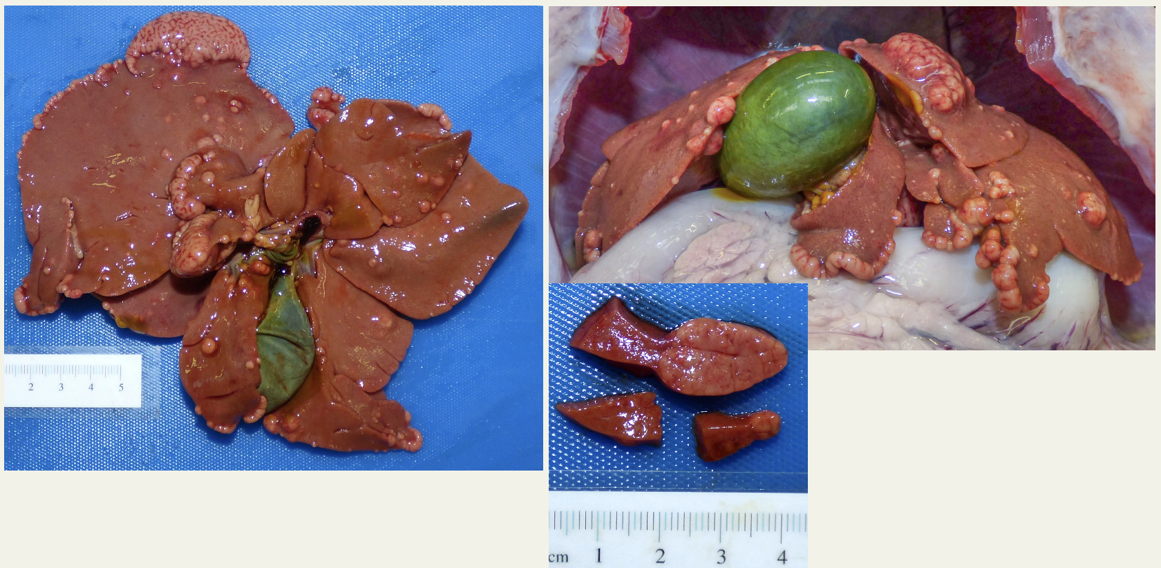
Briefly describe one or two of the main gross pathological features that can be seen affecting this liver.
The liver is tan-brown, and bulging from the capsular surfaces randomly and frequently, along lobe margins, are numerous (50-100) often coalescing, smooth to cerebriform (brain-like), cream to cream-red nodules, ranging from 1-2mm diameter up to approximately 4cm by 2cm diameter. Sectioned surfaces of the liver are smooth and tan-brown with paler brown-cream nodular foci. The gallbladder is moderately distended.
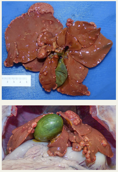
What pathological process or feature might nodular changes represent in this dog's liver?
Nodular regeneration or hyperplasia, neoplasia, or nodules associated with chronic inflammation, e.g. abscesses or granulomas.
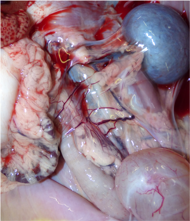
What might these prominent vascular structures in the mesentery represent?
Acquired portosystemic shunts
What pathophysiological change could account for ascites and prominent mesenteric vessels?
Portal hypertension
What liver changes best explain a nodular liver, ascites and prominent mesenteric vessels in a 7-month-old dog?
Chronic hepatitis with fibrosis and nodular regeneration. Fibrosis causes portal hypertension which explains the ascites and suspected acquired shunts.
What single diagnostic test would be most useful to investigate suspected chronic hepatitis with fibrosis and nodular regeneration in a dog?
Liver biopsy for histopathology. It allows the liver's architecture and pathological changes to be examined.

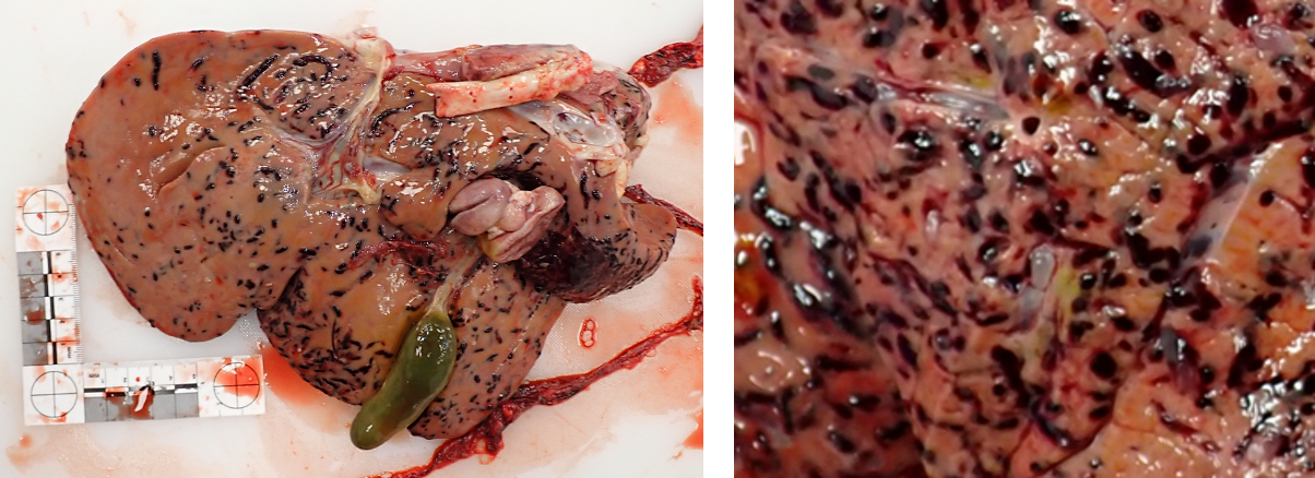
Briefly describe one or two of the main gross pathological features that can be seen affecting this liver.
The liver is light brown with, extensively through the parenchyma, and bulging on the capsular surfaces, innumerable, randomly distributed, dark red-black, round and cylindrical, linear to tortuous foci/tracts, up to approximately 2cm long by 3 to 4mm wide/diameter. Singly and grouped on the capsular surface are approximately 15, piriform (pear-shaped), smooth, translucent, dull white-grey, cystic structures up to approximately 5x3x3mm.

What pathological processes could the dark red-black liver foci and tracts in this lamb represent?
Multifocal random necrosis ± inflammation is most likely. Multifocal haemorrhage , thrombosis or infarction also possible.

Name one parasite that could be a potential cause of the liver lesions in this lamb.
Fasciola hepatica (liver fluke)
If this lamb’s lesions were caused by Fasciola hepatica, would would the disease be classified?
Acute fascioliasis. Immature flukes migrate through the liver parenchyma during the acute phase.
Besides liver fluke, name a parasite of sheep in the UK that can be found in the liver.
Taenia hydatigena. Its larval stage is called Cysticercus tenucollis.
The lambs died in early April, and the liver had surface cysts about 5mm across. The case involved a flock with six farm dogs that had access to the pasture and were not wormed. Based on this context, which parasite is most likely to have caused their liver lesions?
Taenia hydatigena. Early April did not fit the expected timing for acute fascioliasis, which typically follows pasture exposure later in the year. The 5mm surface cysts were also much larger than expected for Fasciola metacercariae. Taenia hydatigena larve migrate through the liver, leave tracts, then form cysticerci on the liver surface or in the peritoneal cavity.
The case involved a flock with six farm dogs that had access to the pasture and were not wormed. What advice could help prevent disease caused by Taenia hydatigena in the flock?
Regularly worm the dogs and reduce contamination of pasture with dog faeces. Dogs are the definitive host of Taenia hydatigena and can shed eggs onto pasture.