MCAT BIO
 Call Kai
 Learn
Learn Practice Test
 Spaced Repetition
 Match
Match Flashcards
 Knowt Play
1/116

There's no tags or description
Looks like no tags are added yet.
Name | Mastery | Learn | Test | Matching | Spaced | Call with Kai | Chat |
|---|
No analytics yet
Send a link to your students to track their progress
117 Terms
what is the cell
a compartmentalized, organized, and well-oiled machine that can undergo various life processes
compartmentalized: split into organelles (eukaryotes)
prokaryotes are a soupy mix of genetic information and life-producing capacity
organized: each process occurs in a controlled manner (positive & negative feed backs)
well-oiled: processes are fluid, flows from one to the next
what is the cell theory
all living things are made up of cells
cells are the basic functional unit of life
cells form from pre-existing cells
cells carry genetic info (DNA!) that they pass onto daughter cells
what makes a virus not living?
it breaks rules 3 and 4 of the cell theory
a virus cant reproduce on their own and need a cell host
a virus cant pass on genetic code to their daughters; but rather, must inject the code into cell hosts which are naturally programmed to run it through the cell cycle
the injected genetic material then either lays dormant within the cell (lysogenic) or spreads throughout the body (lytic)
why do cells only pass on DNA to daughter cells and not RNA
RNA is meta-stable
it closes in on itself by folding and is unable to be used after a while
RNA is unable to be passed on
only viruses can pass it on, not eukaryotes or prokaryotes
what are the main differences between eukaryotes and prokaryotes
EUK (eukarya)
has membrane-bound organelles
can be single (yeast) and multi-cellular (humans)
PRO (bacteria & archaea)
essentially one big organelle
no nucleus
only single celled
what is the nucleus
control center (HQ) of the entire cell
contained inside a semi-permeable double membrane
sign of importance; protection
has nuclear pores which allow for communication and travelling in and out of the cell
has tightly wound DNA inside (negative charged)
this is wrapped around histone proteins
made up of amino acids (basic building blocks of proteins) that have +1 charged R groups
what is the difference between heterochromatin and euchromatin
Heterochromatin
compact/tight form of DNA
Euchromatin
loose/relaxed form of DNA
Histone Acetylation
the act of neutralizing a positive histone protein by attaching an acetyl group to each of its terminals
this undoes the tightly wound DNA which needs to happen before replication of the cell
hetero → eu
what is the mitochondria
the powerhouse of the cell where almost all the effective energy of the cell (ATP) is produced by the electron transport chain (ETC)
has its own genes and is able to self-replicate
has an outer and inner membrane (double protected)
contains cristae
grooved folds of the inner membrane that increase surface area for ETC to work with
each groove fits more proteins for energy (cellular respiration)
coastline paradox
is able to kickstart apoptosis
the controlled programming of cell death when something goes wrong
different from necrosis which is uncontrolled
what are lysosomes
organelles that contain many hydrolytic enzymes used to break down material
lysozymes~ recycling centers
these enzymes are different from vesicles which are like boxes used to transport materials
derived from “lysis” meaning to break
what are the smooth and rough endoplasmic reticulums (SER & RER)
ROUGH
studded with ribosomes
factory machines for protein synthesis
necessary for EVERYTHING
SMOOTH
lacks ribosomes
helps transport proteins from RER to Golgi
synthesizes lipids (fats/oils/waxes/etc.)
detoxes drugs by making them hydrophilic (attracted to water)
primary site for first pass metabolism which is the processes of allowing toxins to be dissolved in the blood in order to be excreted through the kidneys as urine
liver’s job
what is the golgi apparatus
stack of membrane-bound “sacs”
modify proteins to make them more efficient in their roles
“cellular surgery”
packages molecules into vesicles which later merge with the cell membrane during exocytosis
what is the peroxisome
organelle filled with hydrogen peroxide (H2O2) in order to break down material
similar to lysosomes but it doesn’t use enzymes
a natural antibiotic/antiviral/anti-toxin
H2O2 is often used as a disinfectant, antiseptic, or bleaching agent
what is the endomembrane transport
well-oiled, fluid process of the cell:
mitochondria and nucleus work together to give the cell energy and instructions necessary for producing life →
MRNA produced from transcription in the nucleus is send to the RER →
RER translates MRNA into proteins and sends them through SER →
SER transports proteins to the golgi →
Golgi modifies and packages proteins into membrane-bound vesicles →
sacked vesicles fuse into cell membrane and proteins go through exocytosis
Exocytosis
the process by which something inside the cell will leave the cell
Endocytosis
the process by which something outside the cell will come into the cell
what is the difference between microfilaments, microtubules, and intermediate filaments
microfilaments
made of actin whose primary job is to interact with myosin to induce muscle contraction
help with cell movement, contraction, and shape changes
make the cleavage furrow during cytokinesis
the separation of daughter cells during mitosis/meiosis
microtubules
made of tubulin
involved in the synthesis of cilia and flagella
ex: respiratory tract and sperm cells
make up the centrioles and help them move to opposite sides of the cell during cell division
separation creates the mitotic spindle
intermediate filaments
rope-like protein fibers that absorb tension and help anchor organelles
what are tissues
organized sheets of cells
listed in order of increasing regenerative ability:
epithelial
muscle
connective
nervous (neuron)
what are epithelial tissues
covers the body and lines its cavities
cavities= holes (inner ear, nose, etc.)
involved in absorption, secretion, transportation, and sensation
most organs are lined with epithelial tissue
make up the parenchyma
the functional parts of any organ/gland
what are connective tissues
create the framework for other things to rest and function on
most cells here produce and secrete materials like collagen (strength) and elastin (flexibility) which help make up the extracellular matrix (ECM)
complex 3D network of proteins that provides structural support for cells and tissues
ECM lies within connective tissues and gives it the strength and flexibility it needs to do its job
lots of ECM = proper connective tissue
examples: bone/cartilage/tendons, small intestines, blood
even though it transports, blood is made of fluid connective tissue and not epithelial because it’s made of an ECM → plasma
blood vessels (arteries/veins), however, are lined with epithelial tissue
bacteria classifications
cocci
sphere shaped
s. aureus
gram neg or pos
bacilli
rod shaped
myobacterium tuberculosis
mostly gram pos
spirilli
spiral shaped
treponema palidum (syphillus)
only gram neg
what are the kinds of cell walls in prokaryotic cells
thick peptidoglycan layered
gram positive bacteria
stain purple or blue
staphylococcus pneumonae
thin peptidoglycan layered
gram negative bacteria
contains lipopolysaccharide (LPS) which causes much harsher infections in the body due to being immunogenic (activates and flares up the immune system)
stains red or pink
salmonella
key facts about prokaryotic cells
have a cell wall NOT a membrane
often have flagella that operate as motors for transportation
filament at the tip of the flagella rotates from the base and generates torque
have a nucleoid region for compartmentalization
NOT a nucleus
can transfer and acquire DNA through plasmids
small rings of DNA (separate from actual DNA) that hold information in nucleoid region
episomes are autonomous plasmids capable of inserting themselves directly into main bacterial DNA
reproduce asexually
what are the ways prokaryotes can reproduce
Binary Fission
splitting into clones of themselves (SpongeBob)
Horizontal Gene Transfer
the process of bacteria acquiring new genetic material
Genetic Recombination is essential for HGT
cells break down, exchange, and rejoin DNA segments to increase genetic variation and repair damage
crucial for adaptation and evolution
Types of HGT:
Transformation
bacteria picks up elements of another bacteria when it dies and releases its insides
Transduction
when a virus (bacteriophage) accidentally transfers the genetic material from one bacteria to another
Conjugation
bacteria passes on genetic material via a mating bridge which enables the acquirement of new traits like antibiotic resistance
bacteria with fertility factor plasmids (F+ bacteria) can form the conjugation bridge while those without (F-) cannot
this conjugation bridge is a “sex pilus” that allows the transfer of copied plasmids to other bacteria so an F- bacteria can become F+ and continue the cycle!
F+ cannot mate with F+ because it would be ineffective
EVERYTHING has a purpose!!
explain the life cycle of bacteria
bacteria have lag, exponential, plateau, and death phases
they are born into an environment with limited resources (lag)
they must build up their resources (exponential), then eat through them (plateau) until there isn’t any more to consume (death)
the rate of growth = the rate of death in this cycle
example of biological equilibrium
reason for plateau phase
what are the parts of viruses and sub-viral particles
Protein Capsid
protective outer coat made of proteins
encloses and transports viral nucleic acid (DNA/RNA)
can be helical or polyhedral shaped
Genome
either DNA or RNA
if virus has DNA, host cell does all the work and replicates, transcribes, and translates viral genome through cell cycle
if virus has RNA, it must bring its own enzymes:
RNA-dependent RNA polymerase used to help a virus make its own RNA copies for host ribosomes to make into proteins
Reverse transcriptase used by retroviruses to turn their RNA into DNA host can work with
lytic or lysogenic reproduction
Spike Proteins
glycoproteins that facilitate host entry and infection through penetration
determine tropism: method by which a virus recognizes a host cell to infect with the specificity of a key to a lock
help virus evade host immune system
SOME have a lipid envelope
a phospholipid bilayered membrane stolen from host cells
able to enter host cells through fusion
what are viruses and what are the “special” viruses
intracellular parasites that obligate (force) a living host cell for reproduction
Bacteriophages
viruses that only target bacteria
can perform transduction
Retroviruses
use reverse transcriptase to change and integrate themselves into a host cell’s genome
famous retrovirus is HIV
Viroids
tiny viruses that infect plants
lack a protein coat
what is a superinfection
a second, overlapping infection that’s often caused by
microorganisms resistant to treatment given to combat the first infection
antibiotic treatment killing off protective microflora, allowing new pathogens to strengthen
example:
s. aureus is commonly caused by a post influenza infection and extremely dangerous due to immune impairment and lung damage that allows pneumonia to develop quickly
what is endocrinology
study of the endocrine system
a series of glands, hormones, effectors, and pathways that mainly regulate long-distance and long-term communication within the body
essentially acts as a carrier pigeon: gland → circulatory system (bloodstream) → target
the goal is homeostasis: resisting changes to maintain a stable environment
glands
secretory tissues or organs within the body that respond to various stimuli
adrenal glands, pituitary glands, etc.
hormones
a molecule or protein that has the capacity to bind receptors and influence responses on the cellular/tissue/organ level
the thing that is released from glands into the bloodstream
dopamine, cortisol, insulin, etc.
effectors
structures that act in response to stimuli
liver responds to insulin and increases glucose uptake
pathways
complex, interconnected, signalling networks in which glands release hormones into the bloodstream to function
HPT axis, HPG, HPA, etc.
why does the endocrine system exist
we are not single-celled and need a way to communicate between the furthest of cells
the system allows us to:
hijack communication pathways
relay signals to specific places and get them there in time
have the signaled message stay long enough to maintain a response/effect
the body is very needy!
it wants what it wants, when it wants, and in the amounts it wants it in
what is the tropic effect
when a hormone is unable to perform a job on its own and employs a secondary messenger to regulate the response
example:
the growth hormone (GH) exhibits its effects through linear growth (height) and is released from the anterior pituitary gland (APG). In order to release it, the APG is stimulated by the growth hormone releasing hormone (GHRH)
A → B → response
A is tropic, B is direct
what is the hypothalamus
the control center (HQ) of the endocrine system
the pituitary gland serves the hypothalamus as a henchman
split into two main sections: anterior and posterior
posterior:
the hypothalamus produces oxytocin and ADH and sends them via long nerve fibers
anterior:
the hypothalamus produces releasing and inhibiting hormones as signals
anterior hormones are its own!
describe the anterior pituitary gland
front lobe of the pituitary gland that is identified as endocrine acting
this is because it secretes hormones directly into the bloodstream
connects to the hypothalamus through a network of blood vessels
hypophyseal portal system
made up by the hypophyseal portal vein
supplies releasing and inhibiting signals from the hypothalamus and blood to the APG
nicknamed the “master gland”
produces 7 key hormones (GH, FSH, LH, etc.)
describe the posterior pituitary gland
back lobe of the pituitary gland that is identified as neuroendocrine acting
a.k.a neurosecretory~ essentially “one giant neuron”
routinely shocked by the hypothalamus with nerve endings that cross over and drop electric signals
shocks cause hormone secretion
only releases 2 hormones
oxytocin and ADH
what are the types of blood vessels and their functions
Veins
carry oxygen-poor blood toward the heart
Arteries
carry oxygen-rich blood away from the heart
Capillaries
connect veins and arteries
facilitate nutrient and waste exchanges
what is a portal vein
a vein that circulates between two organ systems or tissue beds
helps identify toxins before they enter the bloodstream
carries to the liver for detoxing
contains little hormone molecules
what are the different categories for identifying hormones
By Effect
Direct
does the deed
ex: T4 accelerates cellular metabolism
Tropic
gets a guy (usually an organ) to do the deed
ex: TSH smacks the thyroid to make T4 accelerates cellular metabolism
Double-Tropic
gets a guy to get a guy to do the deed
ex: GnRH stimulates the APG into producing FSH and LH which act on endocrine gonads to produce estrogen, progesterone, and testosterone (1° sex hormones)
By Structure
Steroid Hormones
a derivative of cholesterol via reaction
ex: -one, -ol, -oid family (cortisol, progesterone)
mainly non polar hormones
allows them entrance into cell through membrane so they bind to an intracellular steroid receptor, forming a complex that travels into the nucleus and changes transcription (DNA → RNA) of DNA
complex is a transcription factor
Amino Acid Hormones
very polar and unable to enter through the cell membrane
must bind a receptor from outside, leading to a signal transduction cascade which leads to a hormonal effect
cascade modifies transcription in a similar way to steroid hormones
ex: T3, T4
Peptide Hormones
polypeptides (amino acid chains)
protein-derived
MUST be polar and MUST bind to a receptor
ex: insulin, ghrelin
describe the mitosis cell cycle
a series of phases that eukaryotic cells undergo in order to reproduce
DNA is replicated and split, and the cell divides into 2 identical daughter cells
mitosis only happens to somatic, diploid cells
somatic ~ also called autosomal; body cells with uninheritable mutations
diploid~ have 2 of the necessary genetic material to code for the genome (2n; n=23)
split into 4 phases:
Mitosis
cell is actively dividing
everything else is interphase (preparation for mitosis; G1, S, G2)
individual chromosomes are not visible during interphase due to DNA being in a less condensed state (loose/unwound) to get ready for replication
G1 (pre-synthetic gap)
growth/bulking stage where the cell size increases in preparation for replication
includes a checkpoint before S phase
ensures cell has proper amounts of, type of, and accommodations for the DNA
S (synthesis)
cell replicates genetic material so that each daughter has identical DNA
after replication, each chromosome consists of 2 sister chromatids bound by a centromere
still 1 chromosome
checkpoint occurs before G2 phase to make sure replication is correct
G2 (post-synthetic gap)
another bulking stage with more cell growth, copying of organelles, and protein synthesis
this is the time for any damaged DNA to be repaired before mitosis
another checkpoint
G0
where cells that are not dividing go to carry out functions (chill)
what are the stages of mitosis
Prophase
nuclear envelope dissolves to break the barrier between nucleus and cytoplasm
genetic material condenses again and chromosomes become visible
centrioles form and start to move away
Metaphase
chromosomes line up at the center of the cell (metaphase plate)
centrioles are now at opposite poles of the cell
Anaphase
centrioles begin throwing spindle fibers made of microtubules at the chromosomes, causing sister chromatids to separate from each other
Telophase
spindle apparatus disappears
nuclear membrane reforms
chromosomes uncoil
cleavage furrow begins to form
Cytokinesis
usually begins as mitosis is ending (may have a little overlap)
cleavage of the cytoplasm into 2 daughter cells
what is the evolution of chromosomes during mitosis
chromosomes are counted by centromeres so you can have 1 chromosome with 2 chromatids
chromatid~ 2n, single stranded
sister chromatids~ 2n, double stranded
1 cell with 1 chromosome, 1 chromatid → copying of DNA in synthesis of interphase →
1 cell with 1 chromosome, 2 sister chromatids → chromosome separation in anaphase of mitosis →
2 cells with 1 chromosome, 1 chromatid each after cytokinesis
describe the meiosis cell cycle
a series of phases that non-somatic, eukaryotic cells undergo to reproduce
occurs only in and produces haploid cells
gamete cells (1n; n=23)
sperm or egg (sex) cells
single stranded
enough to code for another genome (23 from mother (XX), 23 from father (XY))
2 haploids = 1 diploid which sustains its life through mitosis
involves crossing over
follows the same general formula as mitosis
what are the stages of meiosis
Meiosis I
starts with prophase I which is the same as mitosis prophase but it involves crossing over/ recombination
the exchange of genetic material between non-sister chromatids of homologous chromosomes by way of touching and fusing with each other
outside of spontaneous mutations, this process is responsible for all the genetic variation in humans
metaphase I is the same as mitosis metaphase
anaphase I is the same as mitosis anaphase but the homologous chromosomes are pulled apart
telophase I is the same as mitosis telophase but the end product is 2 haploid cells with their chromosomes still condensed
Meiosis II
happens right after meiosis I
the exact same as the mitosis cycle but with 1 single chromosome
what is the evolution of chromosomes during meiosis
chromosomes are counted by centromeres so you can have 1 chromosome with 2 chromatids
chromatid~ 2n, single stranded
sister chromatids~ 2n, double stranded
46 double stranded chromosomes, 92 chromatid → pulling apart of homologous chromosomes
23 double stranded chromosomes, 46 chromatid after telophase I → meiosis II
23 single stranded chromosomes having 1 chromatid each as end products
what are non-disjunction errors
problems that arise from incorrect meiosis and lead to of several diseases
create specific changes in sex chromosomes (23rd pair)
typically only affect females
women have all the ova they will ever create at birth (frozen in time since fetuses)
happen primarily during meiosis I when homologous chromosomes fail to separate correctly
leaves a haploid cell with an abnormal # of chromosomes after meiosis is completed
1 extra~ trisomy
missing~ monosomy
what happens in cases of trisomy or monosomy of the 23rd chromosome
Trisomy
when a sperm cell fertilizes an egg with a 3 pairs of the 23rd chromosome, it usually forms a zygote that cannot develop normally
ex: Klinefelter syndrome (XXY) is characterized by tall stature, more breast tissue resulting in a larger chest, sparse hair, small penises and testes resulting in low testosterone and low fertility
Monosomy
when a sperm cell fertilizes an egg with an emply 23rd chromosome, only 22 pairs will form and the zygote will be XO
ex: Turner syndrome is characterized by short stature, low-functioning ovaries, broad chests, puffy hands and feet at birth, webbed necks, and infertility due to rapid loss of eggs within the ovaries
describe the male reproductive system
SEVEN UP ACRONYM:
S
Seminiferous Tubules
specific location of meiosis where spermatozoa (sperm) are created in the testes
E
Epididymus
where sperm are stored until matured, or gain motility
palpable through the skin
V
Vas Deferens
transports sperm from epididymus to ejaculatory ducts before ejaculation occurs
E
Ejaculatory Duct
transports sperm through prostate gland into the urethra during ejaculation
sperm becomes semen as it mixes with seminal fluid from the seminal vesicles, prostate gland, and cowper’s gland during ejaculation
N
N/A
U
Urethra
formed from merged ejaculatory ducts
transports urine and semen to outside of the body
P
Penis
external sex organ that connects to the urethra
enables urination and reproduction
what is seminal fluid
fluid that mixes with sperm to nourish it and help transport it out of the body during ejaculation
produced by the seminal vesicles, prostate gland, and cowper’s gland
this also produces pre-seminal fluid (pre
cum) to clear out the urethral pathway of urine which is a hostile (acidic) environment for incoming sperm
semen is
the mix of seminal fluid and sperm
explain the onset of puberty in men
the sex-determining region in the Y chromosome (SRY) activates in development and stimulates testes formation as well as the formation of other sex organs
acts as a “master switch” during gestation: pregnancy stage
at the onset of puberty, the hypothalamus releases GnRH to stimulate the APG into releasing FSH and LH
FSH enters the testes and helps facilitate spermatogenesis
the production of semen from the primordial germ cells (diploid → haploid)
FSH provides the structural and nutritional needs for sperm development
LH enters the testes to make and release testosterone into the testes and blood
hormone responsible for 2° sex characteristics (body hair, voice, etc.)
testosterone helps mature sperm and has positive feedback on cells that facilitate spermatogenesis
describe the female reproductive system
Ovaries
main reproductive organ that contains thousands of follicles
derived of the same tissue as testes but respond differently to hormones due to differences in development
SRY prevents development of female parts during pregnancy
Follicles
sacs that nourish and protect ova
Ova
plural of ovum
a single egg cell (largest in human body)
One ovum is expelled per month in response to hormonal activity. It is put into the abdominal cavity and sucked by the fimbrae of the fallopian tube
highest point in the fallopian tube is the most common site of fertilization
explain the onset of puberty in women
before puberty, the hypothalamus restricts the synthesis of GnRH
when it finally triggers, GnRH stimulates APG into releasing FSH and LH
FSH
stimulates ovaries to secrete estrogen
forms 2° sex characteristics (body hair, breast development, etc.)
thickens the endometrium
uterine lining designed perfectly to sustain life
LH
stimulates corpus luteum to secrete progesterone
protects and maintains the endometrium in preparation for fertilization
when fertilization doesn’t occur, the super thick endometrium layer (continual estrogen exposure) sheds
this shedding is menses: bleeding
explain the menstrual cycle
Follicular Phase (Days 1-13)
begins with menstrual shedding (flow) in response to the sudden loss of hormones from luteal phase (cycle)
causes stimulation of hypothalamus to release GnRH → APG → FSH & LH
release of FSH leads to the stimulation of follicles
matures 1 egg cell in preparation for the next cycle
secretes estrogen which in turn has a negative feedback effect on GnRH release from the hypothalamus
secreted estrogen stimulates the growth of endometrial lining, glands, and decidua
thick layer of mucus that lines the endometrium; reason for discharge
late into this phase, estrogen levels hit their peak and turn off their negative feedback effect on GnRH
results in a huge influx of FSH and LH release
Ovulation (Day 14)
caused by the spike in LH caused by the turning off of estrogen’s negative feedback
characterized by mittelshmertz
sharp pain in the right abdomen
leads to the release of mature ovum
ovum breaks out of the follicle it resides in, turning it into the corpus luteum: ruptured follicle
the corpus luteum stays behind in the ovary responding to the LH
Luteal Phase (Day 15-28)
preparation of the uterus for fertilization
LH stimulates the corpus luteum to secrete progesterone which:
leads to further development of endometrium layer
has a negative feedback on GnRH production
NO FERTILIZATION:
corpus luteum eventually desensitizes to LH, breaking off the chain of command
sudden loss of progesterone leads to endometrium shedding → menses
negative feedback turns off, restarting the cycle by releasing GnRH again → follicular phase
endometrium is rebuilding as it sheds! fast-healing wound to prevent infections
FERTILIZATION:
formation of a zygote which burrows into the endometrium leading to the production of β-hCG (beta human chorionic gonadotropin)
looks exactly like LH but the corpus luteum cant get tired of it due to shear amounts of progesterone it demands
happens until the placenta is matured and makes the progesterone itself until the end of pregnancy
β-hCG can be detected 2 weeks after implantation of the zygote
determination of pregnancy tests
explain the cause for the negative feedback effect estrogen has on GnRH if it leads to FSH which leads to more estrogen
the negative effect is proportional to ovum maturity in the body
prevents the expulsion of more than 1 ova at the same time
this effect is a homeostatic, fine-tuned dial
balance
happens until only one egg cell is left responding to GnRH
perfect for fertilization or the expulsion of the egg in the next cycle
prevents an early surge of LH which would release the egg before fully maturing
explain menopause
ovaries become less sensitive to FSH and LH with age and eventually atrophy (shrink and lose function), leading to:
less production of estrogen and progesterone
end of menstruation
abundant levels of FSH and LH in the blood
continually trying to stimulate follicles that are no longer responding → menopause
characterized by:
vaginal dryness
hot flashes
sleep problems
mood changes
risk of cardiovascular disease
what is p53
gene modulates DNA sequences so they don’t go haywire and cause cancer
natural tumor suppressor
people who do not properly inherit this gene are predisposed to cancer and usually experience several tumors of various tissues in early adulthood
describe the heart’s role in the cardiovascular system
main player that acts as a glorified pump (among other things)
takes in blood and shoots it back out
main goals:
transfer oxygenated blood throughout the body
maintain adequate perfusion pressure
amount of pressure necessary to maintain blood flow to the body
perfusion~ process by which blood enters the place it needs to go
the pump (heart) accomplishes these goals through pipes (vessels)
Arteries~ away; most carry oxygenated blood
Veins~ toward; most carry de-oxygenated blood
ex: pulmonary artery travels away from the heart and towards the lungs but carries de-oxygenated blood to be later oxygenated in the lungs
what are the general compartments of the heart
Right Atrium
connects to the vena cava
largest vein in the body
divides into 2 parts: superior and inferior (SVC & IVC)
SVC carries blood from upper body (head, neck, arms, chest)
IVC carries blood from lower body (abdomen, pelvis, legs)
carries all blood returning to the heart and lungs for re-oxygenation (venous blood)
this blood drains into the right atrium which then empties into the right ventricle through a valve
Right Ventricle
connects to the pulmonary artery
this artery carries the venous blood that’s just drained into the right ventricle to the lungs to be re-oxygenated
it parts into left and right pulmonary arteries to send the blood to each lung
Left Atrium
connects to the pulmonary veins (4 of them)
these carry oxygenated blood from the lungs into the other side of the heart
veins plug into the left atrium, allowing blood to be drained into the left ventricle through a valve
Left Ventricle
connects to the aorta
largest artery in the body
handles all the oxygenated blood volume that comes out of the heart through the left ventricle and transmits it to the part of the body it needs to go
the right and left sides of the heart are separated by septa~ vertical barriers
interatrial septum separates the left and right atria
interventricular septum separates the left and right ventricles
ventricles are more muscular/beefy than atria
the left ventricle is much bulkier than the right ventricle
atria are low pressure environments, contrary to ventricles which need to generate high pressure to pump out blood through vessels
explain each valve of the heart
valves prevent back flow of blood and open when it is time
act as one-way gates that are triggered by pressure changes
situated at ventricle and atria exits
Tricuspid Valve
a 3-leaf valve that acts as a trap door situated at the bottom of the right atrium
prevents back flow of blood into the right atrium as it drains into the right ventricle
this valve opens passively
Bicuspid Valve
a 2-leaf valve that acts as a trap door situated at the bottom of the left atrium
most commonly called the mitral valve
prevents back flow of blood into the left atrium as it drains into the left ventricle
this valve opens passively
Pulmonary Valve
situated at the bottom of the right ventricle
acts as a gate that restricts de-oxygenated blood into only flowing towards the lungs and not back flowing into the right ventricle
this valve swings open with pressure changes in the ventricle associated with contraction
Aortic Valve
situated at the bottom of the left ventricle
acts as a gate that restricts oxygenated blood into only flowing out of the heart and not back flowing into the left ventricle
this valve swings open with pressure changes in the ventricle associated with contraction
the sound of the heart beat is each valve opening and closing
the tricuspid and bicuspid valves open and close together and are known as the atrioventricular valves because of where they are located
when these shut it is the first heartbeat
the pulmonary and aortic valves open and close together and are known as the semilunar valves because they are shaped like a half moon
when these shut it is the second heartbeat
explain the cardiovascular cycle
the blood involved in this cycle DOES NOT perfuse (sustain) the heart
this details the pulmonary cycle not the systemic cycle (blood → tissues/organs)
All the venous blood in the body comes back to the heart through the vena cava to be reoxygenated and empties into the right atrium
From the right atrium, it drains through the tricuspid valve to go into the right ventricle
The blood then moves through the slightly beefy right ventricle and exits through the pulmonary valve
valve leads to pulmonary artery which transports blood to lungs for oxygenation
Once blood picks up O2 at the lungs, it enters back into the heart through the pulmonary veins to be distributed to the part of the body it belongs
from here on out, all the blood is oxygenated to its greatest capacity unless there is a heart or blood issue
Blood flows into the left atrium and then drains into the very beefy left ventricle via the bicuspid or mitral valve
Left ventricle pumps out blood through the aortic valve which leads to the aorta
Oxygenated blood is distributed from the aorta to the rest of the body until it becomes de-oxygenated again
whole cycle restarts
explain electrical conduction in the heart
the heart also acts as a battery
cardiac myocites (heart muscle cells) have action potentials
only neurons and muscle cells are capable of this
everything the action potential touches contracts!!
except SA and AV nodes
Sinoatrial Node
the heart’s internal pacemaker located at the junction between atria and cardiac sinus
vein complex at the back of the heart (behind the right atrium)
made up of a bundle of cardiac myocites that fires off electrical potentials for the rest of the heart to pick up on
shoots off an an action potential anywhere from 60-100 BPM (beats per minute) = heart rate
the SA node acts solely as a messenger
Atrial Ventricular Node
located at the junction between the atria and ventricles (touches all 4)
in charge of a roughly 0.1 second delay depending on lifestyle habits
this delay is of action potentials or electrical impulse from the atria to the ventricles
gives atria time to empty and ventricles time to fill before contraction is triggered
Process:
SA node fires action potential across both atria causing them to depolarize which leads to them “contracting” in what’s called an atrial kick
Signal reaches the AV node which slows down the electrical impulse, causing a delay
Action potential then swiftly travels down the interventricular septum after the delay using the Bundle of His (BOH) and wraps around the ventricle walls using the Purkinje fibers
ventricle walls contract
BOH and Purkinje fibers together make up the His-Purkinje system
action potential travels fast across atria, then slow at the AV nodes, then super fast down the septum and around the ventricles
explain how the cardiovascular cycle and electrical conduction of the heart happen simultaneously
Tricuspid and bicuspid valves open passively as the heart relaxes after contracting (pulmonary and aortic valves shut)
as this happens, blood is entering the heart following the last pump of the ventricles and drains from the atria into the ventricles due to the agape atrioventricular valves
As the blood is filling up the ventricles after entering the atria, the SA node fires off action potential leading to the atrial kick
depolarized atria “contracts”/shocks and kicks any remaining blood down the trap doors of the atrioventricular valves
Action potential signal hits the AV nodes leading to the delay
delay gives enough time for the atrial kick to get every last drop of blood to the right and left ventricles
Signal then travels down the left and right BOH and is distributed through the Purkinje fibers to the ventricles
this causes ventricles to depolarized and contract, forcing open the semilunar valves and pushing blood out of the heart through them
as the semilunar valves open, the atrioventricular valves shut making first heart beat
Left and right ventricles repolarize, causing the heart to relax and the semilunar valves to close
shutting of semilunar valves produces the second heart beat
heart relaxation and shutting of semilunar valves opens the atrioventricular valves passively
cycle restarts
what does it mean that veins and arteries are umbrella terms
Veins
compliant; “pushed around”
carry de-oxygenated blood back to the heart in systemic circulation and oxygenated blood back to the heart in pulmonary circulation
systemic veins are much further from the heart than the arteriole system = they have much lower pressure than arteries
types:
venules~ smallest levels of veins which receive de-oxygenated blood from capillaries
lead back to larger veins → vena cava
larger veins~ have their own valves to prevent backflow
ex: keeps blood going to the heart instead of gravity → legs
Arteries
elastic and muscular
resist change by “pushing back” which generates pressure
arteriole system has the highest pressure in the body
types:
large arteries~ closest to the heart; have high elastic tissue to withstand high heart pressure
medium arteries~ very muscular; distribute blood to specific body organs/tissues
arterioles (small)~ main source of systemic vascular resistance (SVR) which is majority of the resistance in the body
capillaries (very small)~ final step of arteriole system; lead directly to venules
only 1 cell layer thick- red blood cells travel in a single file
best scenario for RBC picking up O2 and transferring it
explain systemic vascular resistance (SVR)
SVR is the resistance that must be overcome for blood flow to circulate throughout the body
sourced in the arterioles → medium arteries → large arteries → aorta
pressure translates up the arteriole system and to the aortic valve, keeping it pushed closed until the LV contracts enough to force it open (SA/AV nodes)
when action potential shoots down to the ventricles and depolarizes them, they contract until the pressure inside the ventricle is greater than the pressure of the SVR keeping the aortic valve closed
this is how blood pressure is generated in the body
when SVR is overcome by LV, the valve shoots open and blood pumps out the aorta to the rest of the body, leaving pulmonary circulation and entering systemic circulation
what is blood pressure
the amount of force your blood uses to get through your arteries
needs to 2 things to generate BP:
adequate blood volume outside the heart to pump against
adequate blood volume inside the LV for the heart to pump against
measured by systolic pressure / diastolic pressure~
normal range is 100/65 < x < 120/80
systolic pressure
peak pressure of LV contraction against aortic valve to force it open
diastolic pressure
pressure in the arteriole system when heart rests in between beats
lowest pressure in the aorta as the ventricle is refilling after contraction
equation: MAP = HR · SV · SVR
MAP is reflective of average blood pressure (1/3 SP + 2/3 DP)
HR is heart rate
SV is stroke volume or blood volume
HR · SV = CO or cardiac output~ the amount of blood that exists in the heart at any given time
SVR is systemic vascular resistance
describe the arteriole system (systemic circulation)
Oxygen-rich blood leaves the LV and goes through the aorta
Aorta → large arteries → medium arteries → arterioles
Arterioles lead blood to the capillary beds where waste exchange happens
capillaries exchange O2 for CO2 with the cells
O2 and nutrients pass through capillary walls into tissues whole CO2 and waste enters blood
anything to large to pass through capillary walls (like proteins) is not being exchanged
After waste exchange, de-oxygenated blood moves to the venules from the capillaries
Venules → larger veins → vena cava → RA → RV → lungs
** after de-oxygenated blood leaves the capillaries and enters the venous system, it can take a secondary pathway to the vena cava via portal veins where it drops by another organ/tissue bed first
this is the Hepatic Portal System
these portal system veins ensure blood containing toxins and nutrients is filtered, detoxed, and metabolized (nutrients) by the liver before reaching the heart
portal veins are not considered “true veins” because they do not lead to the heart but the liver
describe blood composition
blood is 55% fluid (plasma) and 45% cells:
erythrocytes
red blood cells
job is to carry and drop off O2; return with CO2 and drop that off
biconcave (side profile teaaa) which increases surface area
this allows them to hold more hemoglobin and improve gas exchange
each RBC has 250 million molecules of Hb that each hold 4 O2 molecules
a single RBC carries 1 billion O2 molecules
they lose organelles during maturation which means NO MITOCHONDRIA
mitochondria require O2 for energy which limits efficiency
unable to divide~ life span is 90-120 days
travel to the spleen to die after circulation
leukocytes
white blood cells
platelets
cell fragments
aid in coagulation
blood clotting, or thickening, that prevents excessive bleeding after an injury
describe how blood types work
blood types are determined by alleles (genetics)
ABO Gene (3 alleles)
A (IA) - equally dominant with B
B (IB) - equally dominant with A
O (i) - recessive (need 2)
RH Factor (2 alleles) ~ protein found on the surface of RBC
Rh+ - dominant
can receive Rh+ or Rh- blood
Rh- - recessive (need 2)
Rh- can only receive likewise blood
Hemolytic Disease of a baby is caused by an Rh- mother developing anti-D antibodies against her Rh+ fetus during pregnancy
a person’s immune system attacks whatever blood it doesn’t have (except O-)
AB+ is the universal recipient and O- is the universal donor
what are the all the blood type options
IAIA ~ type A
IBIB ~ type B
IAIB ~ type AB (both show because equally dominant)
IAi ~ type A with a carrier for O
IBi ~ type B with a carrier for O
ii ~ type O
each with ± for Rh
what is the interstitium
spaces that exist outside of capillaries and act as storage sites for interstitial fluid
hold tons of fluid that is made of plasma (55% of blood)
help move nutrients/waste and anchor structures
exert pressure on capillary walls and vice versa
what is hydrostatic and oncotic pressure
hydrostatic
the pressure exerted by plasma on the walls of blood vessels as it travels through them
helps push water, O2, and nutrients out of capillaries and into surrounding tissues so cells can feed
oncotic
the sucking pressure of large proteins on red blood cells to ensure water taken by hydrostatic pressure returns to blood
occurs due to proteins being to large to pass through capillary walls
helps pull CO2 and waste back into capillaries before fluid pools and causes tissue swelling
high hydrostatic pressure at the arterial end
high osmotic pressure at the venous end
fluid balance
describe hydrostatic and oncotic pressures of the capillaries and interstitium
Pc
hydrostatic pressure of the capillary
the force plasma exerts on capillary walls as it travels through
πc
oncotic pressure of the capillary
“sucking” pressure of the capillary from the interstitium
Pi
hydrostatic pressure of the interstitium
the force interstitial fluid exerts on capillary walls
πi
oncotic pressure of the interstitium
“sucking” pressure of the interstitium from the capillary
Pc and πc both originate from the capillary while Pi and πi originate from interstitium
equilibrium is when Pc = Pi and πc = πi
fluid balance
this does NOT mean there is no fluid entering or exiting the capillary but that it is happening at the same rate
equation: π = iMRT
π = oncotic pressure
more salt in the bloodstream = more oncotic pressure because the salt absorbs or “sucks” water from around it
i = van’t hoff factor
M = molarity of the substance present
RT = constant
describe the process of fertilization
after an LH spike, the most mature egg cell breaks from its follicle and gets released into, eventually, the fallopian tube
the “most mature” is the most sensitive to gonadotropins and the only egg cell responding to estrogen’s dialed down signals
it travels to the ampulla
highest point of fallopian tube where fertilization most often takes place (egg meets sperm)
the tip of the sperm, acrosome, binds with the outside of the egg cell creating a cortical reaction
an explosive release of calcium in which the sperm cells expels everything it has in order to inhibit further fertilization (dangerous)
happens only after the acrosome passes the first 2 protective layers of the egg cell and DNA is ready to be implanted inside
corona radiata and zona pellucida
the cortical reaction results in the egg cell having a depolarized membrane, or fertilization membrane
no longer penetrable
what is a zygote
a single fertilized egg
takes about a week to travel through fallopian tube before implanting in the endometrium layer
ectopic pregnancy
when a zygote fails to implant inside the uterus (typically in the fallopian tube)
can lead the fallopian tube to rupture causing life-threatening internal bleeding
embryo
when a zygote becomes more than a single cell
how do twins happen
dizygotic (most common)
two separate fertilization events of 2 eggs by 2 sperm cells at the same time
leads to 2 zygotes that develop 2 organisms no more genetically alike than regular siblings
fraternal
monozygotic (chance)
1 zygote splits itself in half, making 2 copies of itself
leads to 2 embryos that share the same genetic material
identical
characterized by the structures they share during pregnancy (di-di, mono-di, mo-mo)
the more babies you are pregnant with = the earlier you deliver
biggest complication
what is the order of embryonic development
Fifty
Fertilization
Cats
Cleavage
Blast
Blastulation
Into a
Implantation
Gosh
Gastrulation
Darn
Differentiation
Nebula
Neurulation

describe the step of cleavage in embryonic development
rapid cell division event that results in a larger number of smaller cells (unchanging volume)
the first cleavage happens as early as 16 hours after fertilization takes place and divisions continue in increasing rates
zygote → 2-celled embryo → 4-celled embryo → etc.
2 types of cleavage:
indeterminate
results in cells that can still form the entire organism
roles are not yet pre-determined
if separated, each clump of cells is capable of developing into a complete embryo independently
monozygotic twins
determinate
cells are “pre-determined” to be a specific cell type with specific functions
their developmental fate is set, they have a limited capacity, and they are not independent

describe the step of blastulation in embryonic development
after several rounds of cleavage, an embryo becomes a solid ball-like mass of cells called a morula
the morula then undergoes a process of blastulation, meaning it forms a blastula
a hollow ball of cells with a fluid-filled cavity (center) and an inner mass of cells
essentially, a glorified morula that implants the endometrium
morula has no fluid center~ blastocoel
2 parts of a blastula:
inner mass of cells
sits inside the fluid and later becomes the actual organism
much like a fetus in a fluid-filled womb
outer cell mass
trophoblast which eventually becomes the chorion
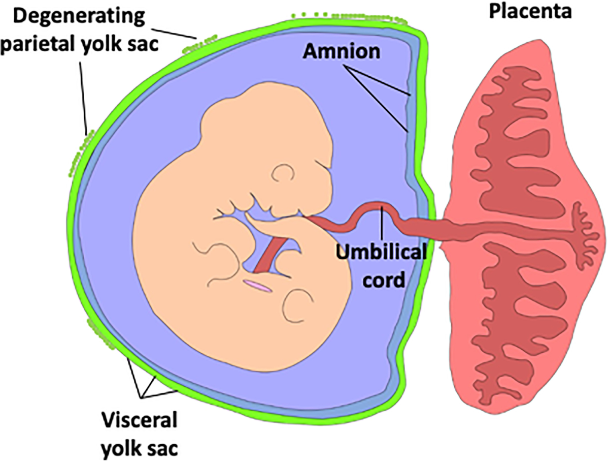
chorion is the outer protective membrane that forms the placenta, while the amnion is the inner protective membrane

describe the placenta’s role
takes over the production of progesterone to protect the endometrium and prevent spontaneous abortions
miscarriages
connects fetal heart and mother’s cardiovascular system and influences her heart functions to ensure the fetus obtains adequate O2 and nutrients
this is why a woman’s blood pressure drops and blood volume expands the further into pregnacy
edema (swelling) typically occurs in the legs due to increased capillary hydrostatic pressure from taking on a lot of blood volume to maintain placenta AND account for low blood pressure
connects to the baby via the umbilical cord
acts as interface, serving as the fetus’ lungs, liver, and kidneys without mixing its blood with the mother’s blood
until the placenta matures and becomes functional, the embryo is supported by the yolk sac (enclosing pouch)
the allantois is an extension of the sac and aids in waste and gas exchange between the yolk sac and the embryo
describe the step of gastrulation in embryonic development
occurs 16 days after fertilization
invagination of the blastula occurs turning it into a gastrula
a process of a surface folding in on itself to form a cavity or tube
outward pressure forms on the outside of blastula to initiate this process
the archenteron is the internal cavity of the gastrula and is connected to the outside of the embryo by a hole/opening called a blastopore
archenteron eventually becomes the lumen (inside) of the gut and the blastopore becomes the anus
gastrulation is one of the most critical steps of development because it primes the body for organogenesis by generating the 3 primary germ layers

what are the 3 primary germ layers
Ectoderm
upper, outermost layer
develops
nervous system, adrenal medulla, epidermis and cavities (skin, hair, nails, eye lens, inner ear, epithelial of nose, mouth, anal canal)
Endoderm
innermost layer
inaccessible from the outside (must enter through ectoderm)
develops
GI stract, gut lining, parts of the liver & pancreas, thyroid, bladder, epithelium of digestive & respiratory tracts
Mesoderm
middle layer
develops
musculoskeletal system, circulatory system, gonads, excretory system, kidneys and their adrenal cortex, muscular and connective tissues of digestive & respiratory systems

difference between cortex and medulla
in any sphere-like object, the cortex is the outer layer closer to the edge at all parts while the medulla is the inner layer removed from the border
in the body,
cortex
responsible for steroid hormone production (endocrine-acting ~ APG)
closer to gonad tissues = comes from mesoderm
medulla
1 big neuron (neuro-secretory ~ PPG)
closer to nervous tissues - comes from ectoderm

what is the difference between specification, determination, and differentiation
specification (freshman)
cells begin their path toward becoming a specific cell type
have unlimited potential and can reverse the process
determination (grad school)
cells commit to being a specific cell type
irreversible
can occur due to morphogens
chemical signals in the embryo that depend on concentration gradient for the strength of their signal (high near the source and low from far away)
based on how far morphogens spread out, the cell decides its fate
differentiation (white coat)
cells undergo the processes necessary to become their cell types
stem cells are cells that have not yet differentiated and can give rise to other cells
types of stem cells
Totipotent
“whole”
can develop into any cell type
Pluripotent
“many”
can develop into numerous cell types except embryonic and placental cells
Multipotent
“several”
can only develop into a handful of cell types
describe the step of differentiation in embryonic development
process by which cells start to look like their type
critically dependent on communication of nearby cells
inducers are cells (or tissues) that emit signals to direct the developmental fate of nearby cells
signalling molecules they send are morphogens which influence adjacent cells using a concentration gradient
foundational for tissue differentiation and organogenesis
relies heavily on paracrine signaling
**in some cases, an inducer can refer to the actual signal, or morphogens, being sent**
responders are competent cells with the proper receptor molecules to respond to inducers/morphogens
explain the types of intercellular communication
Autocrine Signaling
cell signals itself to do something
Paracrine Signaling
cell signal stays local
Juxtocrine (“side-by-side”) Signaling
signals between touching cells; direct contact communication
Endocrine Signaling
cell signals travel far away, through the bloodstream
describe the step of neurulation in embryonic development
occurs after gastrulation
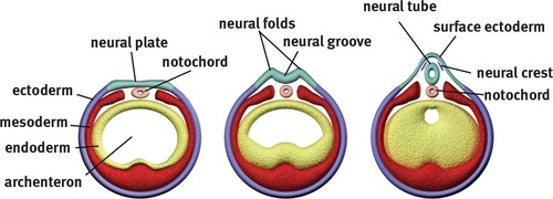
process where the flat ectoderm layer folds and transforms into a tube that later develops into the central nervous system
notochord is a rod of mesodermal cells that help develop the fetus’ nervous system through signalling
overlying ectodermal cells fold in to form horms, neural folds, and the neuro groove lowers down towards the notochord
all of this is a result of cell-to-cell communications
as invagination continues, the 2 neural folds pull up and conjoin together to ft7neural tube
neural crest cells migrate in through here before tube closes completely
neural folds pinch and fold into the neural tubes to make the central nervous system (brain + spine)
as the folds fuse into the tube, specialized cells at the borders detach and develop into the peripheral nervous system, facial structures, and melanocytes (pigment)
neural crest cells
the anterior and posterior ends of the tube remain open temporarily
failure to close creates defects like anencephaly or spina bifida
describe fetal circulation
a fetus’s lungs DO NOT WORK
they are filled with fluid and have high pressure
before birth, the right side of the fetus heart has greater pressure because lungs are shut
when lungs start functioning after birth, the left side has greater pressure because they begin supplying blood to that side and it has the job of supplying to the rest of the body
more blood = greater volume = greater force = greater pressure
the placenta is where fetal blood connects to maternal blood without mixing, gets O2 and nutrients, and removes CO2 and waste
even though it does the job of the digestive system, lungs, liver and kidneys, a little blood still goes to these areas in order to nourish the tissues
umbilical arteries
2 arteries that carry de-oxygenated fetal blood to the placenta
umbilical vein
1 vein that carries oxygenated blood from the placenta back to the fetus
fetuses have a much higher affinity (desire) for oxygen than adults
3 shunts (redirecting passages)
operate simultaneously in the fetus so it is adequately nourished by O2 blood from the placenta
what are the 3 shunts of the fetal cardiovascular system (pulmonary cycle)
Foramen Ovale
means “oval hole”
a hole that exists in the interatrial septum to connect the right atrium to the left atrium
allows fetuses to bypass pulmonary circulation by shunting the blood directly to the left atrium
this hole closes after birth due to increased pressure in the left atrium after the first breath
Ductus Arteriosus
means “arteriol duct”
necessary because some blood misses the foramen ovale and continues to spill into the right ventricle from the right atrium
this duct diverts blood from the pulmonary artery into the aorta, bypassing the non-functional lungs
duct closes after birth due to increased oxygen and a drop in prostaglandins
hormone-like lipids that keep this duct widened and relaxed
in menstruation, they contract the uterus to shed the endometrium lining → cramps
act right when they are produced (unlike hormones)
Ductus Venosus
means “venus duct”
connects the umbilical cord to the IVC to bypass the liver
liver isn’t needed before birth because the placenta processes nutrients for the fetus
IVC → RA → Foramen Ovale → LA → LV → Aorta
IVC → RA → RV → Ductus Arteriosus → Aorta
O2 AND NUTRIENTS:
Mother → Placenta → Umbilical Cord → Umbilical Vein → Ductus Venosus → IVC
CO2 AND WASTE:
Aorta → Umbilical Arteries → Umbilical Cord → Placenta → Mother

what are teratogens
substances that interfere with embryonic development
drugs/medication, viruses, chemicals, alcohol, etc.
vitamin B9 (folate/folic acid) deficiency is teratogenic because it blocks the vitamin’s ability to aid in neural tube development, leading to defects like Spina Bifida
describe gestation
the time from conception until birth
measured in trimesters
each trimester is 12-14 weeks
human gestation is 40 weeks or 10 months
we start counting weeks after implantation, which takes place after 2 weeks after conception (bHCG is detected)
First Trimester
at 3 or 4 weeks, a baby has a heartbeat
at 8 weeks, an embryo becomes a fetus
all major organ systems have formed, but it is still immature
from this point on, its just growing/maturing
Second Trimester
tremendous growth
fetus begins moving
human face can be seen and eyes begin to open
fingers and toes elongate
when a fetus closes its hands, pressure of amniotic fluid creates a unique indent~ fingerprints
Third Trimester
continued rapid growth
lots of central nervous system (CNS) development
fetus has less space → little to no movement
explain birth
NECESSARY POSITIVE FEEDBACK PATHWAY TO KNOW
the natural signal that starts birth is unknown
induction occurs and uterus begins to contract
prostaglandins
as the baby’s head presses against the cervix on the outlet of the vaginal canal, uterine contraction begins
this leads to cervical dilation which signals the hypothalamus to stimulate the PPG into releasing oxytocin which again stimulates uterine contraction
positive feedback loop
this continues happening until the end of childbirth, meaning oxytocin levels in the body are extremely high
newborn effect~ intense emotional bond of a mother to her child after birth
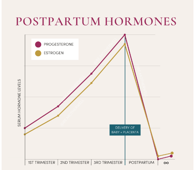
high oxytocin levels suddenly lower after birth, while estrogen and progesterone levels crash leaving a mother with postpartum
loss of placenta after birth
different for all women due to environmental, genetic, and hormonal factors
breastfeeding keeps oxytocin levels relatively high (fluctuates)
women who stop breastfeeding early typically have trouble bonding with their child
describe the different levels of aging
body breaks down
wrinkles, poor posture, slower
accumulation of “insults” to the body
environmental stressors, poor lifestyle choices, etc.
telomeres stop regenerating
non-coding terminals of chromosomes with repetitive nucleotide sequences that exist to protect the ends of DNA during replication
get shorter and shorter over time, eventually leading to cessation of cellular reproduction of that chromosome
types of cell death
apoptosis
controlled
cell breaks into fragments in of itself to be digested by other cells
happens to limit the release of harmful chemicals
necrosis
uncontrolled
cell dies due to environmental factors and explodes
releases a bunch of toxic compounds that can kill other cells
neurons
cells of the nervous system capable of receiving and transmitting specific electrical impulses that the body can recognize and act on
bundles of neurons
CNS: a tract
PNS: a nerve
sensory, motor, mixed, and automatic types
parts of the neuron:
soma
the body of the cell in which the nucleus, ribosomes, and ER are located
within a tract in the CNS, somas collect into nuclei
within a nerve in the PNS, somas collect into ganglia
dendrites
finger-like structures that surround the soma and receive information from the outside
axon
d
axon hillock
boundary between the cell body and the axon
“point of no return” that filters and integrates incoming signals as “inhibitory” and “excitatory”
nerve terminals
signals get spat back out in the form of chemical messengers called neurotransmitters
disspitate into synaptic cleft
summation
adding up of signals to determine if they’re enough to an fire action potential
inhibitory
signals that hyperpolarize membrane and move it further from firing an action potential
negative
excitatory
signals that polarize cell membrane and move it towards firing an action potential
positive
saltatory conduction
signals travelling through axon by jumping between non-insulated points on the axon (outside of myelin sheaths)
these points are called the Nodes of Ranvier
electrical vs chemical transmission
electrical
electricity is used to pass signals down the axon within a single neuron
chemical
neurotransmitters (chemicals) pass signals to subsequent neurons/muscles/glands
effectors
glial cells
also known as neuroglia
surround neurons and play supportive and structural roles for them
can hold them in place, supply O2 and nutrients to them, insulate them from other neurons so electrical impulses are not scattered, destroy pathogens, and remove dead neurons
types
astrocytes
nourish neurons and form the blood-brain barrier (BBB)
layer of cells that surround the brain blood vessels and control the transmission of solutes from blood stream into CNS
essentially blocking pathogens and toxins
ependymal cells
line the ventricles of the brain and produce celebrospinal fluid (CSF)
clear fluid (filtered plasma) that supports and cushions the brain and spine as a “shock absorber”
also form barrier between CSF and the interstitial fluid of the CNS
microglia
phagocytic cells that ingest and breakdown waste and pathogens in the CNS
“specialized macrophages”
mylenating cells
create muelin sheaths around axons as insulation points that fix all electrical signals to destination with maximum influence and more guaranteed responses (prevents scattering of voltage)
in CNS, these cells are called oligodendrocytes
in PNS, these cells are called Schwann cells
signal transmission and action potential of neurons
every cell membrane has a voltage that almost always creates a more negative charge inside the cell than outside
shifts in voltage across a cell membrane create action potential used for signalling of messages to the body
muscle contraction, hormone influence, etc.
Vm = membrane potential
depolarization = raised Vm = more positive inside cell = more AP
hyperpolarization = lowered Vm = more negative inside cell = less AP
resting membrane potential (RMP)
the net electrical potential difference that exists across a cell’s membrane, created by movement of charged molecules
in neurons, this potential is -70 mV
the two ions generating and maintaining this potential are potassium (K+) and sodium (Na+)
these ions keep the potential negative inside the cell by resisting its desire for neutrality
Na+/K+/ATPase
sodium potassium pump
maintains the neurons resting membrane potential by pumping out 3 sodium ions for every 2 potassium ions pumped in
NOKIA- Na outside, K inside, 1 ATP used
a lesser concentration of Na+ is found inside the cell than inside, so ions desire to enter the cell to neutralize this imbalance → 3 ions are pumped out to maintain it
net positive charge (sodium > potassium)
a greater concentration of K+ is found inside the cell than outside, so ions desire to leave the cell to neutralize this imbalance → 2 ions are pumped in to maintain it
net negative charge (potassium > sodium)
1 adenosine-triphosphate (ATP) molecule is required to generate the energy required to transport ions since both are moving against their gradient
potassium leak channels
function to allow slight movement of K+ ions down their gradient (whichever direction that may be)
leaking out (NORMAL): when the cell has too much K+ inside, channels balance the sodium potassium pump
leaking in: restores -70 mV after hyperpolarization, helping pump when the cell becomes too negative inside
sodium leak channels
function to allow slight movement of Na+ ions down their gradient (whichever direction that may be)
leaking out: when the cell has too much Na+ inside, channels balance the sodium potassium pump
leaking in (NORMAL): restores -70 mV after hyperpolarization, helping pump when the cell becomes too negative inside
equilibrium potential of K+ and Na+
equilibrium between the concentration gradient that moves these ions out and the electrical gradient that moves them in → results in no net movement of the ions
Na+ = +60 mV
K+ = -90 mV
threshold
the lowest magnitude of stimulus strength (electrical voltage) that will induce a response (action potential)
membrane at axon hillock summates enough excitatory signals and depolarizes to a level between -55 mV and -40 mV
types of summation
temporal summation
a single presynaptic terminal fires rapidly in succession at dendrites, so each new potential adds to the previous one before it fully decays, building up enough voltage at the hillock
spatial summation
multiple different presynaptic terminals fire at the same time at different locations on the neuron dendrites, combining their individual potentials together at the hillock