RAD PATH UNIT 2 XRAY
 Call Kai
 Learn
 Practice Test
 Spaced Repetition
 Match
 Flashcards
 Knowt Play
1/68

There's no tags or description
Looks like no tags are added yet.
69 Terms

Spina Bfida Occulta
RA of splitting of neural canal
Spina Bfida

Osteopetrosis

Osteopetrosis
RA of symmetric & generalized increase in bone density
Osteopetrosis

Osteogenic Imperfecta

Osteogenic Imperfecta
Flattening of vertebral bodies w/ fractures of multiple ribs & long bones
RA of extremely fragile skeleton; pathologic fractures; very thin cortical density
Osteogenic Imperfecta
RA of progressive interpedicular distance from superior to inferior & scalloping of posterior vertebral bodies
Achondroplasia
CT of spinal narrowing due to spondylosis & vertebral column changes
Achondroplasia
CT/MRI – soft tissue posterior mass with or without spinal cord and nerve routes
Spina Bfida
US – fetal demonstration of location & severity of pathophysiology
Spina Bfida
RA of superior and posterior dislocation of hip
Congenital Hip Dysplasia

Congenital Hip Dysplasia
RA of periarticular soft tissue swelling; symmetric joint destruction & deformity
Rheumatoid Arthritis

Rheumatoid Arthritis

Ankylosing Spondylitis
RA of fusion of intervertebral joint
Ankylosing Spondylitis
RA of irregular narrowing of joint space with small bony spurs
Osteoarthritis

Osteoarthritis

Osteoarthritis
RA of early joint narrowing with focal erosions
Infectious arthritis

Infectious arthritis
RA not visualized well on plain films, calcific tendinitis in 50%
Bursitis

Bursitis
RA of soft tissue swelling with periosteal elevation
Osteomyelitis
US – soft tissue vascularity elevating periosteum;
Osteomyelitis
NM – bone scan most accurate at localizing process & recurrence
Osteomyelitis

Osteomyelitis
RA of loss of bone density, cortical thinning appears as a relatively dense & prominent thin line
Osteoporosis

Osteoporosis
RA of loss of bone density; cortex becomes thin & often indistinct
Osteomalacia

Osteomalacia
RA of cupped & frayed metaphysis in long bones
Rickets

Rickets
RA of joint inflammation & urate crystals in joint space
Gout

Gout
RA of radiolucencies in destructive phase
Paget’s disease
RA of cotton wool appearance in reparative phase
Paget’s disease

Paget’s disease
RA of well defined radiolucent area, homogeneous ground glass density or soap bubble appearance
Fibrous dysplasia
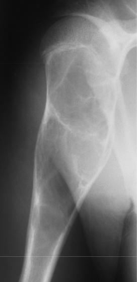
Fibrous dysplasia
RA of tumor runs parallel to long bone & points away from nearest joint
Osteochondroma

Osteochondroma
US – determines cartilage cap thickness
Osteochondroma
MRI – cartilage cap produces a high signal intensity on T2 weighted spin echo images
Osteochondroma
RA of multiple large bubbles separated by strips of bone
Giant Cell Tumors
MRI – demonstrates tumor extension with soft tissue involvement & bone marrow changes
Giant Cell Tumors

Giant Cell Tumors

Giant Cell Tumors
RA of well circumscribed extremely dense round lesions; less than 2 cm in diameter
Osteoma

Osteoma
RA of round or oval lesions with lucent center (nidus), sometimes surrounded by dense sclerotic zone; less than 1 cm
Osteoid Osteoma
CT – contrast enhancement to demonstrate nidus
Osteoid Osteoma

Osteoid Osteoma
RA of mixed destructive/sclerotic lesion with soft tissue mass
Osteogenic Sarcoma
RA of “sunburst” pattern; Codman’s triangle
Osteogenic Sarcoma

Osteogenic Sarcoma
RA of medullary destruction with “onionskin” periosteal reaction
Ewing’s Sarcoma,
RA of multiple punched out lesions
Multiple Myeloma

Multiple Myeloma
RA of punctuate or amorphous calcification in cartilaginous matrix
Chondrosarcoma

Chondrosarcoma
RA of irregular, poorly defined lucent lesions or poorly defined increase densities depending on site of origin
Bone Metastases

Bone Metastases
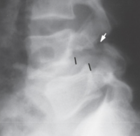
RA of defect in pars articularis without displacement
Spondylolysis

Spondylolysis
RA of forward movement of one vertebra on another
Spondylolisthesis
Spondylolisthesis