Special Senses
 Call Kai
 Learn
 Practice Test
 Spaced Repetition
 Match
 Flashcards
 Knowt Play
1/62

There's no tags or description
Looks like no tags are added yet.
63 Terms
4 Special senses
1. Sight
2. Hearing
3. Taste
4. Smell
Protects eye
cushion of fat
4 Accessory structures of eye
1. Extrinsic eye muscle
2. Eyelids
3. Conjuctiva
4.Lacrimal apparatus
Eyelid
Protects eye anteriorly

Papebral fissure
Space between eyelids when eye is open
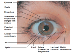
Tarsal glands
Located along edges of eyelids, Responsible for excreting oily substance that assists lubricating eye
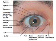
Conjunctiva
1. Lines the eyelids and covers outer surface of eyelids and eye itself
2. Binds with corneal epithelium
3. Secretes mucus, keeping eye moist

Lacrimal apparatus
1. Lacrimal gland
2. Number of ducts draining lacrimal secretions into nasal cavity

Lacrimal glands
1. Located superior laterally to each eye
2. Act to continually release dilute salt solution onto anterior surface of eye

Lacrimal secretions
1. Muscus
2. Antibodies
3. Lysozyme

2 Functions of tears
1. Protect eye
2. Moisten eye
Extrinsic eye muscle
1. 6 total
2. Produce gross eye movements
3. Allow to actively follow moving object
4. Attach to outer portion of eye

3 Layers of the eye
1. Fibrous
2. Vascular
3. Sensory
Internal eye structure
1. Spherical
2. Made of 3 layers
3. Fluid filled center
Fibrous layer of eye
1. Outmost layer
2. Consists of sclera and transparent cornea

Sclera
1. Made of thick, white connective tissue
2. Can be seen anteriorly

Cornea
1. Located in center of sclera
2. Clear area where light enters eye
3. Supplied with nerve endings
4. Vulnerable to damage
5. Ability to repair itself
6. Can be transplanted without rejection

Vascular layer of the eye
1. Middle layer of ear
2. 3 distinguishable regions
3 Regions of vascular layer of ear
1. Choroid
2. Ciliary body
3. Iris
Choroid
1. Most posterior region of vascular layer
2. blood rich nutrient covering
3. Contains dark pigment to prevent light scattering in eye
Ciliary body
Region where lens attaches via ciliary zone
ciliary zone
suspensory ligament
Iris
1. Pigmented area
2. Contains pupil
Pupil
1. Rounded opening in iris
2. Smooth muscle fibers act to open or close
Sensory layer of eye
1. Inner layer of the eye
2. Primarily made of retina
Retina
2 layered structure, Extends anteriorly to ciliary body of eye
Pigmented layer of retina
Reacts similar to choroid, Removes dead cells and photoreceptors, Stores vitamin A
Inner layer of retina
1. Transparent
2. Contains rods and cones or photoreceptor cells
Optic disc
1. AKA blind spot
2. Only place where photoreceptors are not scattered in retina
3. Where the optic nerve exits
Rods
1. Most dense on periphery of retina
2. Number decreases towards center of retina
3. Allow for seeing gray tones in dim light and peripheral vision
Cones
1. Allow for seeing details and color in bright light
2. 3 types in sensory layer
3. Each type sensitive to specific wavelength of visible light
Fovea centralis
1. Lateral to blind spot
2. Tiny area of high cone concentration
3. High visual acuity
Lens
1. Focuses light on the retina
2. Held upright by ciliary zonule
3. Attached to ciliary body
4. divided into 2 chambers providing structure and intraocular pressure
2 Chambers of lens
1. Aqueous
2. Vitreous
Aqueous chamber
1. Anterior
2. contains clear watery fluid called aqueous humor
Vitreous chamber
1. Posterior
2. contains gel-like substance called vitreous humor
Internal eye muscles
1. Controlled b y autonomic nervous system
2. Control pupil size
3. Control lens configuration
External eye muscles
1. Eye movement
2. Controlled by somatic fibers of cranial nerves
Photopupillary reflex
1. Pupil constricts to prevent light from damaging photoreceptor cells
2.Pupils constrict when objects are close for more acute vision
2 Distinct functions of ear
1. Hearing
2. Balance
3 Main parts of ear
1. External ear (hearing)
2. Middle ear (hearing)
3. Internal ear (hearing and balance)

External ear
1. Comprised of auricle (pinna) and external auditory meatus (auditory canal)
Auditory meatus
1. Short narrow chamber
2. Lined with skin and cerminous glands
cerminous glands
Secrete cerumen (ear wax) which traps foreign bodies and repels insects
Ear drum
functions as end of external ear and beginning of middle ear
Middle ear
1. AKA tympanic cavity
2. Small, air-filled mucosa line cavity within temporal bone
Pharyngotympanic tube
1. AKA auditory tube
2. Runs obliquely downward
3. Links middle ear with throat
4. Typically closed but briefly opens
Ossicles
1. 3 bones in the ear
2. Transmit vibration motion of eardrum to fluids of inner ear
3 bones of middle ear
1. Malleus (hammer)
2. Incus (anvil)
3. Stapes (stirrup)
Sound transfer process
1. Ear drum moves transferring motion to malleus
2. Malleus hits incus
3. Incus transfers to stirrup
4. Stirrup press on oval window and sends motion to fluid located in inner ear

Inner ear
1. Maze of bony chambers (filled with perilymph)
2. AKA Osseous labyrinth
3 Distinct parts of osseous labyrinth
1. Cochlea
2. Vestibule
3. Semicircular canals
Membranous labyrinth
1. Suspended in perilymph
2. system of membrane sac that follow shape of bony labyrinth
3. Contains endolymph
Static equilibrium
1. Allows to keep head erect
2. Made of calcium salts
Maculae
1. Located in membrane sacs of vestibule
2. Receptors influencing static equilibrium
3. Report changes in head position with respect to gravity

Dynamic equilibrium
1. Receptors found in semicircular canals
2. receptors respond to angular or rotator movement of the head

Olfactory system
1. Sense of smell
2. based on chemoreceptors
Olfactory receptor cells
neurons equipped with olfactory hairs (long cilia) protruding from nasal epithelium
Olfactory hairs
1. Surrounded by layer of mucus excreted by nearby glands
2. Hairs are stimulated when chemicals dissolve in mucus
3. Signal is sent to olfactory nerve then transported to olfactory part of brain

2 types of papillae on tongue
1. Circumvallate pappilla
2. Fungiform papilla

Circumvallate pappilla
taste buds on the side
Fungiform papilla
taste buds on top
Gustatory hairs
1. Respond to dissolved chemicals in saliva
2. Protrude through taste pores (once stimulated transmit impulses via cranial nerves