Disorder of Vision
 Call Kai
 Learn
 Practice Test
 Spaced Repetition
 Match
 Flashcards
 Knowt Play
1/60

There's no tags or description
Looks like no tags are added yet.
61 Terms
Aqueous humor pathway
Made by ciliary body EP → travel through posterior chamber → enter anterior chamber → 90% enter trabecular meshwork → Canal of Schlemm → episcleral vss; 10% drain into uvea and sclera
Parts of accommodation
Lens accommodation by zonule tension/relaxation, miosis, convergence
Cellular eye layers
Outer = choroid → pigmented EP → rod and cone → Outer nuclear → outer plexiform → inner nuclear → inner plexiform → ganglion cell layer → nerve fiber layer → nearest to vitreous humor
Vascular supply of the occipital cortex
PCA from basilar supplies the medial part of occipital; MCA from ICA supplies the lateral part of occipital
Vascular supply of the eye
ICA clinoid → Opthalmic A → Central retinal that pierce and feed center of eye + posterior ciliary a → short is near optic disc, long supplies ciliary muscle
What part of the eye is spared when there is infarction of retinal supply
Foveola only
Pupillary light reflex pathway
Light enter optic nerve → signal branch out (one cross chiasm one doesn’t) → optic tract → synapse at pretectal area in LGN → Bilat projection into Edinger-Westphal → travel along CN III → synapse at ciliary ganglion → miosis
Mechanisms of reduced vision (non nerve)
Refractive area = light is not doing into retina
Media opacity = light is obstructed bc media is cloudy
Retina = photoreceptor and structures fail
What are refractive errors
Simple bad eyesight fixed with glasses
Myopia → light does not reach retina → fix w concave lens
Hyperopia → light extends past retina → fix w convex lens
Astigmatism → many light refractions → fix w circular lens
What is presbyopia
Impaired accommodation in older people from loss lens elasticity, change in lens curvature and lower ciliary muscle strength
What is pinguecula and pterygium
Fibrovascular growth of conjuctiva
Pinguecula → does NOT involve limbus
Pterygium → grows big enough to reach into cornea causes astigmatism

What is the difference between these two images
Lt → has not reached limbus = pinguecula → likely no vision problem
Rt → crosses limbus to cornea = pterygium → expect astigmatism
Causes of corneal ulcer and scar
Infection (HSV), SJS, Reiter, corneal edema/deposition, keratitis, glaucoma, trauma
What are cataracts
INCREASED opacity of the lens
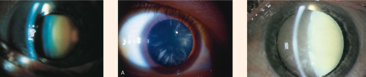
What is the difference between these 3 types of cataracts
Lt → See orange center = senile nuclear
Middle → See white fibers in center with some clear area → Cortical
Rt → white fiber with NO clear area → Mature cortica

What is this
Fully opaque lens and brown deposits at the bottom of lens → Margagnian cataract
Component of glaucoma
Loss of retinal ganglion cell, cupping, visual field defect
What is glaucoma
Change in aqueous humor flow → eg flow of trabecular meshwork = lack of outflow into vss
What is the difference between primary and secondary open angle glaucoma
Primary (POAG) → more frequent in older and black people
Secondary (SOAG) → RBC/WBC etc block trabecular network → higher episcleral venous pressure
Signs of angle closure glaucoma
EMERGENCY → painful red eye, sudden vision loss, halo around light, headache, fixed dilate pupil, nausea vomiting
Types of uvea abnormalities
Keratic precipitate, hypopyon (cell that settle at the bottom), vitritis (blurred optic disc), neuroretinitis (cat scratch, see macular star)
What is diabetic retinopathy
Non proliferative DR → damaged capillary leak of blood/lipid/macular edema
Proliferative DR → chronic hypoxia → neovascular formation → retinal traction

What are the abnormalities found in DR
Green = hard exudate
Purple = microaneurysm
Blue = dot/blot hemorrhage

What is this
Blood in vitreous humor → rupture of ciliary/retinal vss

Seeing this with increased frequency is a sign of what
If you see floaters infrequently is benign → if increased is sign of vitreous detachment common in DM w neovascularization
Types if retinal detachment
- Rhegmatogenous retinal detachment (RRD): retinal breaks
- Tractional retinal detachment (TRD): DR
- Exudative retinal detachment: Uveitis, mass
What is hypertensive retinopathy
Chronic uncontrolled HTN → causes endothelial disruption, fibrinoid necrosis and retinal damage
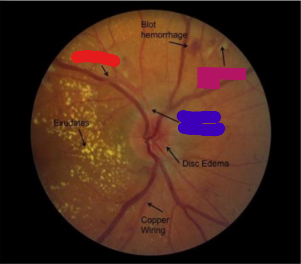
What are all these signs
Signs of HYPERTENSIVE RETINOPATHY
Blue = Arterial narrowing
Red = Arteriovenous (AV) nicking
Pink = Cotton wool spots
Can also present with: Flame-shaped retinal hemorrhages, Microaneurysms, Optic nerve head swelling
What is central retinal artery occlusion (CRAO)
Acute painless monocular vision loss → see retinal WHITENING with CHERRY RED fovea

What is this
CRAO → white retina + cherry red fovea
What is BRAO
Occlusion of a BRANCH of central retina → whitening only in area that branch supplies

Diff these two images
Lt = CRAO → retina is white and general + cherry red fovea
Rt = BRAO → partial white retinal area
What is amaeurosis fugas
Sudden painless vision loss → curtain linked lasting a few seconds → completely back to normal → caused by TIA

What is this
Retinal vein occlusion → Lt is central (CRVO), Rt is branch (BRVO) → see hemorrhage and venous engorgement
What is age related macular degeneration (AMD)
May present with metamorphosia = loss of straight line and scotoma = loss of central vision
What is retinitis pigmentosa
Dystrophy of retina EP and photoreceptor → present as tunnel vision and nyctalopia = trouble seeing in dark

What is this
Green = optic disc pallor
Purple = vss attenuation
Blue = pigmentation with spicule shaped margins
Are all TRIAD of findings for Retinitis pigmentosa
What is leukocoria
Loss of red/orange reflection/glow → replaced with white/gray color found in congenital cataract and retinoblastoma
What is papilledema
High ICP → bilateral optic disc edema → transient visual obscure/bigger blind spot
What is ischemic optic neuropathy
AAION → caused by GCA and NAION
Lower flow to optic nerve head → altitudinal VFD
How to pre chiasmatic from chiasmatic and post chiasmatic lesions
Pre chiasmatic respect HORIZONTAL meridian
Rest respect VERTICAL meridian
Optic nerve lesion sign
Ipsilateral ANOPIA/blindness with positive RAPD/Marcus Gunn
Bitemporal hemianopia is caused by lesion to what area
Optic chiasm → compression from mass (pituitary macroadenoma/craniopharyngioma etc)
What is junctional scotoma
Distal optic nerve lesion and anterior chiasm → Wilbrand’s knee
LGN stroke signs
Lateral posterior choroidal → incomplete, incongruous homonymous
(horizontal) sectoranopia
Anterior choroidal → incomplete quadruple sectoranopia OR incomplete homonymous hemianopia with horizontal sparing

based on this visual field where is the lesion
Lt → Lateral posterior choroidal of LGN
Rt → Anterior choroidal of LGN
What is the difference between visual loss of Meyer and dorsal bundles
Meyers: In temporal lobe causes SUPERIOR quadrant loss from MCA infarct
Dorsal: In parietal lobe causes INFERIOR quadrant loss from PCA infarct
Why is the macular area spared in infarction
Dual supply by PCA and MCA → if either infarct the remaining artery still supply
What is monocular occipitotemporal crescent syndrome
Contralateral lesion of anterior occipital → controls the most PERIPHERAL part of field (60-90 degrees) → lesion causes loss of peripheral sight
PNS control of pupil
Causes CONSTRICTION → neuron at EWN go to ciliary ganglion through CN III → synapse at short ciliary nerve of sphincter pupillary muscle → miosis
Pathway of near pupillary reflex
Primary visual cortex → Supranuclear → Supraoculomotor area In midbrain → Oculomotor nucleus → MR → Convergence → EW nucleus → ciliary ganglion → Short ciliary n. Lens accommodation and miosis
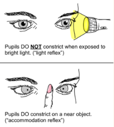
What is this
Argyll Robertson pupil → PRETECTAL defect → No light reflex + spare accommodation
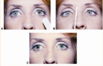
What is this
Adie’s tonic pupil → PSN ciliary ganglion damage → aberrant regeneration → poor light stimulus with better response to near object on affected eye
SNS control of pupil
DILATION → from hypothalamus and synapse at C8-T2 ciliospinal center → travel across lung apex → superior cervical ganglion synapse → plexus ascend ICA → travel by opthalmic artery
Horner syndrome triad
Miosis, anhidrosis, ptosis
Cause of CN III palsy
EOM problem + headache = PComm Aneurism
DM, HT, atherosclerosis
EOM problem + pupil problem = compressive lesion
What is MLF pathway
Frontal eye field → PPRF → abducen nucleus → LR movement → project to contralateral MLF → oculomotor nucleus → movement of MR
What is INO
Lesion of MLF → show abducting eye nystagmus to stimulate CN III; MG can mimic this
Cavernous sinus syndrome signs
Unilateral ophthalmoplegia (cranial nerve III, IV, VI)
- Autonomic dysfunction (Horner syndrome) or
- Sensory CN V1 - CN V2 loss
Parts of the eye
Fibrous tunic: sclera and cornea
Vascular tunic/uvea: iris, ciliary body, choroid
Retina: pigmented and neural layer
Parts of the uvea
Iris → muscles dilatory and sphincter
Ciliary body → zonule suspensory ligaments
Choroid → brown pigmented cells that absorb light