Language of the skin
 Call Kai
 Learn
 Practice Test
 Spaced Repetition
 Match
 Flashcards
 Knowt Play
1/24

There's no tags or description
Looks like no tags are added yet.
25 Terms

the skin revision
• Epidermis: thin outer portion, that is the keratinised stratified squamous epithelium of skin. The epidermis is important for the protective function of skin. Separated from the dermis by the basement membrane. Apocrine Adipose tissue Sebaceous glandArrector pili muscle
• Dermis: thicker inner portion. This is the connective tissue layer of skin. It is important for sensation, protection and thermoregulation. It contains nerves, blood vessels, and adnexa.
• Hypodermis (subcutis): underneath the dermis and merges with it. It mainly contains adipose tissue and sweat glands. The adipose tissue has metabolic functions.


the epidermis
Keratinocytes in different layers are in different stages of development. All keratinocytes derive from stem cells in the deepest layer (stratum basale) and undergo structural and functional changes as they migrate upward toward the surface of the epidermis (stratum corneum). as they migrate they flatten into dead keratinocytes


the epidermis revision
• Stratum basale: single layer of cuboid cells, attached to the underlying dermis by the basement membrane. The stratum basale is constantly producing epidermal cells (keratinocytes) from stem cells. Some cells may display cytoplasmic dark brown pigment (melanin).
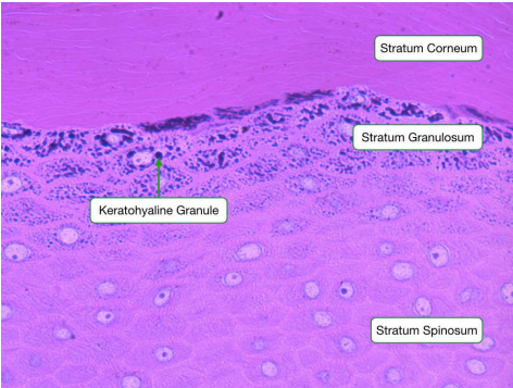
• Stratum spinosum: slightly flattened cells that are responsible for protein synthesis, primarily keratin. This is the thickest layer of the epidermis
• Stratum granulosum: 3–5 layers of flattened cells that contain lipid-rich granules (keratohyalin granules). The intercellular spaces are filled with a lipid-rich material making skin a barrier to both water loss and extrinsic foreign material
• Stratum lucidum: flattened eosinophilic, translucent cells. Provides thickness and strength to withstand friction.
• Stratum corneum: most superficial layer, numerous layers of dead keratinized cells that lost their nuclei and are continuously being shed (desquamation).
cell adherence revision
• Hemidesmosomes: bind the keratinocytes of the stratum basale to the basement membrane.
• Desmosomes: represent major intercellular adhesive junctions (in all epidermal layers) allowing cell-to-cell adhesion of keratinocytes. They are randomly arranged on the lateral sides of plasma membranes.
• Basement membrane: mediates attachment of the epidermis to the underlying dermis.
This combination of tightly adhered filaments and lipidrich envelopes is what gives the skin its ability to serve as both a barrier to loss of water from the body and protection from extrinsic foreign material.

pilosebaceous unit
INFUNDIBULUM
Extends from the opening of the hair follicle to the opening of the sebaceous gland duct
ISTHMUS
Extends from the entrance of the sebaceous duct to the attachment of the arrector pili muscle (bulge)
INFERIOR PORTION
Extends from the bulge to the base of the follicle. This includes the bulb with mitotically active cells.


stages of the hair cycle
kenogen = empty

basic functions of the skin
PRIMARY FUNCTIONS
• Structural and metabolic barrier (stratum corneum, granusolum and spinosum): prevents fluid loss, penetration of pathogens and chemicals, limits physical injury.
• Photoprotective barrier: the thick layer of keratin and epithelium (keratinocytes) reduces the amount of UV light reaching the dermis. Melanin-containing cells (melanocytes) absorb that UV light, protecting the DNA from damage.
• Immunologic barrier: Langerhans cells (antigen-presenting cells) in the epidermis capture antigens and interact with lymphocytes to initiate an immunologic response.
SECONDARY FUNCTIONS:
• Temperature and blood pressure control • Sensory perception
• Antimicrobial action
• Vitamin D production
• Absorptive and excretory function
• Stores nutrients
responses to injury

glossary - epidermal hyperplasia
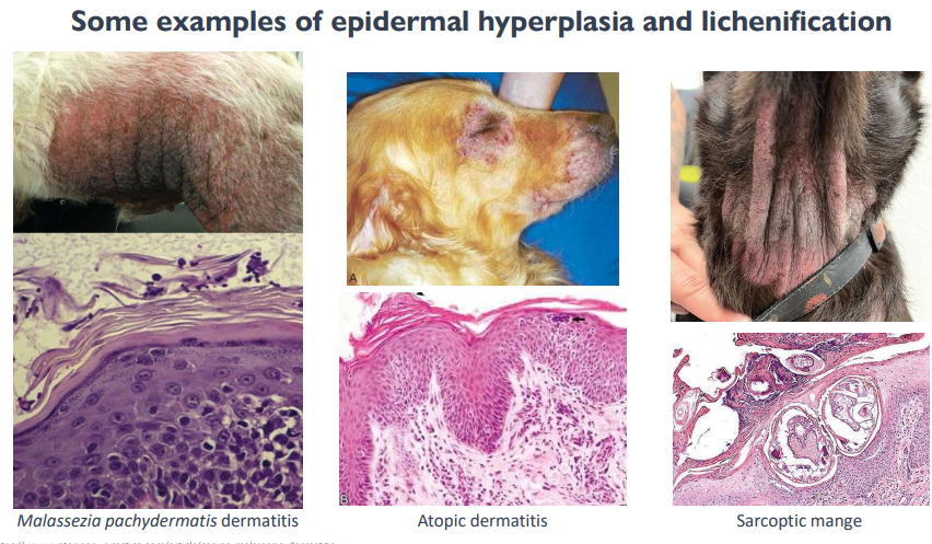
Definition: increased numbers (layers) of keratinocytes in the epidermis, as a response common to a variety of stimuli (often chronic). This represents the earliest reaction of skin to injury
• Acanthosis: increased thickness of the stratum spinosum - often used as synonym to epidermal hyperplasia.

lichenification
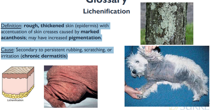
Definition: rough, thickened skin (epidermis) with accentuation of skin creases caused by marked acanthosis; may have increased pigmentation.
Cause: Secondary to persistent rubbing, scratching, or irritation (chronic dermatitis)

hyperkeratosis
Definition: increased thickness of the stratum corneum due to an abnormal amount of keratin. Often associated with epidermal hyperplasia.
• Orthokeratotic: preserved keratinocyte maturation (nuclear loss).
• Parakeratotic: retained nuclei as sign of delayed keratinocyte maturation May also present as scales
calf-diffuse
dog-multifocal (middle)
dog-coalescing/multifocal


callus
Definition: thick, firm, hyperkeratotic, hairless plaque with increased skin folds, wrinkles, or fissures
• In haired skin, follicular plugging and comedones also may develop
• Chronic licking/rubbing
Cause: trauma over bony prominence such as elbow, sternum, or side of digit


vesicles and bullae
Definition: Localized collection of fluid within or beneath the epidermis (“blister”).
• Vesicle: <1 cm in diameter
• Bulla: >1 cm in diameter
Elevated, circumscribed, horizontal, fluid-filled (clear, tan, red) lesions differentiated by size.
Cause: Burn, viral infection, immune-mediated diseases such as bullous pemphigoid and lupus erythematosus.


vesicle in stratum spinosum in epidermis
bulla is in….
some examples of vesiculo-bullous lesions
• Vesicular stomatitis (ruminant, swine and horse)
• Foot and mouth disease (ruminant and swine)
• Swine vesicular disease (swine)
• Vesicular exanthema of swine
• Seneca virus (swine)

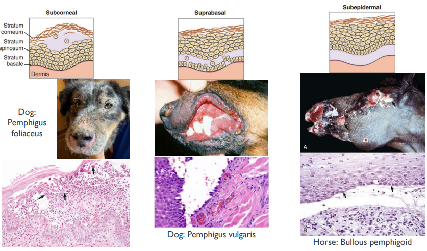
the importance of localization in autoimmune diseases
pustule
Definition: elevated, pus-filled (degenerate neutrophils) vesicle (usually less than 1 cm)
Cause: bacterial infections
distribution is multifocal
wehneevr oyu see neutrophils → acute
fibrin-acute
fibrosis-chronic

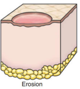
erosion
Definition: partial-thickness loss of epidermis resulting in shallow, moist, glistening depression. The basement membrane remains intact.
Bleeding? No vessels in the epidermis!
Cause: Secondary to vesicle or pustule rupture or secondary to surface trauma
red cos inflammation

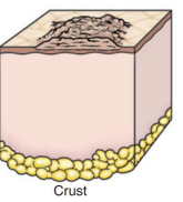
crust
Definition: dried exudate (including fluid, blood, pustular debris, scale, or microorganisms) on the skin surface.
Often the end result of loss of integrity of the epidermis (rupture of vesicles and pustules, ulcerations, erosions, neoplasia).

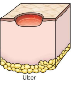
ulcer
Definition: Full-thickness loss of epidermis and basement membrane, and at least a portion of the dermis with depression of the exposed surface. May extend into deeper tissue.
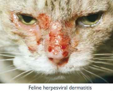
Cause: ischemic lesions resulting from vasculitis, indolent ulcer, feline herpesvirus dermatitis, toxicity, self-trauma.

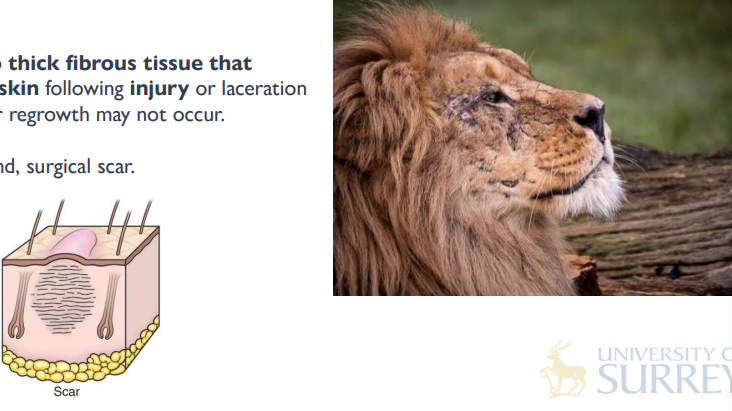
scar
Definition: Thin to thick fibrous tissue that replaces normal skin following injury or laceration to the dermis; hair regrowth may not occur.
Cause: healed wound, surgical scar.

skin nodules
• Macule: flat, circumscribed area (change of colour),
less than 1 cm, without elevation or depression
(e.g., haemorrhage)
• Patch: flat lesion greater than 1 cm, without elevation
or depression
• Papule: elevated, solid lesion less than 1 cm, may
coalesce (e.g., insect bite).
• Plaque: elevated, firm, flat-topped, circumscribed
lesion, usually greater than 1 cm (e.g., eosinophilic
plaque, calcinosis cutis)
• Nodule: elevated, solid lesion greater than 1 cm
(e.g., bacterial or fungal infection, neoplasia)


neutrophils and macrophages surrounding nodules
atrophy
Definition: Decrease in the mass of a tissue due to decreased size and number of cells (after it has reached its normal size)
• Dermal atrophy: thinning of dermal collagen fibrils and decreased dermal thickness
• May be associated with alopecia
Cause: malnutrition, endocrinopathies (hypothyroidism, hyperadrenocorticism [Cushing’s])

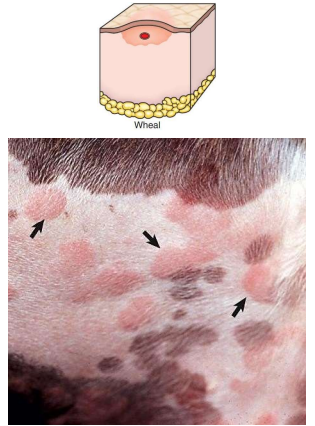
urticaria (wheal)
Definition: smooth, circumscribed, slightly elevated area on skin caused by dermal oedema, solid, usually transient, often pale centrally with an erythematous rim (arrows)
• Angioedema: vascular reaction involving the deep dermis or subcutis and consisting of oedema manifested as giant wheals and caused by dilation and increased permeability of capillaries (deeper version of urticaria)
Cause: Insect bites, allergic reaction
neoplasia
Definition: abnormal mass of tissue, characterized by excessive and uncoordinated growth and persists in the same excessive manner after cessation of the stimuli which evoked the change.

pigmentary changes
Hypopigmentation
• Albinism: inherited disorder that presents with absence of
melanin pigment in the skin
• Vitiligo: immune system attacks melanocytes causing
pigment loss
Hyperpigmentation
• Melanosis: excessive presence of melanin in the skin
• Secondary to skin damage (post-inflammatory) or
endocrinopathy (hypothyroidism).