BIOL 2131_3.3_A
 Call Kai
 Learn
 Practice Test
 Spaced Repetition
 Match
 Flashcards
 Knowt Play
1/29

There's no tags or description
Looks like no tags are added yet.
30 Terms
What is the fundamental path of a vesicle through the endomembrane system, and how do anterograde and retrograde transport differ?
Anterograde transport moves newly made cargo from the endoplasmic reticulum (ER) toward the Golgi and onward to its destination. Retrograde transport returns selected proteins and lipids to an earlier compartment. The full route traces the cargo. Different carriers handle different parts of its journey.
📦 Follow one protein
Package it at the ER. Imagine a protein destined for secretion. It is made at the rough ER and enters the ER lumen (the space inside the ER). At an ER exit site, membrane buds around the protein to form a vesicle, a small sac enclosed by a phospholipid bilayer. Soluble cargo travels inside the sac. Transmembrane cargo stays embedded in its membrane.
Deliver it to the Golgi. In mammalian cells, cargo passes through the ERGIC (ER–Golgi intermediate compartment) and reaches the cis-Golgi, the receiving side. It progresses through the medial and trans regions, where enzymes can modify it.
Sort and deliver it. At the trans-Golgi network, the shipping side, the protein enters a secretory carrier. When that carrier fuses with the plasma membrane, its soluble contents are released outside the cell. Other cargo takes different routes, including one toward endosomes.
🔄 How to tell the directions apart
Cargo movement | Classification | What it accomplishes |
|---|---|---|
Newly made protein moves from ER → Golgi | Anterograde | Advances cargo along the secretory pathway |
An ER-resident protein escapes to the Golgi and returns Golgi → ER | Retrograde | Restores the protein to the compartment where it works |
A Golgi enzyme returns from a later → earlier Golgi compartment | Retrograde | Keeps the enzyme available where incoming cargo needs it |
A cell-surface receptor moves from plasma membrane → endosome | Endocytosis | Brings surface material into the cell. A later endosome → Golgi step would count as retrograde transport |
Exam rule: Identify the starting and receiving compartments. Movement forward along the secretory pathway is anterograde. Return to an earlier compartment is retrograde. Simply moving inward from the cell surface tells you that endocytosis occurred.
📌 Core Concept: Vesicular transport is the regulated movement of membrane-bound phospholipid droplets carrying soluble and transmembrane cargo between organelles or the plasma membrane.
⚙️ Mechanism / Process:
• Anterograde Transport: Moves cargo forward toward the cell membrane (RER → ERGIC → cis-Golgi → medial-Golgi → trans-Golgi → plasma membrane/endosomes).
• Retrograde Transport: Moves vesicles backward away from the cell membrane (plasma membrane/Golgi → ERGIC/RER) to retrieve mislocalized resident proteins.
💡 Biological Significance: Preserves the unique molecular identity, lipid balance, and enzymatic functionality of distinct intracellular compartments.

How do membrane-resident GEFs and small GTPases initiate vesicle coat assembly and membrane curvature?
GEF는 소포를 만들 자리에서 GTPase를 켜고, 켜진 GTPase는 막에 박혀 coat protein을 불러와. 이때 막이 조금씩 휘고, 운반할 단백질이 그 자리에 모여. 소포체에서 출발하는 COPII를 예로 보면 각 단백질의 역할이 보여.
📦 먼저, 소포가 생기는 장면을 그려봐
소포체(ER, endoplasmic reticulum)는 막으로 둘러싸여 있어. 그 막의 작은 부분이 세포질(cytosol) 쪽으로 볼록 튀어나온다고 생각해 봐. 이 볼록한 부분이 싹(bud)이고, 나중에 떨어져 나온 작은 막주머니가 소포(vesicle)야.
이때 위치를 잡아두면 이해가 쉬워. 운반할 물질(cargo)은 주머니 안에 담기거나 막에 끼어 있고, 껍질을 만드는 단백질(coat proteins)은 주머니의 바깥쪽, 즉 세포질 쪽에 붙어.
⚙ 평평한 막이 어떻게 볼록해질까?
GEF가 스위치를 켜. ER 막에는 Sec12라는 단백질이 있어. Sec12는 GEF라는 종류의 단백질이고, 근처에 온 Sar1에서 GDP가 빠져나가도록 도와줘. 빈자리에 GTP가 붙으면 Sar1의 모양이 바뀌어. Sar1처럼 붙어 있는 분자에 따라 상태가 바뀌는 단백질을 small GTPase라고 해. 여기서는 Sar1-GDP = 꺼짐, Sar1-GTP = 켜짐으로 기억하면 돼.
켜진 Sar1이 막 한쪽에 살짝 박혀. 모양이 바뀐 Sar1에서 짧은 나선 부분인 amphipathic helix가 드러나. 이 나선은 한쪽 면이 물과, 다른 쪽 면이 지질과 잘 어울려. 지질을 좋아하는 면이 막의 세포질 쪽 한 층(cytosolic leaflet)에 얕게 끼어들어. 🧩 막은 두 층인데 한 층에만 쐐기가 들어간 셈이야. 그쪽 층이 더 넓은 면적을 차지하려 하면서 막이 볼록하게 휘기 쉬워져. 이 휘어짐이 membrane curvature야.
안쪽 coat가 짐을 골라. 막에 자리 잡은 Sar1-GTP는 Sec23/Sec24를 불러와. 이 둘이 inner coat야. 그중 Sec24가 운반할 단백질의 분류 신호(sorting signal)를 알아봐. ER 내부에 떠 있는 짐은 cargo receptor가 잡아줘. 수용체의 한쪽 끝은 ER 안에서 짐을 잡고, 막을 가로지른 반대쪽 끝은 세포질에서 Sec24와 만나. 그래서 바깥에 있는 coat가 안쪽에 실릴 짐까지 고를 수 있어.
바깥쪽 coat가 휘어진 모양을 잡아줘. Sec13/Sec31이 inner coat 위에 모여 outer coat를 만들어. 여러 단백질이 서로 연결돼 굽은 골격(scaffold)을 만들면, 막이 휘어진 상태로 안정되고 bud가 자라.
💡 원래 문장의 “에너지 없이 조립된다”는 표현은 범위가 너무 넓어. Coat가 모이고 골격을 만드는 데에는 단백질들이 서로 붙을 때의 결합 에너지(binding energy)가 기여해. 한편 Sar1은 GTP가 붙어야 켜지고, 이후 GTP hydrolysis로 GTP가 GDP로 바뀌면 스위치가 꺼지면서 coat가 해체되고 재사용되는 데 기여해.
Clean Answer
물어보는 역할 | COPII에서의 답 |
|---|---|
어디서 조립을 시작할까? | ER 막의 Sec12(GEF)가 그 자리에서 Sar1을 활성화해. |
무엇이 막을 휘기 쉽게 할까? | Sar1-GTP의 amphipathic helix가 막 한쪽 층에 끼어들어. |
무엇이 짐을 고를까? | Sec24가 cargo 또는 cargo receptor의 sorting signal을 인식해. |
무엇이 굽은 골격을 만들까? | Sec13/Sec31이 outer coat를 조립해. |
시험 응용 🔍 Sar1이 GDP 상태에 고정되면 coat 모집부터 어려워져. 반면 특정 cargo의 sorting signal만 손상되면 bud는 형성될 수 있지만 그 짐이 덜 실려. 어느 단계가 고장 났는지 보면 결과를 예측할 수 있어.
📌 Core Concept: Vesicle budding relies on coat proteins (COPII, COPI, clathrin) that spontaneously assemble into polyhedral cages around donor membranes driven by small GTPase activation.
⚙️ Mechanism / Process:
• Membrane-resident GEF exchanges GDP for GTP on small cytosolic GTPase (e.g., Sar1, ARF1).
• Active GTPase inserts amphiphilic helix or fatty acid anchor into outer membrane leaflet.
• Anchored GTPase recruits adaptors and coat proteins, mechanically bending lipid bilayer and capturing cargo.
💡 Biological Significance: Provides physical mechanical force to curve membranes and concentrate cargo without metabolic energy during cage assembly.

What is the step-by-step molecular mechanism of COPII-coated vesicle budding from the RER?
📦 COPII budding은 소포체(ER) 막의 일부가 운반할 단백질(cargo)을 담은 채 세포질 쪽으로 볼록해지다가 떨어져 나오는 과정이야. 분비될 단백질은 rough ER(RER, 거친면소포체)에서 합성되고, 소포는 리보솜이 거의 없는 ER exit site(ER 출구 부위)에서 만들어져.
⚙ COPII 소포가 만들어지는 4단계
Sec12가 Sar1을 켜고, Sar1이 막에 자리 잡아.
ER 막에 있는 Sec12는 GEF(guanine nucleotide exchange factor)야. Sec12가 Sar1에서 GDP가 빠져나가도록 도우면 빈자리에 GTP가 붙어. 활성화된 Sar1-GTP는 짧은 나선인 amphipathic helix를 드러내. 이 나선이 막의 세포질 쪽 한 층(cytosolic leaflet)에 얕게 끼어들면서 Sar1이 고정되고 막이 휘기 쉬워져.Sec23/Sec24가 붙고, Sec24가 짐을 골라.
Sar1-GTP가 Sec23/Sec24를 불러와 막 가까이에 inner coat를 조립해. Sec24는 세포질 쪽으로 나온 단백질 꼬리의 export signal(ER에서 내보낼 짐을 표시하는 신호)을 인식해. 막단백질은 자기 꼬리로 선택될 수 있고, ER 내부에 떠 있는 단백질은 cargo receptor를 통해 선택돼. 수용체의 한쪽 끝이 ER 안에서 짐을 잡고, 반대쪽 꼬리가 세포질에서 Sec24와 만나.Sec13/Sec31이 바깥 골격을 만들고, 소포가 분리돼.
Sec31이 Sec23을 포함한 안쪽 복합체에 결합해. 여러 Sec13/Sec31이 서로 연결되면서 그물 모양의 outer coat가 만들어져. 이 골격이 휘어진 막을 지지하면서 bud(아직 ER에 붙어 있는 막주머니)가 자라. 이어서 bud와 ER을 잇는 좁은 목이 분리되는 사건이 scission이야. 네 노트의 “the cage pinches off the vesicle”은 압축된 표현이야. Sar1의 막 삽입과 coat 조립이 함께 막의 변형과 소포 형성에 기여해.Sar1이 GTP를 분해하면서 coat가 풀려.
Sec23은 GAP(GTPase-activating protein)이기도 해. 여기서 activating은 Sar1이 GTP를 분해하는 효소 활동을 촉진한다는 뜻이야. Sec31이 이 작용을 더 촉진하면 Sar1이 GTP → GDP + 인산(Pi) 반응을 일으켜. 그 결과 Sar1과 coat가 막에서 떨어지기 쉬워져. 이 uncoating(coat 해체)을 거쳐 cargo는 ERGIC(ER–Golgi 중간 구획) → cis-Golgi(골지체의 입구 쪽)로 이동해. GTP 분해와 coat 교체는 bud가 자라는 중에도 일어날 수 있어.
📌 Core Concept: COPII-coated vesicles mediate anterograde transport of newly synthesized proteins from RER exit sites to ERGIC and cis-Golgi.
⚙️ Mechanism / Process:
1. RER GEF Sec12 activates Sar1, anchoring Sar1-GTP into RER membrane.
2. Sar1-GTP recruits Sec23/Sec24 inner coat (Sec24 binds cargo cytosolic tails; Sec23 acts as GAP).
3. Sec23 recruits Sec13/Sec31 outer coat, polymerizing into a polyhedral cage that pinches off vesicle.
4. Sec31 stimulates Sec23 GAP activity to induce Sar1 GTP hydrolysis, triggering uncoating.
💡 Biological Significance: Ensures selective RER export of newly translated proteins and lipids for proper Golgi processing.
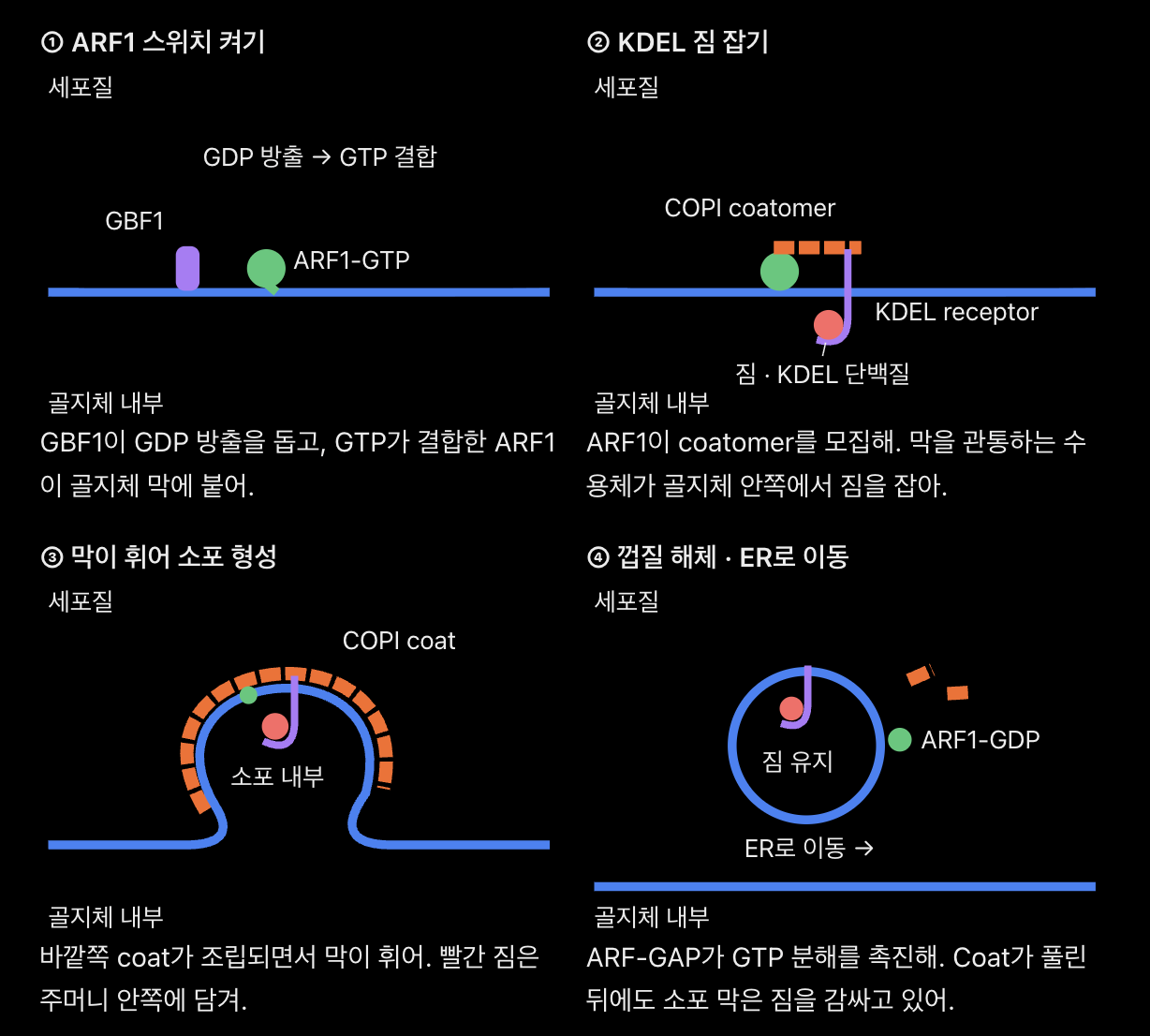
How do COPI coatomer complexes and ARF1 GTPase mediate retrograde vesicular transport?
📦 COPI는 회수용 소포야. 예를 들어, 소포체(ER) 안에서 단백질 접힘을 도와야 하는 단백질이 골지체(Golgi)까지 따라 나왔다고 해 보자. 이 단백질을 골라 ER로 돌려보내는 게 retrograde transport(역행 수송)야. COPI는 골지체의 뒤쪽 구획에서 앞쪽 구획으로 단백질을 돌려보내는 데에도 쓰여.
⚙ COPI 소포가 만들어지는 4단계
골지체 막에서 ARF1 스위치를 켜. 네 노트에서 먼저 고칠 부분은 ARNO → GBF1이야. 포유류의 초기 골지체에서 COPI 조립을 시작하는 대표적인 GEF는 GBF1이야. GBF1이 ARF1에서 GDP가 빠지도록 도우면 GTP가 붙어. 그러면 ARF1의 앞쪽 끝(N-terminus)에 달린 지방산인 myristoyl group과 짧은 나선이 세포질 쪽 막에 자리 잡아. 이제 ARF1-GTP가 그 자리에 coat를 모집할 수 있어.
ARF1-GTP가 coatomer를 한 묶음으로 불러와. 앞에서 배운 COPII는 Sec23/Sec24 → Sec13/Sec31 순서로 조립됐지. COPI에서는 단백질 7개가 이미 coatomer라는 복합체를 이루고 있어. Heptameric은 ‘7개로 구성된’, en bloc은 ‘한 묶음으로’라는 뜻이야.
돌려보낼 짐을 고르면서 막이 휘어져. 여기서는 회수 신호(retrieval signal)가 막의 어느 쪽에 있는지 보면 돼. 골지체 막의 세포질 쪽에 여러 coatomer가 모여 coat를 조립하고, 선택된 짐을 담은 부분이 볼록해져 bud를 형성해.
회수할 짐
신호의 위치
누가 신호를 읽을까?
막단백질
세포질로 나온 꼬리에 KKXX 또는 KXKXX 신호가 있어. K는 lysine, X는 임의의 아미노산이야.
Coatomer의 α-COP와 β′-COP가 이런 dilysine signal을 직접 인식해.
ER 내부에서 일하는 수용성 단백질
골지체 내부 공간(lumen)에 있는 단백질 끝에 KDEL 신호가 있어.
막을 가로지르는 KDEL receptor가 안쪽에서 KDEL을 잡아. 세포질 쪽의 COPI는 이 수용체를 소포에 실어 짐까지 함께 회수해.
ARF1 스위치가 꺼지면서 coat가 풀려. ARF-GAP가 ARF1의 GTP hydrolysis(가수분해)를 촉진해. ARF1에 붙어 있던 GTP → GDP + Pi가 되면 ARF1이 막에서 떨어지기 쉬워지고 coatomer도 해체돼. 이게 uncoating이야. 떨어져 나온 소포는 목적지 막과 융합해 회수한 단백질을 전달해
📌 Core Concept: COPI-coated vesicles drive retrograde transport from Golgi apparatus back to ER and between Golgi cisternae.
⚙️ Mechanism / Process:
• Exchange factor ARNO activates ARF1 GTPase, inserting N-terminal myristoyl group into Golgi membrane.
• ARF1-GTP recruits heptameric coatomer complex (α, β, β′, γ, δ, ε, ζ-COP) en bloc.
• α- and β′-COP bind retrieval tags on cargo or KDEL receptors, polymerizing into a cage.
• ARF-GAP stimulates ARF1 GTP hydrolysis, causing ARF1-GDP and coatomers to dissociate for uncoating.
💡 Biological Significance: Prevents depletion of ER/Golgi resident machinery by continuously retrieving mislocalized proteins.

How does the KDEL receptor retrieve mislocalized RER resident proteins from the Golgi apparatus?
KDEL receptor는 골지체에서 ER 단백질을 붙잡고, ER에 도착하면 놓아줘. 같은 수용체가 장소에 따라 다르게 행동하는 이유는 pH 차이야. 골지체 내부는 ER보다 약간 더 산성이어서 수용체가 KDEL에 잘 결합하고, 중성에 가까운 ER 내부에서는 결합이 약해져. 📦
⚙ BiP 한 분자를 따라가 보자
ER에서 골지체로 빠져나가. BiP는 ER 내부 공간(lumen)에서 새 단백질이 제대로 접히도록 돕는 chaperone이야. 가끔 다른 단백질과 함께 골지체로 딸려 가. BiP의 맨 끝(C-terminus)에는 KDEL이라는 네 아미노산 서열(Lys–Asp–Glu–Leu)이 있어. 이게 ER로 돌려보낼 단백질임을 알려 주는 retrieval signal(회수 신호)이야.
골지체에서 수용체가 KDEL을 잡아. KDEL receptor는 골지체 막을 관통해. 골지체 안쪽을 향한 부분이 BiP의 KDEL 신호를 잡아. 골지체의 약산성 환경에서는 수용체와 KDEL 사이의 binding affinity(결합 친화도)가 높아.
COPI 소포가 둘을 함께 데려가. 여기서 앞 문제와 연결돼. 막 바깥의 세포질 쪽에서는 ARF1-GTP가 COPI coatomer를 모집해. 수용체가 COPI 소포에 실리면, 수용체에 붙은 BiP도 자연스럽게 소포 안쪽에 담겨. BiP는 이동하는 내내 막을 건널 필요가 없어.
ER에서 BiP를 놓아줘. 소포가 ER 막과 융합(fusion)하면 소포 내부가 ER lumen과 이어져. ER의 더 높은 pH에서는 수용체의 결합 친화도가 낮아져서 BiP가 풀려나 다시 단백질 접힘을 도와. 빈 수용체는 골지체로 돌아가 다음 단백질을 회수해.
💡 시험에서 “BiP의 KDEL을 제거하면?” → BiP가 골지체에 도착해도 수용체가 알아보기 어려워져. 따라서 ER로 회수되는 양이 줄고, 더 많은 BiP가 분비 경로를 따라 이동할 수 있어.
📌 Core Concept: KDEL receptor captures escapee soluble RER chaperones containing a C-terminal Lys-Asp-Glu-Leu (KDEL) motif.
⚙️ Mechanism / Process:
• Soluble RER proteins (e.g., BiP, PDI) swept into anterograde flow bind KDEL receptors in Golgi lumen.
• Ligand-bound KDEL receptors engage COPI coatomers via cytosolic domains.
• COPI vesicles transport receptor-bound chaperones back to RER lumen, where neutral pH releases them.
💡 Biological Significance: Maintains critical high concentrations of protein-folding chaperones within RER lumen.


What is the quaternary structure of a clathrin triskelion, and how do adaptor protein complexes (AP1 vs. AP2) link clathrin to cargo?
Clathrin triskelion은 다리가 3개인 격자 조각이고, AP complex는 그 조각을 막에 있는 화물과 연결하는 단백질이야. AP1과 AP2는 서로 다른 막에서 작동하기 때문에, 같은 clathrin을 써도 서로 다른 경로로 화물을 보낼 수 있어. 📦
① 사슬 6개가 어떻게 다리 3개가 될까?
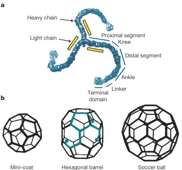
바람개비의 중심을 떠올려 봐. 긴 heavy chain(중쇄) 3개의 C-termini(끝부분)가 중심에서 만나고, 각 heavy chain이 바깥으로 뻗어 다리 하나를 만들어. 짧은 light chain(경쇄)은 각 다리의 중심 가까운 부분에 하나씩 붙어 조립을 조절하고, 세포질에서 격자가 성급하게 형성되는 것을 억제하는 데 기여해.
1 triskelion = 3 heavy chains + 3 light chains = 6개 사슬, 3개 다리. 여러 triskelion의 다리가 서로 겹쳐야 소포를 둘러싸는 polyhedral cage(다면체 격자)가 돼.
② AP는 어떻게 화물과 clathrin을 연결할까?
AP(adaptor protein) complex는 서로 다른 단백질 4개로 된 heterotetramer야. AP가 읽는 신호는 두 종류야. 막의 신호는 어디에서 조립할지 정하고, 화물 꼬리의 신호는 무엇을 실을지 정해.
AP1 | AP2 | |
|---|---|---|
어디에서 작동해? | Trans-Golgi network(TGN)와 endosome | Plasma membrane의 세포질 쪽 |
어떤 막 신호를 읽어? | TGN에서 ARF1-GTP와 막지질 PI4P | 세포막의 막지질 PI(4,5)P₂, 흔히 PIP₂라고 불러 |
대표 경로 | TGN ↔ endosome 사이 화물 분류 | Plasma membrane → endosome으로 들어가는 endocytosis |
AP가 막에 자리 잡으면, 막단백질이나 cargo receptor의 cytosolic tail(세포질 쪽 꼬리)에 노출된 sorting motif(분류 신호)를 인식해. 대표 신호인 YxxΦ에서 Y는 tyrosine, x는 아무 아미노산, Φ는 부피가 큰 소수성 아미노산이야. 또 다른 예는 두 leucine을 포함한 dileucine motif야. AP가 이 꼬리와 clathrin에 결합하면, 선택된 화물이 격자가 자라는 막 부위에 모여.
💡 시험에서 “세포 표면 수용체의 YxxΦ 신호가 손상됐다”고 하면, 세포 표면이라는 단서로 AP2를 고르면 돼. YxxΦ 손상은 그 수용체를 알아보기 어려워졌다는 뜻이므로, 예상 결과는 해당 수용체의 endocytosis 감소야.
Clean Answer
Structure — A clathrin triskelion contains three heavy chains joined at their C-termini and one light chain per heavy chain. Each heavy chain forms one leg.
Cargo linkage — Heterotetrameric AP complexes bind membrane signals, recognize sorting motifs such as YxxΦ or dileucine motifs in cytosolic cargo tails, and recruit clathrin.
AP1 — ARF1-GTP and PI4P recruit AP1 at the trans-Golgi network for trafficking between the TGN and endosomes.
AP2 — PI(4,5)P₂ recruits AP2 at the plasma membrane for clathrin-mediated endocytosis.
📌 Core Concept: Clathrin forms polyhedral cages using 3-legged triskelions, linked to cargo via heterotetrameric Adaptor Protein (AP) complexes.
⚙️ Mechanism / Process:
• Triskelion: 3 heavy chains joined at C-termini + 3 light chains preventing premature cytosolic assembly.
• AP Complexes: Bind PIP2 or ARF1-GTP and capture cargo via cytosolic motifs (YxxΦ or dileucine).
• AP1 vs. AP2: AP1 mediates trans-Golgi network to endosome traffic; AP2 mediates plasma membrane endocytosis.
💡 Biological Significance: Spatially separates post-Golgi export from plasma membrane endocytosis via distinct adaptor signals.
How do dynamin and Hsc70 cooperate to achieve vesicle scission and uncoating during clathrin-mediated endocytosis?
A1.
📌 Core Concept & Definition
Clathrin-mediated endocytosis (CME) is a process in which cells internalize extracellular substances by forming membrane-bound vesicles coated with clathrin, a protein that forms a cage-like structure.
Two proteins perform different but sequential functions:
Dynamin: A GTPase (enzyme that hydrolyzes GTP) that separates the budding vesicle from the plasma membrane (scission).
Hsc70: An ATP-dependent chaperone (protein that helps remodel other proteins) that removes the clathrin coat (uncoating).
Key distinction: Dynamin cuts the vesicle free → Hsc70 removes its protein coat.
⚙ Mechanism & Step-by-Step Process
Give feedback
Step 1. Clathrin-coated pit formation
Adaptor proteins (AP2) recruit clathrin to the plasma membrane and select cargo.
Clathrin triskelions assemble into a curved cage → the membrane bends inward, forming a coated pit.
Step 2. Dynamin-mediated scission (GTP-dependent)
Dynamin assembles into a helical collar around the narrow vesicle neck (the membrane connection between the budding vesicle and plasma membrane).
GTP hydrolysis (GTP → GDP + Pi) drives structural changes in dynamin.
These changes generate mechanical forces that constrict and destabilize the neck → membrane scission releases the coated vesicle into the cytoplasm.
Step 3. Hsc70-mediated uncoating (ATP-dependent)
Auxilin, a co-chaperone, recruits Hsc70 to the newly released clathrin-coated vesicle.
Auxilin stimulates Hsc70 ATP hydrolysis (ATP → ADP + Pi).
Hsc70 binds clathrin and promotes cage disassembly → clathrin proteins are released for recycling.
Step 4. Vesicle transport
- The uncoated vesicle becomes available for trafficking and fusion with an early endosome, where internalized cargo can be sorted.
Sequence to memorize
Clathrin-coated pit
Membrane bends inward
Dynamin + GTP
Neck constriction → scission
Auxilin + Hsc70 + ATP
Clathrin coat disassembly
Uncoated transport vesicle
Trafficking toward an early endosome
💡 Biological Significance & Consequence
Dynamin failure: Coated pits can form but cannot efficiently detach from the plasma membrane → accumulation of membrane-connected coated pits.
Hsc70 uncoating failure: Vesicles can detach but retain their clathrin coats → impaired vesicle maturation, trafficking, and clathrin recycling.
Biological importance: Both proteins ensure efficient receptor and cargo internalization, supporting nutrient uptake and receptor recycling.
🎯 Exam Readiness
Feature | Dynamin | Hsc70 |
|---|---|---|
Enzyme type | GTPase | ATPase / chaperone |
Energy source | GTP | ATP |
Main function | Scission | Uncoating |
Target | Vesicle neck | Clathrin cage |
Key partner | Dynamin oligomers | Auxilin |
Failure | Vesicle remains attached | Vesicle retains coat |
Exam takeaway: Dynamin uses GTP hydrolysis to sever the vesicle neck, whereas auxilin-recruited Hsc70 uses ATP hydrolysis to disassemble the clathrin coat. They act sequentially to produce an uncoated vesicle capable of intracellular trafficking.
📌 Core Concept: Dynamin is a neck-cleaving GTPase, and Hsc70 is an uncoating ATPase essential for releasing functional endocytic vesicles.
⚙️ Mechanism / Process:
• Scission: Dynamin polymerizes into a helical collar around the invaginated vesicle neck.
• Pinching: GTP hydrolysis drives collar constriction, severing the membrane stalk.
• Uncoating: Cytosolic chaperone Hsc70 uses ATP hydrolysis to disassemble the clathrin cage, exposing docking machinery.
💡 Biological Significance: Failure of dynamin or Hsc70 arrests coated pits at plasma membrane, paralyzing endocytosis.
What are the structural components of a Low-Density Lipoprotein (LDL) particle, and how is it recognized by host cell receptors?
Q2. What are the structural components of a Low-Density Lipoprotein (LDL) particle, and how is it recognized by host cell receptors?
A2.
📌 Core Concept & Definition
Low-Density Lipoprotein (LDL) is a spherical lipid–protein particle that transports cholesterol through the bloodstream to peripheral tissues.
Cholesterol is a lipid required for cell membrane structure and steroid hormone synthesis.
Because cholesterol is poorly soluble in water, LDL packages it into a particle that can circulate in blood.
Apolipoprotein B-100 (ApoB-100) is the structural protein that allows LDL to bind to specific cell-surface receptors.
⚙ Mechanism & Step-by-Step Process
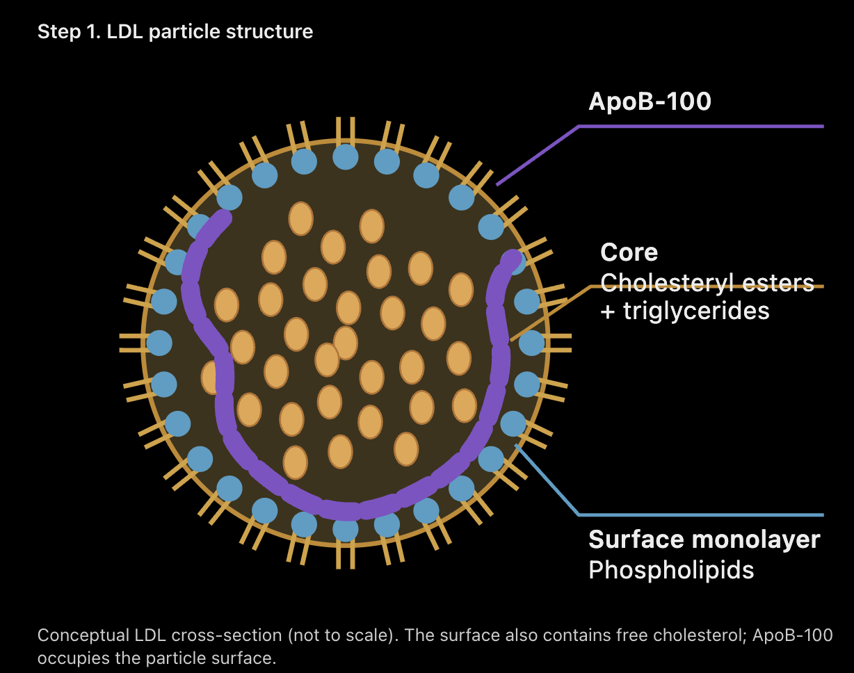
Step 1. LDL particle structure
Conceptual LDL cross-section (not to scale). The surface also contains free cholesterol; ApoB-100 occupies the particle surface.
Outer shell: A phospholipid monolayer containing unesterified (free) cholesterol.
Hydrophilic heads face the surrounding blood plasma.
Hydrophobic fatty acid tails point inward toward the lipid core.
Hydrophobic core: Contains approximately 1,500 cholesteryl ester molecules (cholesterol chemically linked to fatty acids), along with smaller amounts of triglycerides.
Recognition protein: Each LDL particle contains one ApoB-100 molecule embedded across its surface.
Step 2. LDL receptor recognition
LDL receptors (LDLR) are transmembrane proteins located on the cell surface.
The extracellular region of LDLR specifically recognizes and binds ApoB-100 on the LDL particle.
This interaction is noncovalent, meaning LDL and its receptor associate without forming a permanent chemical bond.
Step 3. Receptor-mediated uptake
LDL–LDLR binding → LDL–receptor complexes concentrate in clathrin-coated pits.
Endocytosis → the plasma membrane invaginates and forms an intracellular vesicle.
Endosomal acidification → LDL separates from its receptor.
Receptor recycling → LDLR returns to the plasma membrane.
Lysosomal degradation → cholesteryl esters are hydrolyzed, releasing free cholesterol for cellular use.
💡 Biological Significance & Consequence
Lipid transport: LDL carries water-insoluble cholesterol through the aqueous bloodstream.
Cellular delivery: ApoB-100 allows cells to selectively recognize and internalize LDL.
Cholesterol homeostasis: Cells use LDL-derived cholesterol for membrane synthesis and other metabolic functions.
LDLR dysfunction: Defective LDL receptors reduce LDL clearance from blood → increased plasma LDL cholesterol → greater risk of atherosclerosis (cholesterol-rich plaque accumulation in artery walls).
🎯 Exam Readiness
Component | Location | Main function |
|---|---|---|
Phospholipids | Outer monolayer | Maintain compatibility with blood |
Free cholesterol | Outer shell | Stabilize surface organization |
Cholesteryl esters | Hydrophobic core | Store and transport cholesterol |
Triglycerides | Hydrophobic core | Carry neutral lipids |
ApoB-100 | Particle surface | Bind LDL receptors |
LDLR | Host-cell membrane | Recognize and internalize LDL |
Exam takeaway: LDL consists of a phospholipid monolayer, a cholesteryl ester-rich hydrophobic core, and a single ApoB-100 molecule. ApoB-100 binds LDL receptors noncovalently, triggering clathrin-mediated endocytosis and delivering cholesterol into cells.
📌 Core Concept: Spherical lipid-protein complexes transporting insoluble cholesterol through bloodstream to peripheral tissues.
⚙️ Mechanism / Process:
• Outer Shell: Phospholipid monolayer + unesterified cholesterol with polar heads facing plasma.
• Hydrophobic Core: ~1,500 cholesterol molecules esterified to fatty acids.
• Recognition Protein: Single apolipoprotein B-100 molecule binds transmembrane LDL receptors noncovalently.
💡 Biological Significance: Solubilizes hydrophobic lipids for systemic circulation and receptor-mediated uptake.
What is the complete pathway of clathrin-mediated endocytosis for LDL particles from receptor binding to cytosolic cholesterol release?
A3.
📌 Core Concept & Definition
LDL receptor-mediated endocytosis is a selective process in which cells internalize low-density lipoprotein (LDL) particles and transport them to lysosomes, where cholesterol is released for cellular use.
Key components:
LDL: A particle that transports cholesterol through the bloodstream.
LDL receptor (LDLR): A transmembrane protein that recognizes LDL through ApoB-100.
Clathrin: A protein that forms a cage around developing endocytic vesicles.
Early endosome: An acidic sorting compartment that separates LDL from its receptor.
Lysosome: An organelle containing digestive enzymes that break down LDL and release cholesterol.
⚙ Mechanism & Step-by-Step Process
Step 1. LDL recognition and binding
ApoB-100 on LDL binds noncovalently to the LDL receptor (LDLR) on the plasma membrane.
AP2 adaptor proteins recognize the receptor's cytoplasmic sorting signal and help recruit clathrin.
Clathrin assembly causes the membrane to curve inward → formation of a clathrin-coated pit.
Step 2. Vesicle scission and uncoating
Dynamin (GTPase) assembles around the neck of the budding vesicle.
GTP hydrolysis drives membrane scission → the vesicle separates from the plasma membrane.
Auxilin recruits Hsc70 (ATPase) to remove the clathrin coat using ATP hydrolysis.
The uncoated vesicle delivers LDL and LDLR to the early endosome.
Step 3. Endosomal acidification and receptor recycling
V-type H+ ATPases use ATP to pump H+ ions (protons) into the endosome.
The early endosomal pH decreases to approximately 6.0–6.5.
Increased proton concentration changes the charge and conformation of LDLR → LDL dissociates from LDLR.
Empty LDL receptors are sorted into recycling tubules → transported back to the plasma membrane for reuse.
Step 4. Lysosomal degradation and cholesterol release
LDL remains in the endosomal pathway → transported through late endosomes to lysosomes.
Lysosomal acid lipase (LAL) hydrolyzes cholesteryl esters into free cholesterol and fatty acids.
NPC2 and NPC1 proteins facilitate cholesterol movement out of the lysosomal compartment.
Free cholesterol becomes available to the cell for membrane synthesis, metabolism, or storage.
📌 Core Concept: Selective receptor-mediated internalization delivering LDL to lysosomes for cholesterol extraction.
⚙️ Mechanism / Process:
1. ApoB on LDL binds LDL receptors clustered in clathrin-coated pits via AP2.
2. Dynamin pinches off vesicle; coat disassembles via Hsc70; vesicle fuses with early endosome.
3. Acidic pH (~5.0) causes ligand-receptor dissociation; empty receptors recycle to plasma membrane.
4. Free LDL moves to secondary lysosome where acid hydrolases release free cholesterol into cytosol.
💡 Biological Significance: Supplies essential membrane cholesterol while clearing circulating plasma lipids.
Biological Significance & Consequence
Cholesterol supply: Provides cholesterol needed for cell membranes and other essential cellular functions.
Receptor recycling: Allows LDL receptors to be reused repeatedly, reducing the need for continuous receptor synthesis.
Blood cholesterol regulation: Removes LDL particles from circulation, helping maintain cholesterol homeostasis.
LDLR dysfunction: Impaired uptake → increased circulating LDL → higher risk of atherosclerosis.
NPC1/NPC2 dysfunction: Cholesterol accumulates inside lysosomal compartments instead of reaching other cellular membranes.
Exam takeaway: ApoB-100 binds LDLR → AP2/clathrin-coated pit → dynamin-mediated scission → Hsc70-mediated uncoating → early endosomal acidification → LDLR recycling + LDL delivery to lysosomes → LAL-mediated cholesterol release → NPC1/NPC2-dependent cholesterol export.
Critical distinction: The LDL receptor is recycled, whereas the LDL particle is degraded to release cholesterol.

How does early endosomal acidification via V-type H+ ATPases cause ligand-receptor dissociation and receptor recycling?
A4.
📌 Core Concept & Definition
Early endosomal acidification is the process by which V-type H+ ATPases lower the pH inside early endosomes, allowing internalized cargo to separate from its receptors.
Early endosome: An intracellular sorting compartment that determines whether molecules are recycled or sent for degradation.
V-type H+ ATPase: An ATP-powered proton pump that transports H+ ions into endosomes.
Ligand: A molecule that binds a receptor, such as LDL binding to the LDL receptor (LDLR).
Protonation: The addition of an H+ ion to a molecule, which can change its electrical charge and structure.
Key principle: Acidification changes receptor–ligand binding → cargo separates → receptors can be recycled.
⚙ Mechanism & Step-by-Step Process
Step 1. Proton pumping and acidification
V-type H+ ATPases are embedded in the endosomal membrane.
They hydrolyze ATP → ADP + Pi to pump H+ ions from the cytosol into the endosomal lumen.
H+ concentration increases → endosomal pH decreases to approximately 6.0–6.5 in early endosomes.
The lumen becomes progressively more acidic during maturation, reaching approximately pH 4.5–5.0 in lysosomes.
Step 2. Ligand–receptor dissociation
Increased H+ concentration causes protonation of specific amino acid residues in the receptor.
Protonation alters electrostatic interactions (attractions between charged groups) and can change the receptor's conformation (three-dimensional shape).
In the LDL–LDLR complex, acidic pH promotes a conformational change in LDLR that weakens ApoB-100 binding.
LDL dissociates from LDLR → the receptor and its cargo can be sorted separately.
Step 3. Endosomal sorting and receptor recycling
The early endosome develops distinct tubular and vacuolar regions.
Tubular regions: Concentrate LDL receptors and transport them back to the plasma membrane through recycling pathways.
Vacuolar regions: Retain LDL cargo, which proceeds through late endosomes → lysosomes.
At the plasma membrane's near-neutral pH, recycled LDLR can bind another LDL particle.
💡 Biological Significance & Consequence
Receptor reuse: LDL receptors can undergo many rounds of uptake and recycling, reducing the need for new receptor synthesis.
Efficient cargo sorting: Separates receptors destined for recycling from LDL particles destined for lysosomal degradation.
Cholesterol homeostasis: Supports continuous LDL clearance and cellular cholesterol delivery.
Failure of acidification: Inhibition of V-type H+ ATPases disrupts LDL–LDLR dissociation and endosomal sorting → impaired receptor recycling and cholesterol processing.
🎯 Exam Readiness
Feature | Early endosome | Lysosome |
|---|---|---|
Approximate pH | 6.0–6.5 | 4.5–5.0 |
Main function | Ligand dissociation and sorting | Cargo degradation |
LDL receptor | Recycled | Normally avoids degradation |
LDL particle | Separated from LDLR | Degraded |
Exam takeaway: V-type H+ ATPases hydrolyze ATP to pump protons into early endosomes → lower pH changes receptor protonation and conformation → LDL dissociates from LDLR → receptors enter recycling tubules and return to the plasma membrane, while LDL continues toward lysosomal degradation.
Critical distinction: Acidification does not directly digest LDL. It primarily enables ligand–receptor dissociation in early endosomes; lysosomal enzymes subsequently degrade LDL.

What is the molecular cause and clinical outcome of inherited familial hypercholesterolemia(고 콜레스테롤 혈증)?
🧬 Familial Hypercholesterolemia (가족성 고콜레스테롤혈증)
Q. What is the molecular cause and clinical outcome of inherited familial hypercholesterolemia?
① 📖 이해용 — 콜레스테롤 수거 시스템이 고장 난 이야기🌍 1. Big Picture: 우리 몸에는 콜레스테롤 청소 시스템이 있어!
일단 Cholesterol(콜레스테롤)이 무조건 나쁜 물질은 아니야.
우리 몸은 콜레스테롤을 이용해서 Cell Membrane(세포막)을 만들고, Steroid Hormones(스테로이드 호르몬)도 합성해.
그런데 문제가 하나 있어.
콜레스테롤이 혈액 속에 지나치게 많아지면 혈관에 쌓일 위험이 커진다는 거야.
그래서 우리 몸에는 혈액 속 콜레스테롤을 회수하는 시스템이 존재해.
여기서 등장하는 주인공이 세 명이야.
LDL Cholesterol and Heart Disease
① LDL (Low-Density Lipoprotein)
콜레스테롤을 혈액 속에서 운반하는 입자야. 쉽게 말하면 콜레스테롤 배달 트럭이지.
The structure of bad cholesterol comes into focus
② LDL Receptor (LDLR)
세포 표면에서 LDL을 인식하고 붙잡는 단백질이야. 특히 간세포에서 혈액 속 LDL을 제거하는 데 핵심적인 역할을 해.
Lipoprotein, low density from human plasma CAS No.308068-14-6 Sigma
③ ApoB (Apolipoprotein B-100)
LDL 표면에 있는 단백질이야. LDL Receptor가 LDL을 알아보고 결합할 수 있게 해주는 일종의 신분증이지.
🎬 2. 정상적인 상황: LDL 수거 시스템
간세포가 혈액을 돌아다니는 LDL을 발견했다고 상상해 봐.
이 과정을 Receptor-Mediated Endocytosis(수용체 매개 세포내섭취)라고 해.
왜 굳이 Receptor가 필요할까?
세포는 주변의 모든 물질을 무작정 집어삼킬 수 없어. 따라서 LDL Receptor로 특정 LDL 입자를 선택하고, Clathrin(클라트린)이라는 단백질이 세포막을 소포 형태로 만드는 데 도움을 주는 거야.
🚨 3. 그런데 Familial Hypercholesterolemia에서는?
어느 날 LDL 수거 시스템의 설계도인 Gene(유전자)에 Mutation(돌연변이)이 생겼어.
대표적으로 두 가지 문제가 발생할 수 있어.
Case A — LDLR Gene Mutation
LDL Receptor 자체가 제대로 만들어지지 않거나 작동하지 않아.
그러면 LDL이 Receptor에 결합하지 못하거나, 결합하더라도 세포 안으로 들어가는 과정이 제대로 이루어지지 않을 수 있어.
Case B — ApoB Gene Mutation
이번에는 LDL Receptor가 멀쩡해도, LDL 표면의 ApoB가 변형되어 Receptor에 잘 결합하지 못하는 거야.
즉, 신분증이 잘못 만들어진 셈이지.
결국 두 경우 모두 결과는 비슷해.
Gene Mutation
Defective LDL Binding or Internalization
Reduced LDL Clearance
Elevated Plasma LDL Cholesterol
Increased Atherosclerosis Risk
여기서 중요한 것은 LDL이 아예 움직이지 못하는 게 아니라, 혈액 속에서 충분히 제거되지 못해 농도가 높아진다는 것이야.
❤ 4. 왜 LDL이 많아지면 심장마비까지 발생할까?
혈액 속 LDL이 오랫동안 높은 상태로 유지되면 일부 LDL이 Arterial Wall(동맥벽) 안으로 들어가 축적될 수 있어.
그러면 염증반응이 일어나고, 면역세포가 LDL에서 유래한 지질을 흡수하면서 Foam Cells(거품세포)가 생겨.
시간이 지나면 Atheroma(죽종, 동맥벽에 형성되는 지질성 병변)가 커지고 혈관이 좁아질 수 있어.
특히 Coronary Artery(관상동맥, 심장근육에 혈액을 공급하는 혈관)에 문제가 생기면 위험해.
Atherosclerosis(죽상동맥경화증): 동맥벽에 죽상경화반이 형성되는 질환
Coronary Artery Disease(관상동맥질환): 관상동맥이 좁아지거나 막혀 심장으로 가는 혈류가 감소하는 질환
Myocardial Infarction(심근경색): 심장근육으로 가는 혈액 공급이 급격히 차단되어 심장근육 일부가 손상되거나 죽는 상태
특히 Atherosclerotic Plaque(죽상경화반)가 파열되면 Blood Clot(혈전)이 형성되어 관상동맥을 갑자기 막을 수 있어.
그래서 Familial Hypercholesterolemia 환자는 비교적 젊은 나이부터 심혈관질환이 발생할 위험이 높아지는 거야.
🧬 5. 왜 Autosomal Dominant일까?
Autosomal Dominant(상염색체 우성 유전)는 부모에게서 물려받은 두 개의 유전자 사본 중 하나에 질병을 일으키는 변이가 있어도 질환이 나타날 수 있다는 뜻이야.
LDLR 관련 FH에서는 정상적으로 작동하는 수용체의 양이 부족해지면 LDL 제거 능력이 떨어질 수 있기 때문이야.
다만 모든 환자가 동일하게 심각한 것은 아니야. 유전자 변이 종류와 기능 손상 정도에 따라 콜레스테롤 수치와 발병 위험도 달라져.
📌 Core Concept:
Familial hypercholesterolemia:
an autosomal dominant ( 상염색체 우성, 부모에게서 물려받는 유전자 사본 중 하나에 질병을 일으키는 변이가 있어도 질환이 나타날 수 있는 유전방식) metabolic disorder caused by mutations in genes such as LDL receptor or ApoB genes that impair LDL clearance from the blood.
⚙ Mechanism / Process:
Mutations in LDL receptor or ApoB reduce LDL binding to its receptor. Some LDL receptor mutations also prevent receptors from entering clathrin-coated pits (LDL-수용체 복합체가 클라트린으로 덮인 세포막 함입 부위에 모이는 과정).
As a result, receptor-mediated endocytosis is impaired, reducing LDL uptake by cells and increasing LDL levels in the blood.
Elevated LDL cholesterol promotes the accumulation of cholesterol in arterial walls, leading to atheroma formation (동맥 죽종 형성, 동맥벽에 지질과 염증세포 등이 축적되어 병변이 형성되는 과정).
💡 Biological Significance:
Familial hypercholesterolemia increases the risk of premature atherosclerosis (조기 중증 죽상동맥경화증, 비교적 젊은 나이에 동맥벽에 죽상경화반이 심하게 형성되는 질환), coronary artery disease coronary artery disease (관상동맥질환, 심장근육에 혈액을 공급하는 관상동맥이 좁아지거나 막히는 질환), and myocardial infarction (조기 심근경색, 비교적 젊은 나이에 심장근육의 혈액 공급이 차단되어 조직 손상이 발생하는 상태) at a young age.

How do pathogenic viruses like SARS-CoV-2 highjack host receptor-mediated endocytosis to gain entry into host cells?
① 📖 먼저 이해하기 — 바이러스가 세포 안으로 들어가는 방법
세포에는 원래 외부 물질을 내부로 가져오는 Receptor-Mediated Endocytosis(수용체 매개 세포내섭취) 시스템이 있어.
그런데 SARS-CoV-2는 이 시스템을 이용해 세포 안으로 들어갈 수 있어.
핵심은 세 가지야.
Binding: 바이러스 표면의 Spike Protein(스파이크 단백질)이 세포 표면의 ACE2 Receptor(ACE2 수용체)에 결합해.
Endocytosis: 일부 세포에서는 바이러스와 수용체의 복합체가 Clathrin-Coated Pit(클라트린 피복 함입부)을 통해 세포 안으로 들어가.
Membrane Fusion: Endosome(엔도솜)의 산성 환경에서 Cathepsin(단백질을 절단하는 효소)이 Spike Protein을 활성화하면 바이러스 막과 엔도솜 막이 융합하고 Viral RNA가 Cytoplasm(세포질)으로 방출돼.
그런데 왜 바이러스가 세포 안으로 들어왔다고 바로 감염이 성립하는 건 아닐까?Endocytosis는 바이러스를 막으로 둘러싸인 공간에 넣는 과정일 뿐이야. 바이러스가 복제하려면 Viral RNA가 세포질에 도달해야 해. 그래서 Membrane Fusion이 추가로 필요한 거지.
참고로 SARS-CoV-2는 모든 세포에서 이 경로만 사용하는 건 아니야. TMPRSS2라는 효소가 있는 세포에서는 세포 표면에서 직접 막 융합을 통해 들어갈 수도 있어
Viruses such as SARS-CoV-2 can use host cell receptors and endocytic pathways to enter cells.⚙ Mechanism / Process:
The viral spike glycoprotein binds to ACE2 receptors on the host cell membrane.
In clathrin-mediated entry, the virus-receptor complex is internalized through clathrin-coated pits, which involve adaptor proteins such as AP2.
The virus enters an endosome, where acidic conditions allow endosomal proteases to activate the spike protein. This enables membrane fusion and release of viral RNA into the cytoplasm.
💡 Biological Significance:
This process allows viral RNA to enter the host cell cytoplasm, where viral replication can begin.
How do secretogranin II and chromogranin B (granins) enable high-concentration cargo packaging in the trans-Golgi network?
🧬 Granins: Secretogranin II & Chromogranin B① 📖 이해용 — Golgi의 단백질 압축 포장 시스템🌍 1. Big Picture: 세포는 어떻게 호르몬을 한꺼번에 분비할까?
상상해 봐. 어떤 세포가 호르몬을 만들어서 외부로 내보내야 해.
그런데 문제가 있어.
호르몬을 조금씩 내보내는 게 아니라, 특정 신호가 들어왔을 때 많은 양을 빠르게 분비해야 하는 상황이야.
예를 들어 Endocrine Cells(내분비세포)는 호르몬을 저장해 두었다가 필요할 때 분비해.
그럼 세포는 두 가지 문제를 해결해야 해.
수많은 단백질을 어떻게 작은 Vesicle(소포)에 넣을까?
저장해 둔 단백질을 어떻게 필요할 때 한꺼번에 방출할까?
이때 등장하는 단백질이 바로 Granins(그래닌 단백질군)이야.
Granins에는 Secretogranin II와 Chromogranin B가 포함돼.
얘네는 쉽게 말해서 여러 분비 단백질이 한곳에 모여 고농도로 저장되도록 도와주는 포장 담당자라고 생각하면 돼.
🎬 2. 그런데 왜 그냥 소포에 넣으면 안 될까?
단백질들이 물에 녹아 여기저기 흩어져 있다고 생각해 봐.
그 상태로 작은 소포를 만들면 담을 수 있는 단백질의 양이 제한적이겠지?
그래서 세포는 단백질들을 특정 장소에 모이게 만들어.
그 장소가 바로 Trans-Golgi Network (TGN, 골지체의 출구 쪽에서 단백질을 분류하는 영역)야.
여기에는 특별한 환경이 있어.
Low pH: 약산성 환경
High Ca²⁺ concentration: 비교적 높은 칼슘 이온 농도
이 두 조건에서 Secretogranin II와 Chromogranin B는 Aggregation(응집, 여러 분자가 서로 모여 덩어리를 형성하는 현상)을 일으킬 수 있어
그런데 왜 Low pH와 Ca²⁺가 중요할까?
이 조건들은 단백질의 전하 상태와 분자 사이의 상호작용을 바꿔서 Granins가 서로 모이기 쉽게 만들어.
이렇게 만들어진 응집체는 다른 분비 단백질과 구분되어 Regulated Secretory Pathway(조절성 분비 경로)로 들어가는 데 도움을 줘.
그 결과 작은 소포 안에 높은 농도의 분비 물질을 저장할 수 있어.
이 과정을 Regulated Exocytosis(조절성 세포외배출)라고 해.
소포가 열리면 저장된 분비 물질이 세포 밖으로 방출돼.
세포 밖은 TGN보다 pH가 높고 일반적으로 유리 Ca²⁺ 농도는 낮기 때문에, 일부 Granin 응집체는 분산되거나 용해될 수 있어.
다만 모든 단백질이 반드시 Active Monomer(활성 단량체)로 바뀌는 것은 아니야. 단백질 종류에 따라 성숙 과정에서 절단이나 다른 처리가 필요할 수도 있어.
전체 원리는 다음과 같아.
TGN에서 응집 → 고농도 포장 → 분비 과립에 저장 → 자극 시 Exocytosis → 세포 밖으로 방출
🚀 4. 저장한 단백질은 언제 나올까?
세포가 특정 자극을 받으면 저장된 Secretory Granule(분비 과립)이 Plasma Membrane(세포막)과 융합해.
📌 Core Concept:
Granins, including secretogranin II and chromogranin B, promote the aggregation and concentration of secretory proteins into dense-core granules for regulated secretion.
⚙ Mechanism / Process:
The low pH and high Ca²⁺ concentration in the trans-Golgi network promote the aggregation of secretogranin II and chromogranin B, along with certain secretory cargo proteins.
These concentrated protein aggregates are packaged into regulated secretory granules for storage.
Upon exocytosis, the granules release their contents into the extracellular environment, where changes in pH and Ca²⁺ concentration can promote aggregate dissociation.
💡 Biological Significance:
This mechanism allows cells to store high concentrations of secretory proteins and release them rapidly in response to specific signals.

How do Rab GTPases and tethering proteins establish initial vesicle docking specificity at target membranes?
🧬 Rab GTPases & Tethering Proteins — 소포가 정확한 목적지를 찾는 방법① 📖 이해용 — 세포 안의 택배 배송 시스템🌍 1. Big Picture: 세포 안에는 수많은 배송지가 있어!
세포 안에는 ER, Golgi, Endosome, Lysosome처럼 여러 Organelle(세포소기관)이 있어.
그런데 이 기관들은 서로 물질을 주고받아야 해.
그래서 Vesicle(소포)이라는 작은 운반 주머니에 단백질이나 지질을 담아서 이동시켜.
문제는 소포가 어떻게 자신이 가야 할 목적지를 정확하게 알아보느냐는 거야.
예를 들어 Golgi로 가야 할 소포가 Lysosome에 잘못 도착해서 내용물을 전달하면 곤란하겠지?
그래서 세포는 두 가지 단계를 거쳐 소포가 올바른 막과 융합하도록 조절해.
Rab GTPase + Tethering Protein: 목적지를 인식하고 소포를 붙잡는 역할
SNARE Proteins: 두 막을 가까이 끌어당겨 실제 Membrane Fusion(막 융합)을 일으키는 역할
핵심은 Tethering(초기 포획)과 Fusion(막 융합)은 서로 다른 과정이라는 거야.
🎬 2. Step-by-Step: 택배가 목적지에 도착하는 과정
Step 1. Rab-GTP가 소포 표면에 있어.
Rab GTPase(라브 GTP 가수분해효소)는 세포 내 막 수송을 조절하는 작은 단백질이야.
Rab에는 두 가지 상태가 있어.
Rab-GTP: 활성 상태
Rab-GDP: 비활성 상태
Rab이 GTP와 결합해 활성화되면 특정 Effector Protein(효과기 단백질)과 결합할 수 있어.
여기서 중요한 Effector가 바로 Tethering Protein(계류 단백질)이야.
Step 2. Tethering Protein이 소포를 붙잡아.
목적지 막에는 긴 끈처럼 뻗어 있는 Tethering Protein이 있을 수 있어.
소포가 가까이 오면 Tethering Protein이 소포 표면의 Rab-GTP를 인식하고 결합하는 거야.
이걸 택배 배송에 비유하면, 배송지 직원이 도착한 택배를 붙잡는 것과 비슷해.
그런데 왜 긴 단백질이 필요할까?
아직 소포가 목적지 막에 완전히 가까워지지 않았더라도, 멀리서 소포를 포획할 수 있기 때문이야.
Step 3. 붙잡힌 소포가 목적지 막에 가까워져.
Tethering Protein이 소포를 포획한 뒤, 소포가 Target Membrane(표적막)에 접근하도록 도와.
그러면 두 막에 존재하는 SNARE 단백질들이 서로 만날 수 있는 거리가 돼.
Step 4. SNARE가 다음 단계를 담당해.
이제 두 종류의 SNARE가 등장해.
v-SNARE: Vesicle membrane(소포막)에 있는 SNARE
t-SNARE: Target membrane(표적막)에 있는 SNARE
서로 호환되는 SNARE들이 결합하면 SNARE Complex(복합체)를 형성해.
이 복합체가 두 막을 더 가까이 끌어당기고, 결국 막 융합을 유도하는 거야.
💡 3. 왜 Rab과 SNARE가 둘 다 필요할까?
둘은 하는 일이 달라.
Rab + Tethering | SNARE | |
|---|---|---|
역할 | 초기 인식 및 포획 | 막 융합 |
시점 | 소포가 막에 접근할 때 | 두 막이 가까워진 뒤 |
핵심 기능 | 올바른 수송 경로 선택 | 두 막을 밀착시키고 융합 유도 |
만약 Rab이나 Tethering Protein이 제대로 작동하지 않으면?
소포가 올바른 Target Membrane에서 포획되지 못할 수 있어. 그러면 SNARE가 작용할 기회도 줄어들겠지.
반대로 Rab과 Tethering Protein이 소포를 붙잡았더라도, SNARE가 작동하지 않으면 정상적인 막 융합이 진행되지 않을 수 있어.
즉, Rab은 목적지를 찾고, Tethering Protein은 소포를 붙잡으며, SNARE는 최종 융합을 담당해.
📌 Core Concept:
Rab GTPases(Rab GTP 가수분해효소, 소포의 표적막 인식을 조절하는 단백질) and tethering proteins(계류 단백질, 소포를 표적막 근처에서 포획하는 단백질) help vesicles(소포) recognize and attach to the correct target membranes(표적막) before fusion(막 융합).
⚙ Mechanism / Process:
Active Rab-GTP(활성 상태의 Rab 단백질) on the vesicle membrane(소포막) binds to specific tethering proteins associated with the target membrane.
Tethering proteins capture(포획하다) the vesicle and help bring it closer to the target membrane.
This allows compatible(서로 결합할 수 있는) v-SNAREs(소포막의 SNARE 단백질) and t-SNAREs(표적막의 SNARE 단백질) to interact and form SNARE complexes(SNARE 복합체), which promote membrane fusion.
💡 Biological Significance:
Rab GTPases and tethering proteins improve vesicle targeting specificity(소포가 올바른 표적막을 인식하는 정확성) and reduce inappropriate membrane fusion(잘못된 막 융합), ensuring accurate delivery of cargo(운반 물질) to the correct cellular compartment(세포 내 구획).

Describe the "zippering" mechanism of v-SNAREs and t-SNAREs in driving membrane hemifusion and full fusion.
🧬 SNARE Zippering Mechanism — 두 세포막이 하나로 합쳐지는 원리① 📖 이해용 — SNARE는 어떻게 두 막을 강제로 붙일까?🌍 1. Big Picture: 소포는 목적지에 도착했다고 바로 융합할 수 없어!
앞에서 Rab GTPases와 Tethering Proteins가 소포를 정확한 Target Membrane(표적막)으로 데려오는 역할을 한다고 배웠지?
그런데 여기서 문제가 하나 생겨.
소포가 표적막 바로 앞까지 왔다고 해서 두 막이 저절로 합쳐지는 것은 아니야.
왜 그럴까?
세포막은 Phospholipid Bilayer(인지질 이중층)로 만들어져 있어.
인지질에는 물을 좋아하는 Hydrophilic Head(친수성 머리)와 물을 싫어하는 Hydrophobic Tail(소수성 꼬리)이 있어.
두 막이 융합하려면 막 사이의 물 분자들을 밀어내고, 지질 분자들이 재배열되어야 해. 이 과정에는 Energy Barrier(에너지 장벽)가 존재해.
그래서 막을 서로 가깝게 끌어당기는 단백질이 필요해.
바로 SNARE Proteins야.
🎬 2. SNARE는 마치 지퍼처럼 작동해!
소포막과 표적막에 각각 단백질이 하나씩 연결되어 있다고 상상해 봐.
v-SNARE, Vesicle SNARE, Synaptobrevin, 소포막에 위치
→
t-SNAREs, Target SNAREs, Syntaxin + SNAP-25, 표적막에 위치
이 단백질들이 서로 만나면 나선형 구조인 Alpha-Helix(알파 나선)들이 결합하기 시작해.
여기서 중요한 것은 단백질 4개가 아니라, Alpha-Helix 4개가 모인다는 것이야.
Protein | Alpha-Helix 수 |
|---|---|
Synaptobrevin (v-SNARE) | 1 |
Syntaxin (t-SNARE) | 1 |
SNAP-25 (t-SNARE) | 2 |
Total | 4 |
이 네 개의 나선이 서로 감기듯 결합하면서 Four-Helix Bundle(4중 나선 다발)을 형성해.
그런데 왜 이게 지퍼일까?
처음에는 두 막에서 멀리 떨어진 단백질 끝부분부터 결합하고, 결합이 점점 막 가까운 방향으로 진행되기 때문이야.
이 과정을 Zippering(지퍼링)이라고 불러.
⚙ 3. Zippering → Hemifusion → Full Fusion
Step 1. Zippering — 단백질 지퍼가 잠겨
Synaptobrevin과 Syntaxin, SNAP-25의 Alpha-Helix가 결합하면서 Four-Helix Bundle을 형성해.
결합은 Membrane-Distal End(막에서 먼 쪽)에서 시작해서 Membrane-Proximal End(막에 가까운 쪽)로 진행돼.
왜 이 방향이 중요할까?
단백질들이 결합할수록 소포막과 표적막을 연결하는 부분이 가까워져서, 두 막 사이의 거리가 줄어들기 때문이야.
Step 2. Hemifusion — 두 막의 바깥쪽 층이 먼저 합쳐져
두 막이 충분히 가까워지면 지질이 재배열되기 시작해.
그런데 세포막은 이중층이잖아?
그래서 두 막이 한 번에 완전히 합쳐지기보다는, 먼저 서로 마주 보는 Leaflets(막의 단일 지질층)가 연결되는 중간 단계가 나타날 수 있어.
이것을 Hemifusion(반융합)이라고 해.
이때는 아직 소포 내부와 표적막 반대편 공간이 연결되지 않았어. 따라서 Cargo(운반 물질)가 통과할 수 있는 통로도 없는 상태야.
Step 3. Full Fusion — 드디어 통로가 열려!
SNARE가 만드는 힘과 지질 재배열을 통해 Fusion Pore(융합공)가 형성돼.
이제 소포 내부와 표적막 반대편 공간이 연결돼.
이것이 Full Fusion(완전 융합)이야.
소포가 세포막과 융합하는 경우에는 내부에 있던 Cargo가 세포 밖으로 방출될 수 있어.
💡 4. 왜 SNARE의 Zippering이 중요한가?
SNARE가 없으면 두 막이 충분히 가까워지더라도 효율적인 융합이 어려워져.
반대로 SNARE가 정상적으로 결합하면 단백질 결합 과정에서 방출되는 에너지가 두 막을 밀착시키고 막 융합의 에너지 장벽을 극복하는 데 기여해.
즉, SNARE는 단순히 두 막을 연결하는 단백질이 아니라, 막 융합을 실제로 진행시키는 기계적 힘을 제공하는 단백질인 거야.
📌 Core Concept:
v-SNAREs(소포막 SNARE 단백질) and t-SNAREs(표적막 SNARE 단백질) form a four-helix bundle(4개의 알파 나선으로 이루어진 단백질 복합체) that pulls the vesicle and target membranes together, promoting membrane fusion(막 융합).
⚙ Mechanism / Process:
One v-SNARE (synaptobrevin) combines with three helices(나선 구조) from t-SNAREs (one from syntaxin and two from SNAP-25) to form a four-helix bundle.
The SNARE complex(복합체) assembles through a process called zippering(지퍼처럼 단백질들이 순차적으로 결합하는 과정), starting from the membrane-distal ends(막에서 먼 쪽 끝) and progressing toward the membrane-proximal ends(막에 가까운 쪽 끝).
This pulls the two membranes closer together, promoting hemifusion(반융합), where the contacting lipid leaflets(서로 마주 보는 지질층) merge first.
A fusion pore(융합공, 두 막 사이에 형성되는 통로) then forms, connecting the two compartments(구획) and allowing vesicle contents to pass through.
💡 Biological Significance:
SNARE zippering provides the force needed for membrane fusion, allowing vesicles to deliver their contents to the target compartment.

What is the function of the hexameric AAA-type ATPase NSF in post-fusion SNARE recycling?
📌 Core Concept: NSF is an ATP-driven chaperone that disassembles stable post-fusion cis-SNARE complexes.
⚙️ Mechanism / Process:
• Post-fusion v-SNARE and t-SNARE remain locked in an energetically stable cis-complex on target membrane.
• NSF, assisted by α-SNAP adaptors, binds the 4-helix bundle.
• NSF hydrolyzes ATP to unwind coiled-coils, freeing monomeric SNAREs for subsequent transport cycles.
💡 Biological Significance: Recharges fusogenic machinery; without NSF, all SNAREs become locked and fusion halts.
What is polarized secretion, and how does calcium influx trigger neurosecretory vesicle fusion at presynaptic axon terminals?
📌 Core Concept: Directional exocytosis confined to specific membrane domains, triggered by localized Ca2+ spikes.
⚙️ Mechanism / Process:
• Synaptic vesicles primed at presynaptic active zones by partial SNARE complexing.
• Action potential depolarizes terminal, opening voltage-gated Ca2+ channels.
• Ca2+ influx binds synaptotagmin sensor, releasing fusion clamps and completing full SNARE zippering.
💡 Biological Significance: Enables ultrafast, millisecond-scale neurotransmitter release across the synaptic cleft.
What is the molecular mechanism of tetanospasmin (tetanus toxin), and how does it cause spastic paralysis (lockjaw)?
📌 Core Concept: Clostridium tetani metalloprotease cleaving synaptobrevin in central inhibitory interneurons.
⚙️ Mechanism / Process:
• Cleaves synaptobrevin (v-SNARE) in Renshaw spinal inhibitory interneurons.
• Blocks vesicular exocytosis of inhibitory neurotransmitters (GABA and glycine).
• Disinhibited motor neurons fire continuously, dumping acetylcholine at neuromuscular junctions.
💡 Biological Significance: Causes persistent, violent skeletal muscle contractions, lockjaw, and spastic paralysis.
How does the mechanism of action of botulinum toxin (BOTOX) differ from tetanus toxin, and how is it applied clinically for cervical dystonia and cosmetics?
📌 Core Concept: Clostridium botulinum neurotoxin cleaves SNAREs in peripheral cholinergic motor neurons, causing flaccid paralysis.
⚙️ Mechanism / Process:
• Cleaves synaptobrevin, SNAP-25, or syntaxin at peripheral motor nerve terminals, blocking ACh release.
• Cervical Dystonia: Local injections into overactive neck muscles relax sustained involuntary contractions.
• Cosmetics: Injected into facial muscles to relax contractions, smoothing dynamic wrinkles.
💡 Biological Significance: Provides localized, temporary flaccid muscle relaxation (3–4 months) until new SNAREs synthesize.
What are acid hydrolases, and why are primary lysosomes maintained at an acidic pH (pH ~5.0)?
📌 Core Concept: Digestive enzymes exclusively active in acidic environments, contained safely inside lysosomes.
⚙️ Mechanism / Process:
• V-type H+ ATPases pump protons into lumen, sustaining pH ~5.0 against neutral cytosol (pH ~7.2).
• Acidic environment denatures substrates and optimizes hydrolase catalytic conformations.
💡 Biological Significance: Safety mechanism preventing accidental cytoplasmic autolysis if lysosome accidentally ruptures.
How does heavy glycosylation of lysosomal membrane proteins protect the organelle from self-digestion?
📌 Core Concept: Dense luminal oligosaccharide chains on integral membrane proteins form a protective glycocalyx shield.
⚙️ Mechanism / Process:
• LAMPs and LIMPs acquire extensive N-linked and O-linked glycosylation in RER/Golgi.
• Dense carbohydrate layer physically obstructs proteases from reaching polypeptide backbones and lipid bilayer.
💡 Biological Significance: Prevents acid proteases from digesting the lysosome's own boundary membrane from the inside.
Trace the two-step enzymatic tagging process in the cis- and trans-Golgi that targets soluble hydrolases to lysosomes via Mannose-6-Phosphate (M6P).
📌 Core Concept: Two-step oligosaccharide modification sorting soluble lysosomal enzymes away from constitutive secretion.
⚙️ Mechanism / Process:
1. cis-Golgi: GlcNAc phosphotransferase recognizes hydrolase signal patch, transferring phospho-GlcNAc to mannose.
2. trans-Golgi: Phosphodiesterase (uncovering enzyme) cleaves terminal GlcNAc, exposing active M6P tag.
💡 Biological Significance: Distinguishes lysosomal enzymes from standard secretory proteins lacking the signal patch.
How do Mannose-6-Phosphate (M6P) receptors transport acid hydrolases to late endosomes and recycle back to the trans-Golgi network?
📌 Core Concept: Transmembrane sorting receptors packaging M6P-tagged cargo into endosomal clathrin vesicles.
⚙️ Mechanism / Process:
• M6P receptors bind tagged enzymes in trans-Golgi (pH ~6.5) and recruit AP1/clathrin coats.
• Vesicles fuse with late endosomes; lower pH (~5.0) causes hydrolase dissociation into lumen.
• Empty receptors bud into retromer-coated vesicles and cycle back to trans-Golgi network.
💡 Biological Significance: Efficient delivery of destructive enzymes to lysosomes while preserving receptor pools.
What is the molecular defect causing Inclusion-cell (I-cell) disease, and why does it lead to constitutive secretion of lysosomal enzymes?
📌 Core Concept: Severe lysosomal storage disease caused by defective GlcNAc phosphotransferase activity in the cis-Golgi.
⚙️ Mechanism / Process:
• Inability to attach phospho-GlcNAc prevents formation of M6P targeting signal.
• Without M6P, hydrolases fail to bind M6P receptors in trans-Golgi.
• Enzymes dump into default constitutive secretory pathway and enter extracellular fluids/bloodstream.
💡 Biological Significance: Lysosomes become empty of enzymes, causing massive cytotoxic accumulation of undigested inclusion bodies.
How do primary, secondary (endolysosomes), and tertiary (residual bodies) lysosomes differ in structure and function?
📌 Core Concept: Progressive functional maturation states from newly budded storage vesicles to post-digestive debris containers.
⚙️ Mechanism / Process:
• Primary: Newly budded vesicle from trans-Golgi with inactive acid hydrolases; no substrate.
• Secondary (Endolysosome): Formed by primary fusion with endosome/phagosome; actively digesting contents at pH 5.0.
• Tertiary (Residual Body): Post-digestive vesicle containing indigestible debris; either stored or exocytosed.
💡 Biological Significance: Segregates inactive storage from active destruction, maintaining metabolic homeostasis.
What general enzymatic failure causes Lysosomal Storage Diseases (LSDs), and what specific substrate accumulates in Tay-Sachs disease?
📌 Core Concept: Genetic loss of specific hydrolytic enzymes leading to massive intracellular buildup of undigested macromolecules.
⚙️ Mechanism / Process:
• Mutation eliminates activity of a single lysosomal hydrolase, stalling degradative cascade.
• Undigested substrates progressively enlarge lysosomes, disrupting cellular architecture.
• Tay-Sachs: Deficiency of β-hexosaminidase A blocks breakdown of GM2 gangliosides (sphingolipids).
💡 Biological Significance: Toxic neuronal lipid accumulation causes severe, progressive central nervous system neurodegeneration.
How does recombinant DNA technology provide enzyme replacement therapy (laronidase / Aldurazyme) for Mucopolysaccharidosis I (MPS-I)?
📌 Core Concept: Exogenous administration of functional recombinant enzyme taken up via host M6P-mediated endocytosis.
⚙️ Mechanism / Process:
• MPS-I lacks α-L-iduronidase, accumulating glycosaminoglycans (heparan/dermatan sulfate).
• Recombinant laronidase containing M6P oligosaccharide tags is infused intravenously.
• Host cells bind enzyme via cell-surface M6P receptors, internalizing it to lysosomes to clear debris.
💡 Biological Significance: Halts multi-organ storage pathology and restores metabolic balance without gene editing.
How do plant cell vacuoles function as specialized lysosomes to maintain cell turgor pressure?
📌 Core Concept: Multifunctional lysosomal compartments regulating lytic waste breakdown and osmotic hydrostatic pressure.
⚙️ Mechanism / Process:
• Vacuole lumen contains acid hydrolases for macromolecular turnover.
• V-type ATPases and antiporters accumulate solutes in vacuole, driving water influx by osmosis.
• Generates outward hydrostatic turgor pressure against rigid plant cell walls.
💡 Biological Significance: Keeps non-woody plant stems and leaves upright and rigid while providing lytic recycling.
What secretory functions do modified lysosomes perform in cytotoxic T-cells, mast cells, and melanocytes?
📌 Core Concept: Secretory lysosomes combine degradative storage with regulated, stimulus-triggered exocytosis.
⚙️ Mechanism / Process:
• Cytotoxic T-Cells: Exocytose perforin and granzymes to punch holes and induce apoptosis in target cells.
• Mast Cells: Exocytose histamine and proteoglycans driving immediate inflammatory reactions.
• Melanocytes: Transfer melanin pigment granules (melanosomes) to surrounding keratinocytes.
💡 Biological Significance: Re-purposes standard lysosomal organelles for specialized immune killing and UV photoprotection.
How does lysosomal exocytosis contribute to rapid plasma membrane repair following cellular injury?
📌 Core Concept: Emergency resealing response where intracellular lysosomes fuse with disrupted plasma membranes.
⚙️ Mechanism / Process:
• Membrane lesions trigger massive influx of extracellular Ca2+ into cytosol.
• Local Ca2+ elevation induces rapid lysosomal mobilization and exocytosis at wound boundary.
• Lysosomal membrane patches incorporate into plasma membrane, sealing the mechanical disruption.
💡 Biological Significance: Prevents lethal cytoplasmic loss and restores cellular integrity within seconds of injury.