L04 - pk2 Pressure and Compliance
 Call Kai
 Learn
Learn Practice Test
 Spaced Repetition
 Match
Match Flashcards
 Knowt Play
1/45

There's no tags or description
Looks like no tags are added yet.
Name | Mastery | Learn | Test | Matching | Spaced | Call with Kai | Chat |
|---|
No analytics yet
Send a link to your students to track their progress
46 Terms
environmental and genetic factors affecting lung volume
Height (taller > smaller)
Sex ( males > females)
Age
Race ( European>Asian)
Respiratory Disease
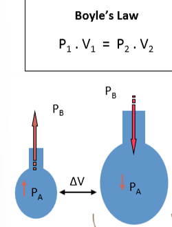
Boyle’s law
altering the thoracic volume leads to a change in alveolar volume which results in air flow
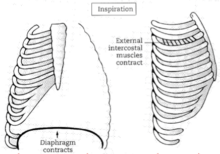
diaphragm and internal/external intercostal muscles during inspiration
external intercostals
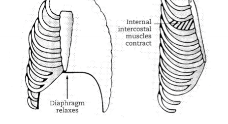
diaphragm and internal/external intercostal muscles during expiration
internal intercostals
respiratory muscles do work to: (2)
to stretch the elastic components of the respiratory system
to overcome the resistance to flow
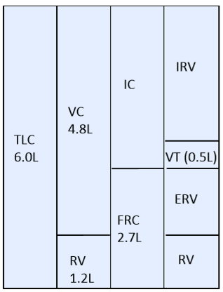
key indicator of lung function
vital capacity
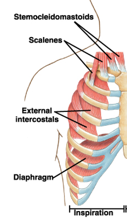
accessory muscles of forced inspiration
Sternocleidomastoids
Scalenes
External intercostals
Contraction of these muscles results in an increase in the lateral and anterior-posterior dimension of the thoracic cage -> greater decrease in PA
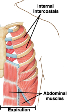
accessory muscles of forced expiration
Internal intercostals
Abdominal muscles
abdominus rectus
abdominal obique muscles
external
internal
transversus abdominis
oxygen consumption by respiratory muscles at rest
Respiratory muscles use 5% of oxygen consumption at rest
what is between the parietal and visceral pleura
pleural space containing a small amount of cohesive, lubricating and nonexpanding, pleural fluid.
how do the lungs adhere to the chest wall (simple version)
Each lung has its own pleural covering
Each lung acts separately from each other in terms of this coupling
Lung adheres to the chest wall via negative pressure in the intrapleural space.
Lung-thorax coupling
Each lung acts separately from each other in terms of this coupling
Lung adheres to the chest wall via negative pressure in the intrapleural space.
Negative pressure (lower than atmospheric pressure) is created in the pleural space:
At every lung volume healthy lung tends to recoil inwards (collapse) and at most lung volumes the thoracic cage tends to recoil outwards and pull away from the lungs.
[Explanation: As a result, they are trying to increase the volume of the pleural cavity which in turn results in a decrease intrapleural pressure → negative]
This creates negative intrapleural pressure (which is exerted on the pleural fluid) which allows for mechanical coupling of the lungs and thorax (the visceral & parietal pleura push against one another)
Therefore, if the chest wall pulls outwards the lung will come with it
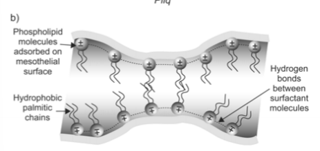
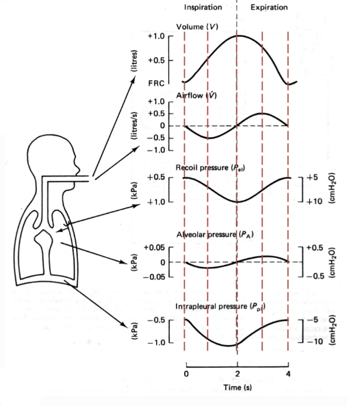
Explain the following image in terms of lung-thorax coupling
Actual touching between opposing pleurae does not occur because of repulsive forces between several layers of phospholipids adsorbed on mesothelial surfaces carrying charges of the same sign
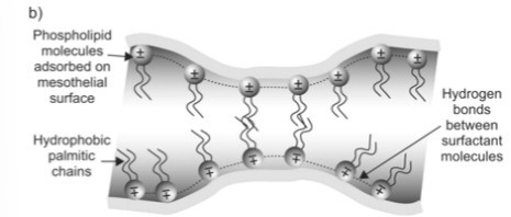
What is occurring at FRC
At FRC, when respiratory muscles are relaxed, the outward and inward recoils are equal and opposite - there is a negative intrapleural pressure but the respiratory system is at equilibrium
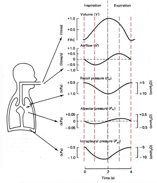
Explain what is happening at t=0
comment on lung volume, air flow, intrapleural pressure
Lung volume = FRC
Air flow = 0 as alveolar pressure is 0 relative to barometric pressure
Negative intrapleural pressure -> allows for coupling of lungs and thorax (-0.5kPa)
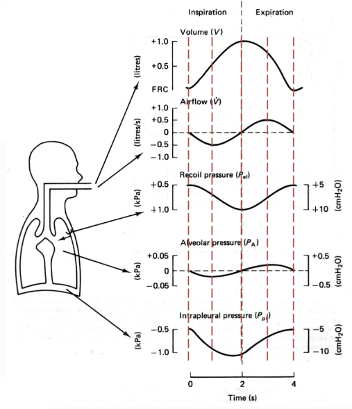
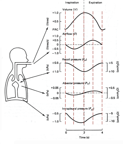
explain what is happening at the 2nd line
Half way through inspiration
peak inspiratory air flow → increase in lung volume
Air flow is at its peak here as we have the greatest difference between alveolar pressure and barometric pressure
Intrapleural pressure becomes more negative during inspiration:
The chest wall moves outwards and the lungs recoil pressure becomes greater → as a consequence, this makes intrapleural pressure more negative (coupling lung more strongly to chest wall)
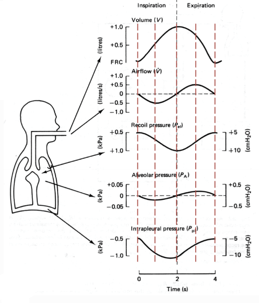
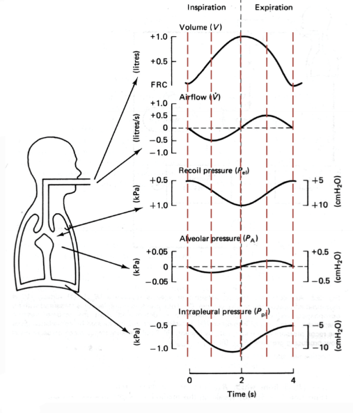
explain what is happening at the 3rd line t=2
Air flow = 0
as alveolar pressure is now equal to barometric pressure
Max lung volume
Intrapleural pressure is now at its most negative:
As Lung is at its greatest recoil → greatest volume and greatest tendency to collapse.
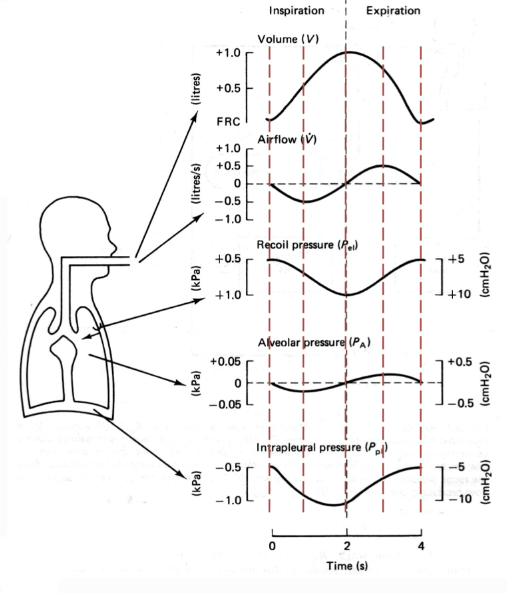
explain what is happening at the 4th line
half way through expiration
Peak expiratory air flow → results in an decreasing in lung volume
Air flow is at its peak here as we have the greatest difference between alveolar pressure and barometric pressure
Intrapleural pressure also rises back to resting FRC value
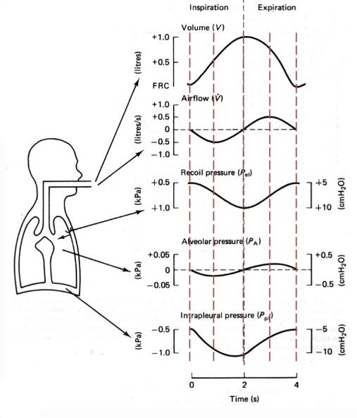
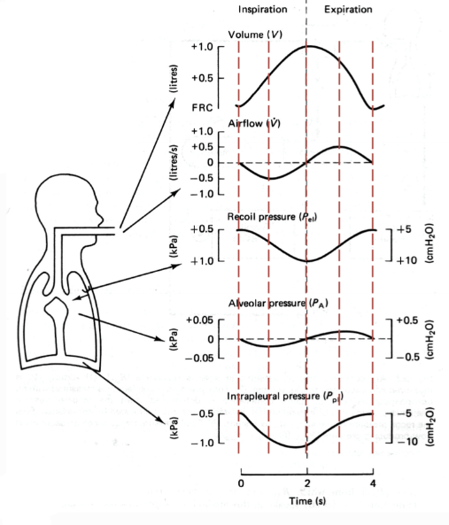
explain what is happening at the 5th line
Lung volume = FRC
Air flow = 0 as alveolar pressure is 0 relative to barometric pressure
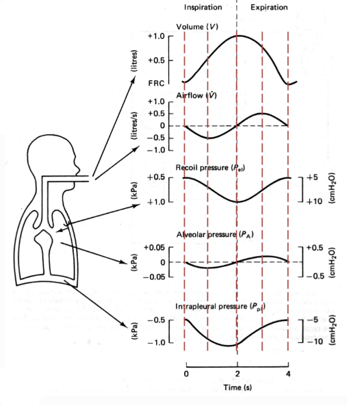
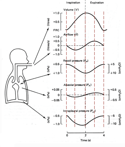
1. Airflow profile follows PA
2. During expiration, both Ppl and PA rise
3. PA is always > Ppl
4. In quiet breathing, Ppl is always negative whilst PA is negative in insp and positive in exp
5. Airflow ceases when PA is zero
6. At high ventilation, Ppl and PA changes are increased.
7. Ppl can be +ve in forced expiration (e.g. +8kPa in coughing/sneezing)
due to increased compression into pleural space
what is compliance
and what is meant by high compliance
A measure of the ‘distensibility’ of an elastic structure
High compliance = lungs can stretch easier for smaller pressure changes. High change in volume, for a fixed change in pressure
Elastic recoil of the lungs brings it back
how to measure compliance
equation and conditions
In order to measure the compliance of lung or Thoracic cage we need to remove air way and air flow resistance.
Therefore, compliance is measured during static manoeuvres – when air is not moving.

distending pressure calculation
Distending pressure = Pin - Pout
Positive Distending pressure: Pin > Pout so this the structure is being distended
Negative distending pressure: Pin < Pout so the structure is being compressed.
calculating distending pressure for the lungs
Pressure in = PA
Pressure out = Ppl
Therefore, distending pressure is PA- Ppl
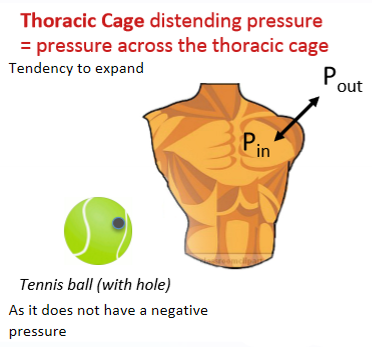
calculating distending pressure for the thoracic cage
Pressure in = Ppl
Pressure out = PB
Therefore, distending pressure is Ppl - PB
thoracic distending pressure
lung distending pressure
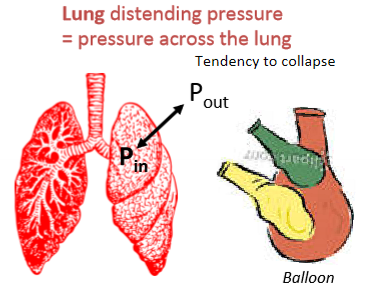
can lung volume reach 0
It is not possible for lung volume to reach 0 due to the presence of the residual volume.
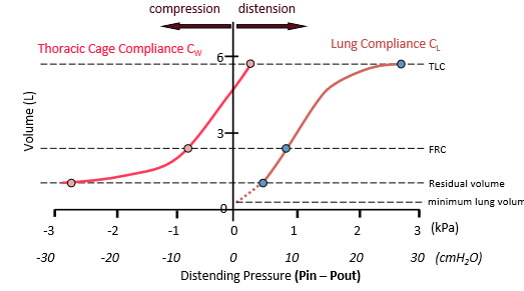
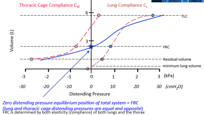
lung and thoracic compliance curve
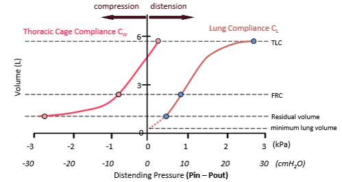
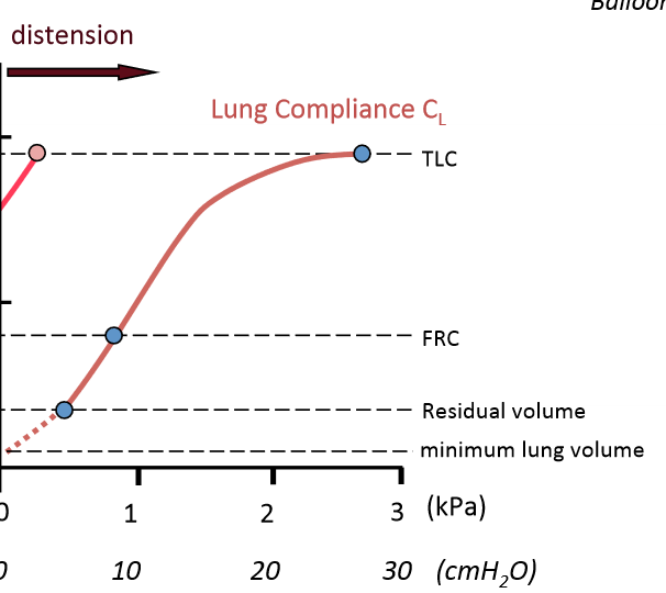
explaining the lung compliance curve
As distending pressure increases the volume of the lungs also increases
This continues to TLC – where we reach elastic limit of the lungs is reached.
NB: A lung volume of 0L occurs when there is no distending pressure.
This is the equilibrium volume of the lung (=0L)
Equilibrium volume of structure is found at zero distending pressure – this is where elastic structure would like to be is where there is not distending pressure on it. Where elastic structure is attempting to recoil towards.
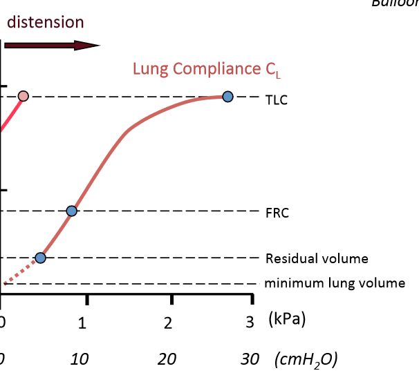
explaining the thoracic cage compliance curve
At high volumes the thorax is being distended.
If we take a deep breath in to TLC at this point we stretch thoracic cage beyond its equilibrium volume so it will now have the tendency to collapse.
The chest walls equilibrium volume is around 5L.
When we are breathing in the chest wall is moving towards where it wants to go (being helped) but after large inspiration chest wall wants to collapse. (at the point where it crosses the y axis)
specific compliance and its importance
specific compliance = compliance/FRC
Lungs of a baby and adult have similar compliances but different specific compliances
Flatter lung = low compliance
at what point is the equilibrium volume reached
Equilibrium volume found at zero distending pressure
Only thoracic cage compliance curve crosses zero distending pressure
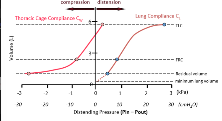
Lung compliance lowest towards TLC
if you take a deep breath in and try to breathe again it is very difficult to breathe.
Normal compliance ca. 1.5L.kPa-1
Lung and thoracic cage compliance similar around FRC
slope of line is identical → so chest wall and lung are similar in there stretchiness.
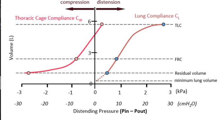
Compliance (slope) greatest around FRC
why is this convenient
This is convenient as this is where the region where we breathe
So this is the region that is easiest to breathe – requires the least amount of energy.
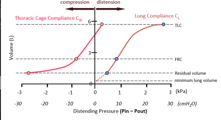
why is total system compliance considered
The lungs lie within the thoracic cage and both need to be stretched together during inspiration - so we need to consider the ‘total’ system compliance.
calculating total system compliance

what is FRC in terms of distending pressure
why does a pneumothorax occur
Occurs if the lung perforates or there is a penetrating wound of the chest wall, air enters and fills the pleural space from the lung or from the atmosphere.
Distending pressures lost
Ppl = PB = 0 kPa
The lung collapses & the thoracic cage expands with both structures moving towards their respective equilibrium volumes
result of pneumothorax on coupling
As a result, breathing becomes uncoupled (lungs no longer move as chest wall moves) → thoracic cage can still move if innervation intact but breathing is painful, difficult or impossible.
types of pneumothorax (4)
primary spontaneous
secondary
traumatic
tension
primary spontaneous pneumothorax
signs/symptoms + treatment
most common
Occur often in tall thin young individuals
Small blebs on lung → ruptures → internal lung perforations
outpatient treatment
seconadary pneumothorax
associations + treatment
Associated with respiratory disease (COPD, asthma)
requires intercostal tube drainage
traumatic pneumothorax
cause + treatment
Following blunt or penetrating chest trauma or mechanical ventilation
Requires intercostal drainage
Tension Pneumothorax
cause and result
Mechanical ventilation or trauma
“Mediastinal shift” - shock
movement of heart & trachea due to air rushing in – great veins are blocked off
Tension Pneumothorax
treatment
Medical emergency - immediate drainage with 14-G needle
allows air to escape from pleural space
so much air flowing into pleural space; wound acts as a valve preventing outflow of air