Red Meat Pathology 1: Sheep
 Call Kai
 Learn
 Practice Test
 Spaced Repetition
 Match
 Flashcards
 Knowt Play
1/25

There's no tags or description
Looks like no tags are added yet.
26 Terms
Most lesions in sheep are usually seen in the ____.
Lungs
Describe the classic appearance of enzootic pneumonia.
Local Rejection of Lungs
Cranioventral consolidation - caused by mycoplasma ovipneumoniae

Describe the classic appearance of fibrinous pneumonia.
Local Rejection of lungs
Bilateral or unilateral - caused by mannheimia haemolytica

Describe the classic appearance of ovine pulmonary adenocarcinoma.
Pink areas of central consolidation
Local rejection of lungs
Also find frothy fluid in airway
Caused by JSRV: Jaagsiekte Sheep Retrovirus

Describe the classic appearance of verminous pneumonia - muellaris capillaris.
Common agent is muellarius capillaris - multifocal lesions on the dorsal aspect of the lungs
Also causes lead shot lesions - chronic lesions
Tend to coalesce and get larger
Acute lesions: hemorrhagic, very congested
Larvae in interstitium - adults in bronchi + bronchioles
di

Describe the classic appearance of verminous pneumonia - dictyocaulus filaria.
Adults found in bronchi - larger airways

Describe the classic appearance of lung abscesses.
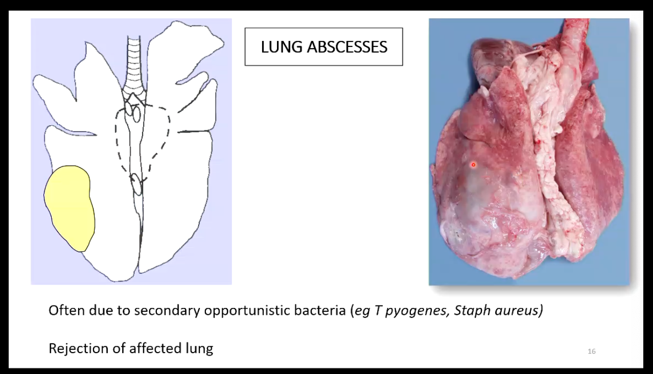
Describe the appearance of hydatidosis.
• regional incidence
• intermediate stage Echinococcus granulosus
• cysts usually in lung
other major organs possible eg liver
unilocular, fluid-filled, thick capsule
• REJECT pluck and any affected organs, carcass goes through
• zoonosis - check farmer and family

Describe the appearance of caseous lymphadenitis.
• Corynebacterium pseudotuberculosis
• lungs only = LOCAL
• check rest of carcase though!

Describe the appearance of maedi visna.
Viral Pneumonia - overinflated lungs (Retrovirus)
Lungs palpate - rubbery

Describe the appearance of blood splashing.
• sudden blood pressure increase due to electrical stunning
ecchymotic or petechial haemorrhages
lung, heart and occasionally skeletal muscle
• if numbers increase
• check
stunning procedure
amperage may be too high

Describe the appearance of pericarditis.
• part of the pasteurellosis complex
• acute pericarditis
fibrinous exudate
RARE in abattoir
• residue lesions more common
fibrous adhesions between layers of pericardial sac

Describe the appearance of cysticercosis or sheep measles.
CYSTICERCOSIS (C. ovis) also called Sheep Measles
• dog tapeworm
• Taenia ovis
• sheep ingest dog faeces
Intermediate host
• larvae develop in sheep muscles
Cysticercus ovis
• check heart, diaphragm and other skeletal muscles including the heart, diaphragm, and skeletal muscles
**NB: MOC: Meat infected with cysticerci shall be declared unfit for human consumption. However, where the animal is not generally infected with cysticercus, the parts not infected may be declared fit for human consumption after having undergone a cold treatment. Individual/low numbers of cysts found = local. Generalised = total.

Describe the appearance of osteomyelitis.
Osteomyelitis is a serious infection and inflammation of the bone and bone marrow, usually caused by bacteria (such as Staphylococcus aureus)

What species account for the main rejections or causes of liver damage in sheep?
Fasciolosis and cystericosis
Describe the main features of fasciolosis.

Describe the pathology caused by the migratory phase of fasciolosis.
**Possible sequela - vessel rupture - massive haemorrhage

Describe the pathology caused by chronic fasciolosis.

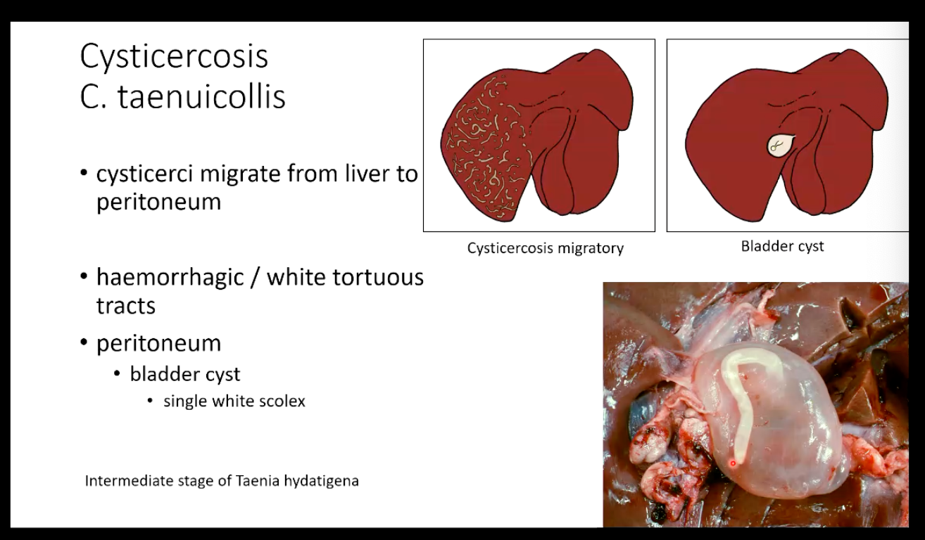
Describe the main features of cysticercosis c. taenuicollis.
Causes serpentine lesions in livers as well
Describe the main pathological features of grid in sheep.
Coenurus cerebralis = larval form of dog tapeworm Taenia multiceps occasionally infests other ruminants
Larva enters CNS by haematogenous route zoonosis
Clinical Signs: relate to space occupying lesion in brain or cord (blindness, nystagmus, deviation of head, eyes, ataxia, collapse)
Diagnosis: Parasitic induced cerebral cysts
Cause: Coenurosis due to Coenurus cerebralis, the immature stage of the tapeworm seen as the adult Taenia multiceps in the canids.

What are the main control measures to minimize the incidence of taenia spp. infections in sheep?
• don't feed farm dogs with uncooked meat / raw diet
• remove carcasses of dead sheep as soon as possible
• dispose of sheep carcasses as ABP category 2
• regular dosing of dogs with anthelmintic (praziquantel)
at least every three months
• farmer to put notes in public footpaths running through sheep fields explaining the disease risks and encouraging dog walkers to worm their dogs regularly
Describe some common kidney pathologies.

Describe the main pathology caused by Johne’s bacteria.
Most affected area is the ileum
Check mesenteric lymph nodes
Mycobacterium avium subsp. paratuberculosis

Describe the main features of small intestinal adenocarcinoma.
“Kissing tumour”

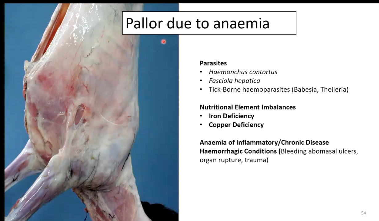
What are the main causes of pallor (Due to anemia) in sheep?
What are the main causes of carotenoid pigment accumulation in adipose tissue?
