3) Visual Fields
 Call Kai
 Learn
 Practice Test
 Spaced Repetition
 Match
 Flashcards
 Knowt Play
1/48

There's no tags or description
Looks like no tags are added yet.
49 Terms
Detect pathology
Monitor disease progression
Evaluate treatment
Assess visual ability

Where is the physiological blind spot located?
15° temporal to fixation
lies 2/3 lies below the horizontal meridian
Function: used to verify reliability of VF test

What are the 12 factors affecting the visual field?
VA
Refractive status (high myopia)
Education, attentiveness, cooperation
Stimulus size
Distance from eye
Stimulus duration
Background illumination
Stimulus intensity
Contrast
Color
Patient anatomy (dermatochalasis)
Light/dark adaptation
Visual sensitivity at that location
At fixation/fovea, where sensitivity is greatest

Sensitivity drops to 0
→ logarithmic unit used to express stimulus intensity/sensitivity
0 dB = maximum brightness
↓ dB stimulus = brighter light
↑ dB stimulus = dimmer light
20–30 dB
What is the limit of foveal vision?
40 dB - dimmest stimulus you can present & pt can see it
What is the absolute blindess threshold?
-10 to -20 dB
What does a high dB vs low dB sensitivity value indicate?
High dB = Good sensitivity - pt detects dimmer stimuli
Low dB = Poor sensitivity - pt requires brighter stimuli
What is the difference between Visual Sensitivity and Visual Threshold?
Visual Sensitivity = minimum perceivable brightness
Visual Threshold = minimum quantity of stimulus that can be detected

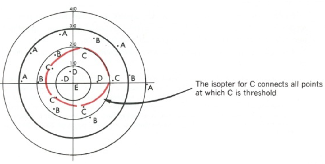
What is an Isopter?
→ line connecting VF points with the same threshold for a specific stimulus
Boundary between the area of visibility and area of non-visibility for a particular stimulus
particular stimulus can’t be seen anywhere outside the isopter
particular stimulus can be seen anywhere inside the isopter
What is a scotoma? What’s the difference between a relative vs absolute scotoma?
→ area of visual field loss surrounded by relatively normal VF
Relative scotoma: area where dim/small stimuli can’t be seen but brighter/larger ones can
Absolute scotoma: area where no stimulus can be detected, even at max brightness

0 dB
What is a depression?
→ Generalized reduced sensitivity without a surrounding normal area
e.g., Cataracts and uncorrected RE
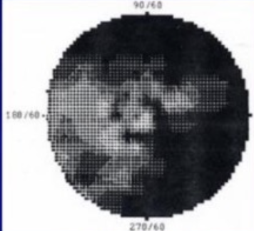
How is the hill of vision made?
→ by plotting multiple isopters representing diff sensitivity levels to (done to determine the prescence or absence of an absolute scotoma)
2-3 isopters are tested
hill peak = area where dim, small stimuli are visible
lower hill = where a stimulus must be made more brigther or larger to be seen

What type of VF is this?

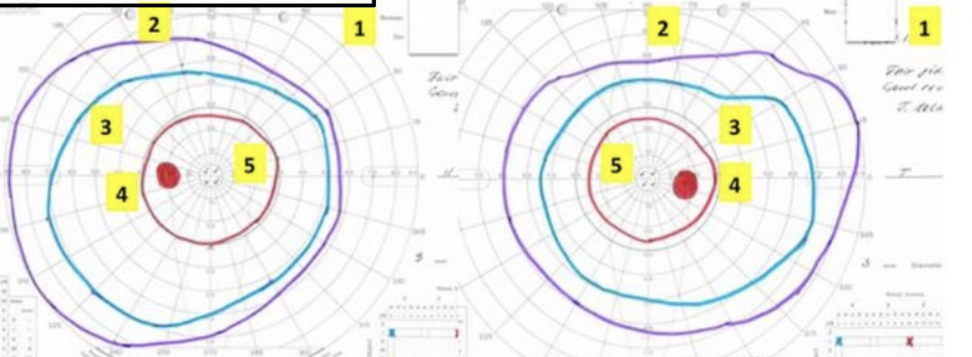
Goldmann VF plot - isopters are represented at rings
How are sensitivity differences represented on Humphrey VF plots?
via numerical sensitivity values (dB)
What are the 4 manual visual field testing methods?
Amsler Grid
Confrontation VF
Tangent Screen
Goldmann Perimetry
What are 3 automated VF testing methods?
Humphrey VF
Octopus Perimetry
FDT
Screening and monitoring central VF/macular abnormalities by testing the central 20° total, or 10° in each direction from fixation
monocular test

How does a scotoma vs metamorphopsia appear on Amsler grid?
Scotoma - missing or blurry area
Metamorphopsia - straight lines appearing wavy

Suprathreshold
List the major limitations of the Amsler Grid.
Suprathreshold stimulus
No fixation monitoring
Poor scotoma mapping/repeatability
Need for good near vision
Poor peripheral sensitivity
What is the confrontation VF testing?
→ Quick screening for VF defects
performed static or kinetic
kinetic is less sensitive to picking up threshold defects → underestimate VF defect
simultaneous stimulation can be used to detect subtle field defects & visual extinction (e.g., parietal-occipital lesions)
temporal and nasal field can be compared (i.e., red desaturation to check for chiasmal lesions)
What are the limitations of confrontation VF testing?
suprathreshold stimulus
pt cooperation
subtle defects may be missed
What is the Tangent Screen (Bjerrum Screen) test?
→ Manual kinetic monocular test
black felt screen with subtle lines sewn in (marking degrees from fixation)
stimulus is moved from non-seeing to seeing area & pt reports when the target first appears/disappear

List the indications for performing Tangent Screen.
used to map 25 - 30o
used to map any suspected VF defect if other methods of testing are not available
used in suspected malingerers and non- organic VF loss (e.g., hysterical fields/functional field loss/tubular field)
determine visual impairment can’t be explained by a pathology/structural abnormality → Diagnosis of exclusion

a field that fails to expand appropriately when testing distance increases
What should happen to the physical size of a normal VF plot when moving from 1 m to 2 m vs a tubular field (e.g., hysterical VF loss)?
Normal = should expand
Tubular field = remains same physical size
No - functional visual loss and malingering are different, and further evaluation is needed
→ systematic measurement of the visual field using a perimeter
detects, quantifies, localizes, and monitors VF damage
List the indications for performing Perimetry.
Look for damage in:
Anterior segment
Retina
Optic nerve
Chiasm
Retrochiasmal pathways

What are the 2 main ways to classify perimetry?
Manual vs. automated
Automated = Goldmann, Humphrey, Octopus, FDT
Static vs. kinetic
What’s the difference b/w static vs kinetic perimetry?
Static: tests a stationary stimulus at a fixed location while varying brightness to determine threshold
measurement of threshold values
better for detecting small sensitivity changes
Kinetic: moving stimulus from a non-seeing area into a seeing area
measurement of isopters
better for mapping peripheral field boundaries
What is Goldmann perimetry?

→ Manual kinetic perimetry
3 stimulus factors that can be changed:
Size (I - V)
Luminance (1-5)
Filter (a-e)
move stimulus from non-seeing to seeing (this marks the outer perimeter of the VF)
List the indications for Goldmann perimetry.
Can’t perform static perimetry
Non-organic disorders (similar to Tangent Screen)
Pt w/ true VF defect but not making sense
Peripheral visual field defects
Difficult patients
Which meridian is especially important when evaluating glaucoma vs neurological lesions?
GLC = horizontal meridian
Neurological lesions = vertical meridian
How is Goldmann perimetry modified for glaucoma suspects, and why?
→ Plot more points along the nasal edge of the isopter
g/c GLC causes nasal steps due to damage to arcuate nerve fibers
Test every 3–5° (instead of standard 15°)
Test 15° above and below the horizontal raphe → GLC defects respect the horizontal meridian b/c ause superior and inferior retinal nerve fiber bundles don’t cross the horizontal raphe

On both sides of the vertical meridian
Which 3 Goldmann stimuli are repeated for glaucoma and neurological suspects?
I-4e (peripheral)
I-3e (intermediate)
I-2e (central)
pt name
date
Rx
pupil size
eye tested
fixation/cooperation
stimulus sizes
What is used to distinguish different isopters on Goldmann plots?
Different colored lines
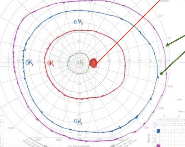
How do scotomas appear on Goldmann plots?
Filled in
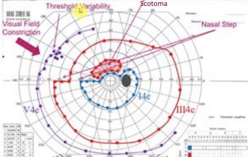
What is the purple arrow pointing to?

Nasal step - GLC suspect
physiological blind spot is always temporal to fixation (think of glasses!)
→ abnormal kinetic plot where successive responses spiral inward or outward (hysterical/functional)
suggests non-organic/functional VF loss

tubular visual field