mock eOSPE 2025
 Call Kai
 Learn
 Practice Test
 Spaced Repetition
 Match
 Flashcards
 Knowt Play
1/10

There's no tags or description
Looks like no tags are added yet.
11 Terms

A 65-year-old female. Fell down flight of steps. Deformity to left shoulder, query fracture or dislocation.
Antero-posterior left shoulder and lateral scapula views.
Abnormal.
There is an unstable fracture-dislocation, of the left
glenohumeral joint – involving anterior dislocation of the
humeral head.
The humeral head is displaced medially and inferiorly - in a sub-coracoid position.
There is also an associated, displaced, intra-articular fracture extending through the humeral head traversing obliquely across the posterolateral aspect of the anatomical neck and through the greater tuberosity (Hill-Sachs deformity) bisecting the biciptal grove.
There is also a lipohaemarthrosis of the affected glenohumeral joint.
No other bony or soft tissue abnormality present

A 71-year-old female, fell in the nursing home.
Anterio-posterior pelvis and both hip joints
Abnormal.
There is an oblique sclerotic band bisecting the
junction of the left femoral head/neck, disrupting the bony cortex and trabeculae.
The appearances are consistent with an impacted/overlapping intracapsular fracture of the proximal femur.
Incidental note is made of degenerative features to the acetabula and lower vertebrae and the presence of phleboliths, none of which is of any consequence.

A 7-year-old male. Inversion injury to foot when transferring from wheelchair. Bony tenderness and swelling base of right 5th MT. Query fracture?
Dorsiplantar and dorsiplantar oblique views right foot.
Abnormal
There is evidence of a polyostotic, sclerosing bone dysplasia– typical of aberrant osteoblastic activity.
There is a lack of differentiation between the cortices, trabeculae and medullary cavities of the visualised bones.
The right cuboid, lateral cuneiform and calcaneus exhibit a
‘bone-in-bone’ appearance. The metatarsals and phalanges, along with the distal tibia and fibula, assume a ‘club-like’ shape with some metaphyseal splaying. The appearances are consistent with osteopetrosis (aka ‘Marble Bone Disease’ or ‘Albers-Schonberg Disease’ ).
Despite these appearances, there is no acute bone or joint abnormality identified., and no signs of pathological fracture.
The skeletal age is consistent with the patient’s chronological age (7 years).

A 35-year-old female complaining of mid thoracic back pain.
Anteroposterior and left lateral views of thoracic spine.
Normal.
The vertebrae appear normal with no signs of lytic or
sclerotic involvement.
Vertebral alignment is unremarkable, and there is no stepping or interruption seen in the anterior vertebral, posterior vertebral or spino-laminar lines.
Normal intervertebral disc spacing is evident, no evidence of significant degenerative changes.
No soft tissue or bony abnormality seen.

An 85-year-old male, with known pneumonia. Admitted to ward and post nasogastric tube insertion.
Antero-posterior erect chest.
Abnormal.
The nasogastric tube is incorrectly sited and must be removed!
The tube is seen right of midline within the thoracic inlet, and then deviates down the left main bronchus, instead of crossing the carina. It fails to pass below the diaphragms , in the midline, and does not terminate 10cm beyond the gastro-oesophageal junction. The tip is situated in the left lower zone.
There is also Ill-defined shadowing in the right mid and lower zones and associated (peri-bronchovascular) thickening likely due to infection or aspiration.
There is also a small right pleural effusion

normal or abnormal
Lateral skull x-ray
Abnormal
Osteopetrosis, also known as “Albers-Schönberg” disease or “Marble Bone Disease”, is a rare hereditary disorder that results from defective osteoclasts. Bones become sclerotic and thick, but their abnormal structure actually causes them to be weak and brittle.
Two sub-types infantile autosomal, and adult autosomal – Infantile sub-type often having more severe ramifications
Plain radiographic features
• Patients generally have a weak, and dense skeleton, which may have multiple healed fractures.
• Metaphyseal splaying may be apparent.
• Bone in bone appearance
• paranasal sinuses: poorly pneumatised (ethmoid sinuses least severely affected
• "hair-on-end" appearance: areas of increased haematopoietic activity

A 76-year-old male, admitted from nursing home after three-day history of constipation and increasing abdominal distension. Admitted with abdominal pain and vomiting.
Anteroposterior / supine abdomen X-ray
Abnormal
Multiple dilated bowel loops are evident both centrally
and peripherally, involving both the small and large
bowel.
The double wall / Rigler’s sign is evident, as well
acute black triangular intersections, indicating the
presence of free gas within the peritoneum confirming
abdominal perforation (pneumoperitoneum).
There is stool present in the rectum and no evidence of
obstruction.
Degenerative changes and a mild scoliosis, concave to the right, are noted as incidental but unrelated findings.

A 42-year-old male. Fell through a roof, fell 6 metres onto a concrete floor. Conscious.
Computed tomography head scan, axial slices. Non contrast. Bone windows.
Abnormal.
Comminuted base of skull fracture. The fractures extend through the right antero-lateral margins of the clivus and into the posterior cranial fossa, interrupting the bony cortices of the body of the sphenoid and the posterior aspect of the sphenoid sinus.
The paranasal sinuses appear dense, with small loculated
collections of air, and menisci, in the context of this injury it must be assumed that this represents blood from the afore mentioned fractures.
The appearances are consistent with an open fracture of
the cranial vault, due to a trans-sphenoidal basilar skull fracture.
A 43-year-old male. Complaining of chronic right flank pain, and haematuria, AXR equivocal, query renal calculus/obstruction.
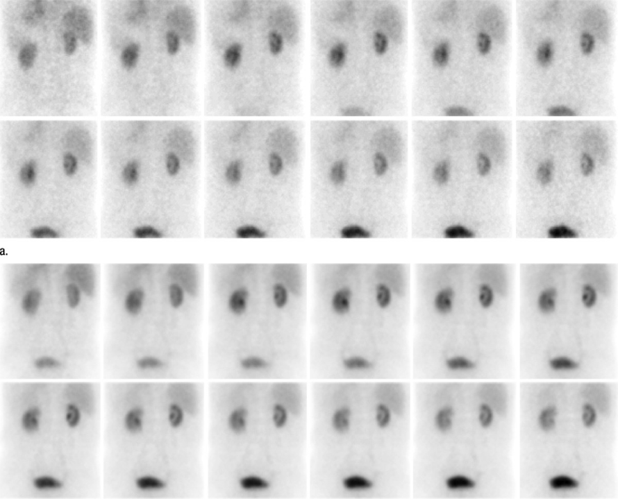

Technetium 99m MAG-3 renogram
Normal.
There is normal perfusion and uptake in both renal moieties, with peak uptake achieved 4 minutes post injection, demonstrating normal renal artery blood flow.
Differential uptake is 48% left kidney and 52% right kidney
which is within normal limits, Normal excretion commences
prior to the administration of diuretics, (at the 10-minute
stage), and a normal excretion curve is demonstrated.
No evidence of delayed emptying or renal obstruction, normal bladder activity, although there are no post micturition images.
Time activity curves show normal renal uptake and excretion of tracer.
A 53-year-old female. Complaining of left flank pain and haematuria.


Abdominal / Renal ultrasound longitudinal/sagittal planes
Abnormal
A large, dilated, central anechoic fluid-filled renal sinus is seen surrounded by an echo dense pelvicalyceal region, in turn surrounded by a smooth homogenous renal parenchyma and cortex.
The images are consistent with a moderately dilated, left sided, pelvi-calyceal system - synonymous with renal obstruction /hydronephrosis.
The ureter also appears dilated, indicating
hydroureter, but there is no sign of the obstruction on these views.
Identify 5 different veins of the upper limb that are suited to intravenous cannulation for the introduction of intravenous radiographic contrast media:
1. Antebrachial vein
2. Basilic vein
3. Cephalic vein
4. Dorsal venous arch
5. Median cubital vein