Anatomy Sem 1 Exam 3
 Call Kai
 Learn
 Practice Test
 Spaced Repetition
 Match
 Flashcards
 Knowt Play
1/308

There's no tags or description
Looks like no tags are added yet.
309 Terms
What are the 3 types of muscle tissues?
1. Skeletal
2. Cardiac
3. Smooth
4 Main Characteristic of Muscle tissue
1. excitability
2. contractability
3. extensibility
3. elasticity
4 main functions of muscle tissue
1. produce movement
2. maintain posture
3. stabilize joints
4. generate heat
Each skeletal muscle, as well as each muscle fiber, is covered in _________ tissue
Connective
Epimysium
surrounds entire muscle

Perimysium
surrounds fascicles within a muscle

Fascicles
Bundles of muscle fibers wrapped in perimysium
Endomysium
Surrounds individual muscle fibers

Skeletal muscle fibers
Long, cylindrical cells that contain multiple nuclei
Sarcolemma
Sarcoplasm
Sarcolemma= muscle fiber plasma membrane
Sarcoplasm= muscle fiber cytoplasm
What are the modified organelles of skeletal muscle?
Myofibrils
Sarcoplasmic reticulum (SR)+ (T Tubules)
Myofibrils
(4 main parts)
Densel packed, rod-like elements
that make up a muscle fiber
A single muscle fiber can contain thousands (80% of muscle volume)
4 parts:
1. Striations
2. Sarcomeres
3. Myofilaments
4. Molecular composition of myofilaments
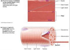
Myofilament striations consist of what three things?
Dark A bands alternating with lighter I bands
Single contractile units called sarcomeres

A Bands
Thick filament regions
Lighter central H Zone which contains no thin filaments

I Bands
Thin filament regions
Bisected by Z Disc proteins called connectin, anchoring elastic and thin filaments

Sarcomere
The smallest contractile unit of a muscle fiber
From one Z disc to the next is a sarcomere

Myofilaments
The contractile proteins, actin and myosin, of muscle cells

Actin myofilaments
Thin filaments
Two strands of actin subunits
Extend across I bands and partway in the A band
Anchored to the Z Discs

What regulatory proteins do Actin contain?
Troponin= bound to actin ad tropomyosin- has Ca2+ bonding site
Tropomyosin
Troponin/tropomyosin complex regulates interaction between active sites on actin and myosin heads

Myosin Filaments
Thick filaments
Several myosin molecules (golf club shape)
Heads bond active sites on actin to form cross bridge
Heads are offset, resulting in staggered cross bridge locations
can bend/straighten at the hinge region
Extend the length of A bands
connected at the M line
ATPase activity, energy used for movement

What are the other (2) structures that support the myofilaments?
1. Elastic filaments= composed of protein titin/// holds thick filaments in place, provides recoil after stretch, resists excessive stretching
2. Dystrophin proteins= links filaments to proteins of the sarcolemma

Duchenne's muscular Dystrophy
caused by a dystrophia gene mutation (dystrophin protein)
Sarcolemma tears, increased apoptosis of muscle fibers, loss of muscle mass
sarcoplasmic reticulum= SR (smooth ER modification)
Network around each myofibril
Dilated end sacs (terminal cisternae) store calcium
Has Traverse t-tubules and triads

Transverse T-Tubules of the SR
Tunnel like in-folding structures
Penetrate the cell//carry electric current to the cell interior

Triads of the SR
Contains: T-Tubule and 2 terminal cisternae

Triad role in SR
T tubule contains integral membrane proteins that act as voltage sensors (sensors change shape in response to electrical current)
SR cistern membranes also have integral membrane proteins that control opening of calcium channels in SR cisterns
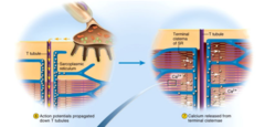
**When an electrical current impulse passes by, T Tubule proteins change shape, causing SR proteins to change shape, causing release of calcium into cytoplasm
sliding filament model
Actin myofilaments sliding over myosin to shorten sarcomeres
>actin and myosin do not change in length
>shortening sarcomeres responsible for skeletal muscle contraction
In short= myosin heads "walk" along actin filaments, pulling actin towards line

Explain how muscle contraction is voluntary
Controlled by the CNS:
The brain makes the decision to move
Signal transmitted down spinal chord to motor neurons
Motor neurons activate muscle fibers
Neurons and muscle cells are excitable cells, can generate action potentials
AP can cross from neurons to muscle cells through ion channels

neuromuscular junction (NMJ)
The area where the motor neuron meets the muscle
What is the axon terminal separated from the muscle fiber by?
The synaptic cleft

What is at the Axon Terminal?
What part of Sarcolemma is related to this?
Axon terminal contains synaptic vesicles of acetylcholine (Ach)
Junctional folds on sarcolemma contain Ach receptors.

Big Picture of Muscle Fiber Contraction: 4 Steps biggest to smallest
1. Events at the NMJ
2. Muscle Fiber Excitation
3. Excitation-contraction coupling
4. Cross bridge cycling
Events at the NMJ
Largest Step of muscle fiber contraction:
1. AP arrives at axon terminal
2. Voltage gated calcium channels open, calcium enters motor neurons
3. Calcium entry causes release of Ach neurotransmitter into synaptic cleft
4. Ach diffuses across Ach receptors on sarcolemma (the junctional folds)
5. Ach bonding to receptors opens gates, allows Na+ to enter local muscle fiber, resulting in the end plate potential EPP
6. Acetylcholinesterase degrades Ach

Muscle fiber excitation
(3 steps)
Second step down of muscle fiber contraction:
1. Resting sarcolemma is polarized (RMP)= voltage exists across membrane-- inside=negative
2. Action potential is caused by changes in electrical charges
Occurs in 3 steps:
1. Generation of EPP
2. Depolarization
3. Repolarization

Excitation-contraction coupling
Third step down of muscle fiber contraction:
1. Events that transmit Action potential across sarcolemma (excitation) are coupled to sliding of myofilaments (contraction)
2. AP is sent along sarcolemma and down into t tubules, where voltage-sensitive proteins stimulate Ca2+ release from SR
3. AP is brief and ends before contraction is seen
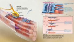
Cross Bridge Cycling
Fourth step down of muscle fiber contraction
Voltage sensitive proteins in T-tubules change shape, causing sarcoplasmic reticulum to release Ca2+ into cytosol

Pre- Cross bridge cycling:
What does muscle fiber look like (tropomyosin)?
What happens that causes the cross-bridge cycle to begin?
Low intracellular Ca2+ concentration:
-tropomyosin blocks active sites on actin
-myosin heads cannot attach to actin
-muscle fiber remains relaxed
Voltage-sensitive proteins in T-tubules change shape, casuing SR to release Ca2+ into cytosol
Four Steps of Cross-Bridge Cycle
1. Cross bridge formation: high-energy myosin head attaches to actin thin filament active site
2. Working (power) stroke: myosin head pivots and pulls thin filament toward M line
3. Cross bridge detachment: ATP attaches to myosin head, causing cross bridge to detach
4. Cocking of myosin head: energy from hydrolysis of ATP "cocks" myosin head into high-energy state
>>>This energy will be used for power stroke in next cross bridge cycle

muscle relaxation
Original nerve stimulus stops
ACh breakdown in synaptic cleft
Ca2+ uptake into SR
Tropomyosin returns to blocking the active sies on actin
ATP required to break cross bridge (detachment)

Rigor Mortis
stiffness of the body that sets 3-4 hours after death
Deteriorating sarcoplasmic reticulum releases calcium
Calcium still actives cross bridging
Muscle contracts but cannot relax because muscle relaxation requires ATP (ATP no longer produced after death)
fibers remain contracted until myofilaments decay
Motor Unit
a motor neuron and all the muscle cells it stimulates
How do motor units sustain long term contraction?
They take turn resting
Allows for posture control
Motor Units: Fine control
Small motor units contain as few as 20 muscle fibers per nerve fiber (eye muscles)
Motor Units: Strength Control
Gastrocnemius muscle (calf) has 1000 fibers per nerve fiber
Muscle Twitch
simplest contraction resulting from a muscle fiber's response to a signle actionpotential from the motor neuron
Muscle fiber contracts quickly, then relaxes
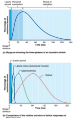
Myogram
a chart of the timing and strength of a muscle's contraction

3 Phases of Muscle Twitch
1. Latent period= events of excitation- contraction coupling
>>> no Muscle tension seen
2. Period of contraction= cross bridge formation (myosin attaching to actin)
>>>tension increases
3. Period of relaxtion= Ca2+ reentry into the SR
>>>Tension declines to zero
Muscle contracts faster than it relaxes
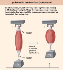
Isotonic Contraction
muscle shortens because muscle tension exceeds load
Isotonic contraction (concentric)
Muscle shortens and does work
(biceps brachii while picking up a book)
Isotonic contraction (eccentric)
Muscle lengthens and generates force
(biceps brachii while laying down a book)
Isometric contraction
Tension increases but does not exceed load, no change in muscle length

ATP supplies the energy needed for the muscle fiber to...
(4 reasons)
1. Move and detach cross bridges
2. Pump calcium back into the sarcoplasmic reticulum
3. Pump Na+ out of and K+ back into the cell after excitation-contraction coupling
4. Contractile acitvites
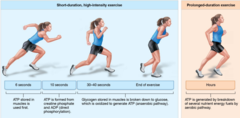
Available stores of ATP deplete in _____
4-6 seconds
ATP is regenerated quickly by 3 mechanisms:
1. Direct phosphorylation of ADP by creatine phosphate (CP)
2. Anaerobic pathway= glycolysis and lactic acid formation (only two ATP per glucose)
3. Aerobic pathway= respiration w/ oxygen; makes way more ATP

Aerobic Endurance
Length of time muscle contracts using aerobic pathways
Light to moderate activity which can continue for hours

Anaerobic Threshold
Point at which muscle metabolism converts to the anaerobic pathway
Fatigue
(3 possible causes)
Physiological inability to contract despite continued stimulation
Ionic Imbalances= Levels of K+, Na+, and Ca2+ can change, disrupting the membrane potential of the muscle cell
Decreased ATP and increased magnesium (unimportant)
Decreased glycogen= stored in liver and skeletal muscle
Note that Lack of ATP is rarely a reason for Fatigue
EPOC
Excess Postexercise Oxygen Consumption
Recovery requires extra oxygen ("oxygen debt")
For a muscle to return to its pre-exercise state:
>>Oxygen reserves are replenished
>>Lactic acid is reconverted to pyruvic acid (pyruvate)
>>Glycogen stores are replaced
>>ATP and creatine phosphate reserves are resynthesized
The force of contraction depends on the number of cross bridges attached, which is affected by 4 factors:
1. Number of muscle fibers= simulated (recruitment)= the ore motor units recruited, the greater the force
2. Relative size of fibers= the bulkier the muscle, the more tension can develop (hypertrophy with exercise)
3. Frequency of stimulation (temporal/wave)= the higher the frequency, the greater the force
>>stimuli are added together
4. Degree of muscle stretch (length-tension relationship) = muscle fibers with sarcomeres that are 80-120% their normal resting length generate more force

If sarcomere is less than 80% resting length...
filaments overlap to much and force decreases

If sarcomere is greater than 120% of resting length...
Filaments do not overlap enough and force decreases

Hypertrophy with muscles
Muscles increase in size with regular exercise
How fast a muscle contracts (velocity) and how long it can stay contracted (duration) is influenced by
3 things
1.muscle fiber type
2.Load
3. Recruitment
Slow oxidative/slow twitch fibers/type I fibers
(where at?)
more mitochondria capillaries "dark meat"
Adapted for aerobic respiration and resistant to fatigue
Postural muscles of the back (100msec/twitvh)
Fats glycolytic/ fast twitch fibers/ type II fibers
(where at?)
rich in enzymes for glycogen-lactic acid system "white meat"
SR releases calcium quickly, so contractions are quicker (7.5 msec/twitch)
Extraocular muscles, biceps, brachii
How are proportions of slow oxidative vs fast glycolytic muscle fibers determined?
Genetically determined
Can be modified slightly
Load (muscle contraction/duration)
Muscles contract fastest when no load is added
The greater load...
the shorter duration of contraction
the slower the contraction

Recruitment (muscle contraction/duration)
The more motor units contracting, the faster and more prolonged the contraction

What are the four stages of cellular respiration?
1. glycolysis
2. pyruvate oxidation (the transitional phase)
3. citric acid cycle (Kreb's cycle)
4. oxidative phosphorylation
>>>ETC
>>>chemiosmosis

Where is smooth muscle found?
walls of hollow organs
Respiratory, digestive, urinary, reproductive, circulatory (except in smallest blood vessels)
NOT IN HEART

Varicosities
In smooth muscle cells:
Bulbous swellings of nerve fibers (instead of skeletal NMJ)
Store and release Neurotransmitters
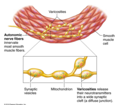
What are the varicosities of smooth muscle innervated by ?
The autonomic nervous system (rest and digest) not the CNS
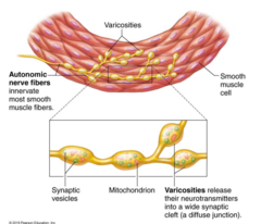
Smooth muscle fibers are usually connected ________________________________________ whereas skeletal muscle fibers are ________________.
Smooth muscle fibers are usually electrically connected via gap junctions whereas skeletal muscle fibers are electrically isolated
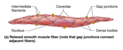
Gap Junctions in Smooth Muscle Cells
Specialized cell connections that allow depolarization to spread from cell to cell
Smooth Muscle SR
(Two Important details)
Less Elaborate SR and no T-Tubules
SR does store Ca2+, but most of the calcium comes from extracellular origins

Caveolae
Pouch-like infoldings in Smooth muscle cells
Caveolae contain numerous Ca2+ channels that open to allow rapid influx of extracellular Ca2+

Smooth muscles have no ____________ or __________, but they do contain ____________________________________.
no striations and no sarcomeres, but they do contain overlapping thick and thin filaments

calmodulin
Protein in smooth muscle that binds to Ca2+
No troponin complex in smooth muscle
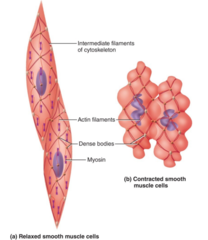
How are thick and thin filaments arranged in smooth muscle ?
Thick and thin filaments arranged diagonally
Myofilaments are spirally arranged, causing smooth muscle to contract in corkscrew manner
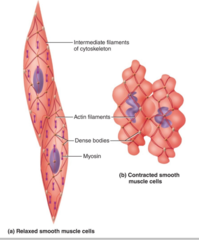
Structural Framework of Smooth Muscle Cells:
Intermediate filament-dense body network
Non contractile intermediate filaments resist tension
Dense bodies= proteins that anchor filaments to sarcolemma at regular intervals
>>>correspond to Z discs of skeletal muscles
During contraction, areas of the sarcolemma between dense bodies bulge outward
>>>Makes the smooth muscle cell appear puffy by microscopy

What happens to a smooth muscle cell during contraction? (Dense Bodies)
Areas of the sarcolemma between dense bodies bulge outward
• Makes the smooth muscle cell appear puffy by microscopy

Some smooth muscle cells are self-excitatory- what does this mean?
They depolarize without external stimuli
Act as pacemakers for sheets of muscle
Contraction in smooth muscle is different from skeletal muscle in the following ways:
(4 ways)
Some Ca2+ still obtained from SR, but mostly comes from extracellular space
Ca2+ binds to calmodulin, not troponin
Activated calmodulin then activates myosin kinase (myosin light chain kinase)
Activated myosin kinase phosphorylates myosin head, activating it
>>>Leads to crossbridge formation with actin

7 criteria for naming muscles
1. Location
2. Shape
3. Size
4. Direction of fibers/fascicles
5. # of origins
6. Location of attachments
7. Action
(any criteria can be combined!)

Rectus Muscle fibers
fibers that run straight
Transversus muscle fibers
fibers that run at right angles
Oblique muscle fibers
fibers that run at angles to imaginary defined axis
Flexion
a movement that decreases the angle between two body parts.
extension
a movement that increases the angle between two body parts
Abduction
Movement away from the midline of the body
Addcution
Movement toward the midline of the body
A muscle that crosses on the anterior side of a joint produces ___________
flexion
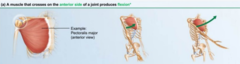
A muscle that crosses on the posterior side of a joint produces _____________
extension

A muscle that crosses the lateral side of a joint produces______________
Abduction

A muscle that crosses on the medial side of a joint produces ____________
adduction

What muscle produces a smile?
Zygomaticus major

What muscle produces a pucker?
Orbicularis Oris

What muscle produces a pout?
Mentalis

What muscle produces angry eyebrows?
Corrugators Supercilli

What muscle produces a blink?
Orbicularis Oculi
