PBL 3: Enzyme-Coupled and G-Protein Receptors
 Call Kai
 Learn
 Practice Test
 Spaced Repetition
 Match
 Flashcards
 Knowt Play
1/37

There's no tags or description
Looks like no tags are added yet.
38 Terms
What are GPCRs:
cell-surface proteins that span the plasma membrane seven times and transmit signals from outside the cell to inside via heterotrimeric G proteins
Structure of GPCRs
a single polypeptide chain that threads back and forth across the lipid bilayer seven times, forming a cylindrical structure, often with a deep ligand binding site in its core
They also all use G proteins to relay their signal into the cell interior
What happens when a ligand binds to the GPCRs
the receptor undergoes a conformational change that enables it to activate a heterotrimeric GTP-binding protein (G protein)
This couples the receptor to enzymes or ion channels in the plasma membrane
G Protein Structure
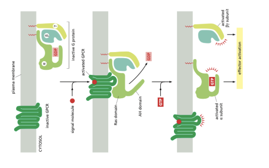
G proteins are composed of three subunits: α, β, and γ.
In the unstimulated state, the α subunit is bound to GDP and the G protein is inactive

What happens when GPCR is activated
When the GPCR is activated, it acts as a guanine nucleotide exchange factor (GEF) and induces the α subunit of the G Protein to release its bound GDP, allowing GTP to bind in its place
GTP binding causes a conformational change, releasing the G protein from the receptor and triggering dissociation of the GTP-bound Gα subunit from the Gβγ pair
Both of which then interact with various targets, such as enzymes and ion channels in the plasma membrane, which relay the signal onward
What is the alpha subunit and what happens after it is activated
The α subunit is a GTPase and becomes inactive when it hydrolyses its bound GTP to GDP
The α subunit then binds to a second protein, which can either be a specific target protein or a specific regulator of G protein signaling (RGS)
RGS: act as an α-subunit-specific GTPase-activating proteins (GAPs), which shuts off G-protein mediated responses
What is Adenylyl Cyclase
an enzyme that is activated by a stimulatory alpha subunit that produces cyclic AMP (cAMP)
Adenylyl cyclase has its catalytic domain on the cytosolic side of the membrane and is regulated by either G proteins or Calcium (Ca2+)
What is Cyclic AMP
Cyclic AMP (cAMP) acts as a second messenger in some signalling pathways
An extracellular signal increases cAMP concentration
Cyclic AMP is synthesised from ATP by the enzyme adenylyl cyclase and destroyed by phosphodiesterases
Many extracellular signals work by increasing cAMP concentrations inside the cell, How does this Work?
These signals activate GPCRs that are coupled with stimulatory G proteins (Gs),
Gs has a Gαs subunit, which directly activates adenylyl cyclase (AC) when bound to GTP, increasing cAMP concentration
Many extracellular signals work by decreasing cAMP concentrations inside the cell on different GPCRs, How does this work?
These signals activate inhibitory G proteins (Gi) which inhibit adenylyl cyclase
Gi has a Gαi subunit, which directly inhibits adenylyl cyclase (AC) when bound to GTP, decreasing cAMP concentration
How does the heart respond to an increase in cAMP concentration?
The heart activates adenylyl cyclase in response to epinephrine leading to an increase in heart rate and force of contraction
What does Cyclic AMP activate
cAMP mainly activates cyclic-AMP-dependant protein kinase (PKA)
This kinase phosphorylates specific serine or threonine on selected target proteins, including intracellular signaling proteins and effector proteins, thereby regulating their activity
What is the structure of cyclic-AMP-dependant protein kinase and the function of this structure?
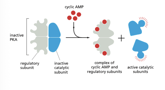
In the inactive state PKA consists of a complex of two catalytic subunits and two regulatory subunits
The binding of cAMP to the regulatory subunits alters their conformation, causing them to dissociate from the complex
The released catalytic subunits are thereby activated to phosphorylate specific target proteins
The regulatory subunits of PKA are important for localising the kinase inside the cell
How is PKA activity controlled and localized within the cell?
AMP activates PKA, but PKA should not be activated everywhere in the cell. Special A-kinase anchoring proteins (AKAPs) help ensure that PKA acts in specific locations
How do Special A-kinase anchoring proteins work (AKAPs)
1. AKAP anchors PKA to a specific location
AKAP (A-kinase anchoring protein) binds to the regulatory subunits of PKA.
It attaches PKA to a particular location in the cell, such as near the nucleus.
This ensures that PKA can phosphorylate specific target proteins in that area.
2. AKAP also binds phosphodiesterase (PDE)
The same AKAP can bind a phosphodiesterase, an enzyme that breaks down cAMP into AMP.
Since cAMP activates PKA, breaking down cAMP reduces PKA activation and phosphorylation of target proteins.
What is the Negative Feedback Loop of cAMP-mediated PKA signalling
Among the target proteins that PKA phosphorylates, is the adjacent phosphodiesterase, which rapidly lowers the cAMP concentration
This negative feedback loop converts what might otherwise be a prolonged PKA response into a brief, local pulse of PKA activity
Whereas some responses mediated by cAMP occur within seconds, others depend on changes in the transcription of specific genes and take hours to develop fully.
Explain the process of slow responses mediated by cAMP
1. PKA is activated by cAMP
An increase in cAMP activates protein kinase A (PKA).
The active catalytic subunits of PKA can enter the nucleus through nuclear pores.
2. PKA phosphorylates CREB
Inside the nucleus, PKA phosphorylates a transcription factor called CREB (cAMP response element-binding protein).
This converts inactive CREB into its activated, phosphorylated form.
3. Activated CREB recruits CBP
Phosphorylated CREB recruits CREB-binding protein (CBP).
CBP acts as a transcriptional coactivator, helping CREB stimulate gene transcription.
4. CREB and CBP activate gene transcription
CREB binds to a specific DNA sequence called the cAMP response element (CRE) near the target gene.
Together, phosphorylated CREB and CBP promote transcription of the target gene into mRNA.

How does the Heart Muscle Contract
Ca²⁺ is released from the sarcoplasmic reticulum into the cytoplasm.
Ca²⁺ binds to troponin C, causing tropomyosin to move away from the myosin-binding sites on actin
Myosin heads bind to actin, forming cross-bridges between the thick and thin filaments
Myosin heads perform a power stroke, pulling the actin filaments toward the centre of the sarcomere.
ATP binds to myosin, causing the myosin heads to detach from actin.
ATP is hydrolysed, providing energy to reposition the myosin heads for another power stroke.
This cycle repeats as long as Ca²⁺ and ATP are available, causing the sarcomere to shorten and the muscle to contract.
Muscle relaxation: Ca²⁺ is pumped back into the sarcoplasmic reticulum, causing tropomyosin to block the myosin-binding sites on actin again.
What is Myosin, Troponin and Tropomyosin
Myosin: Forms the thick filaments in muscle cells, Contains myosin heads, which can bind to actin. Myosin heads use energy from ATP hydrolysis to generate force
Troponin: a complex of three polypeptides, troponins T, I, and C.
Tropomyosin: the elongated protein that binds along the groove of the actin filament helix
What is the Process of norepinephrine binding to the receptor
The norepinephrine ligand binds to a Beta-1 Agrenergic Receptor (GPCR) which activates a Gs
Stimulatory G proteins contain a Gαs subunit
The activated Gs causes Gαs to exchange GDP to GTP through GEF
Gαs-GTP separates functionally from the Gβγ complex.
Gαs-GTP activates adenylyl cyclase, increasing cAMP production
cAMP activates protein kinase A (PKA).
PKA phosphorylates L-type Ca²⁺ channels, increasing Ca²⁺ entry into the cardiac muscle cell.
The incoming Ca²⁺ triggers additional Ca²⁺ release from the sarcoplasmic reticulum through ryanodine receptors (calcium-induced calcium release).
Ca²⁺ binds to troponin C, causing tropomyosin to move away from the myosin-binding sites on actin.
Myosin binds to actin, allowing cross-bridge cycling and muscle contraction.
Wha is the Process of Acetylcholine binding to the receptor
Acetylcholine (ACh) binds to an muscarinic receptor (GPCR), which activates an inhibitory G protein (Gi).
Inhibitory G proteins contain a Gαi subunit.
The activated receptor acts as a GEF, causing Gαi to exchange GDP for GTP.
Gαi-GTP separates functionally from the Gβγ complex.
Gαi-GTP inhibits adenylyl cyclase (AC), decreasing cAMP production.
Decreased cAMP reduces protein kinase A (PKA) activation.
Reduced cAMP signalling decrease ion-channel activity, including Ca²⁺ currents.
Meanwhile, the Gβγ complex activates GIRK potassium (K⁺) channels, allowing K⁺ to leave the cell.
The opening of these channels makes it harder to depolarise the cell and thereby contributes to the inhibitory effect of acetylcholine on the heart
What are Enzyme Coupled Receptors and Structure
transmembrane proteins with their ligand-binding domain on the outer surface of the plasma membrane
Their cytosolic domains has either intrinsic enzyme activity or associates directly with an enzyme
They have one transmembrane segment, and share lots of signalling pathways with GPCRs
What Happens to the Structure of Receptor Tyrosine Kinase once Signal Protein Binds?
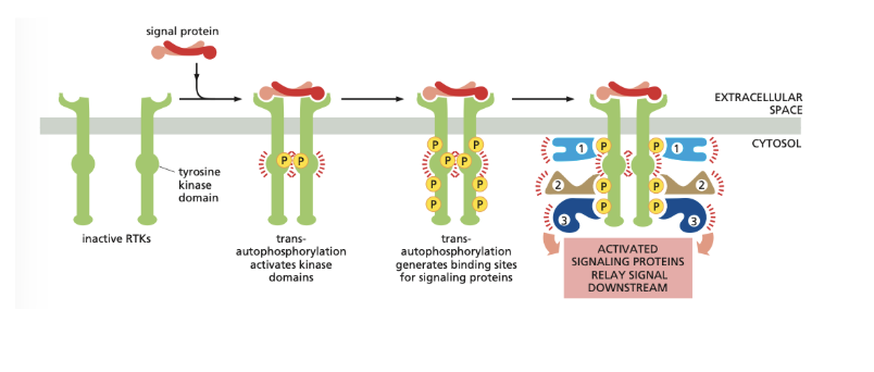
The binding of a signalling protein (ligand) to the extracellular ligand-binding domain of an RTK activates the tyrosine kinase domain on the cytosolic side.
This activation usually involves dimerisation, where two receptor molecules come together, bringing their cytoplasmic kinase domains close to each other.
What Occurs after Dimerisation?
Dimerisation positions the two kinase domains in a stable orientation, allowing them to phosphorylate each other on specific tyrosines in their kinase domains.
This phosphorylation causes conformational changes that fully activate both kinase domains.
The activated kinase domains then phosphorylate additional tyrosine residues on the cytosolic portions of the receptors.
What are the Two Other Types of Receptor Tyrosine Kinases
Some RTKs exist as dimers in the absence of ligands, and ligand binding re-orients the extracellular domains to bring the internal kinase domains into a closer position for phosphorylation to promote kinase activation
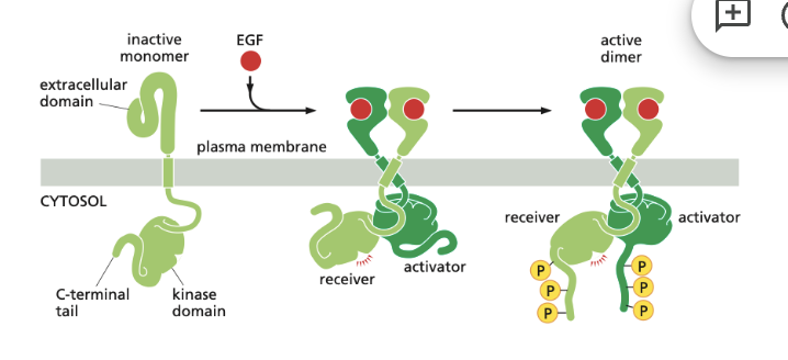
Other cases, the kinase is not activated by phosphorylation but by conformational changes brought about by interactions between the two kinase domains outside their active site (photo below)
What happens once additional tyrosine sites have been phosphorylated?
This phosphorylation creates high-affinity docking sites for specific intracellular signalling proteins.
Each signalling protein binds to a particular phosphorylated site on the activated receptors because proteins contain a specific phosphotyrosine-bonding domain
The signaling protein recognizes both the phosphotyrosine and the surrounding amino acid sequence on the receptor
Different Types of Signalling Proteins on RTKs
Once bound to the activated RTK, a signalling protein may become phosphorylated on tyrosines and thereby activated.
In some cases however phosphorylation is not necessary to activate the docked protein, and the binding alone may be sufficient.
This by either inducing a conformational change or simply bringing it near the protein that is next in the signalling pathway
Structures of Signalling Proteins
Intracellular signalling proteins have different structures and functions but often contain conserved phosphotyrosine-binding domains, such as SH2 domains.
These domains recognize specific phosphorylated tyrosines and allow signalling proteins to bind to activated RTKs.
They can also bind to other tyrosine-phosphorylated intracellular signalling proteins, allowing signals to be relayed downstream.
What is the Other Interaction Domain of Signalling Proteins?
allows them to interact specifically with other proteins as part of the signalling process
These domains include the SH3 domain, which binds to proline-rich motifs in intracellular proteins
What are Adaptor Proteins?
Some signalling proteins are composed almost entirely of SH2 and SH3 domains and function as adaptors to couple tyrosine-phosphorylated proteins to other proteins that do not have their own SH2 domains
Adaptor proteins of this type help to couple activated RTKs to the important signalling protein Ras
Structure and Function of Ras
a monomeric GTPase that in turn can activate various downstream signalling pathways that relays signals from cell-surface receptors
Ras contains one or more covalently attached lipid groups that help anchor the protein to the cytoplasmic face of the plasma membrane (like many monomeric GTPases), from where it relays signals to other parts of the cell
What does Mutated Ras Lead to:
Ras is often required when RTKs signal to the nucleus to stimulate cell proliferation or differentiation (both of which require change in gene expression)
Hyperactive mutant forms of Ras are resistant to Ras GAPs and are therefore locked in the GTP-bound active state,which contribute to the uncontrolled proliferation of cancer cells
What are the Two Classes of Signalling Proteins that Regulate Ras
Two classes of signaling protein regulate Ras activity by influencing its transition between active and inactive states
Ras guanine nucleotide exchange factors (Ras GEFs) stimulate the disassociation of GDP and the binding of GTP from the cytosol, which activates Ras
Ras GTPase-activating proteins (Ras GAPs) stimulate the hydrolysis of bound GTP and the binding of GDP from the cytosol, which deactivates Ras
How do RTKs activate Ras
RTKs normally activate Ras through the indirect binding of the activated receptor to a Ras GEF
The Ras GEF is called Son-of-Sevenless (Sos)
An adaptor protein called Grb2, helps to link the Sev receptor (RTK) to the Sos protein (Grb2 is the indirect binding)
The SH2 domain of Grb2 adaptor binds to the activated receptor, while one or both of its SH3 domain of the Grb2 adaptor bind to Sos
Sos then promotes Ras activation by releasing its bound GDP, allowing GTP to bind in its place
Once activated, Ras activates various other signalling protein to relay the signal downstream

How are Ras and RTK Inactivated?
Both the tyrosine phosphorylations and the activation of Ras triggered by activated RTKs are usually short-lived
Tyrosine-specific protein phosphatases reverse the phosphorylations, and Ras GAPs induce activated Ras to inactivate itself by hydrolysing its bound GTP to GDP
Why should signalling events be converted into long lasting onces and what is a key mechanism for this?
To stimulate cells to proliferate or differentiate, these short-lived signaling events must be converted into longer-lasting ones that can sustain the signal and relay it downstream to the nucleus to alter the pattern of gene expression
One of the key mechanisms used for this purpose is a system of proteins called mitogen-activated protein kinase module (MAP kinase module)
Components of Mitogen-activating Protein Kinase Model (MAP)
The three components of this system are all protein kinases, which mainly phosphorylate serines and threonines
The final kinase in this series is called MAP kinase (MAPK)
Also known as Raf
The next one upstream from this is MAP kinase kinase (MAPKK)
Its function is that it phosphorylates and thereby activates MAP kinase
Also known as Mek
The next about that is MAP kinase kinase kinase (MAPKKK)
It receives activating signals from Ras and it phosphorylates and thereby activates MAPKK
Also known as Erk
How does the Mitogen-Activating Protein Kinase Model lead to cell proliferation and differentiation
Once activated, the MAP kinase relays signals downstream by phosphorylating various proteins in the cell including transcription regulators and other protein kinases
MAPK enters the nucleus and phosphorylates one or more components of a transcription regulatory complex
This activates the transcription of a set of immediate early genes
These early genes turn on within minutes after an RTK activation
Some of these genes encode other transcription regulators that turn on other genes, a process that requires both protein synthesis and more time.
Outcome: The Ras–MAPK pathway alters gene expression, including activating genes involved in cell proliferation, such as genes encoding G1 cyclins.
