chapter 27: disorders of cardiac function, heart failure, and circulatory shock
 Call Kai
 Learn
 Practice Test
 Spaced Repetition
 Match
 Flashcards
 Knowt Play
1/103

There's no tags or description
Looks like no tags are added yet.
104 Terms
heart is enclosed within the:
pericardium!
serous pericardium
thin membrane with 2 layers: parietal and visceral
pericardial cavity is potential space btwn parietal and visceral layers of serous pericardium
heart wall consists of 3 layers:
endocardium
myocardium
epicardium
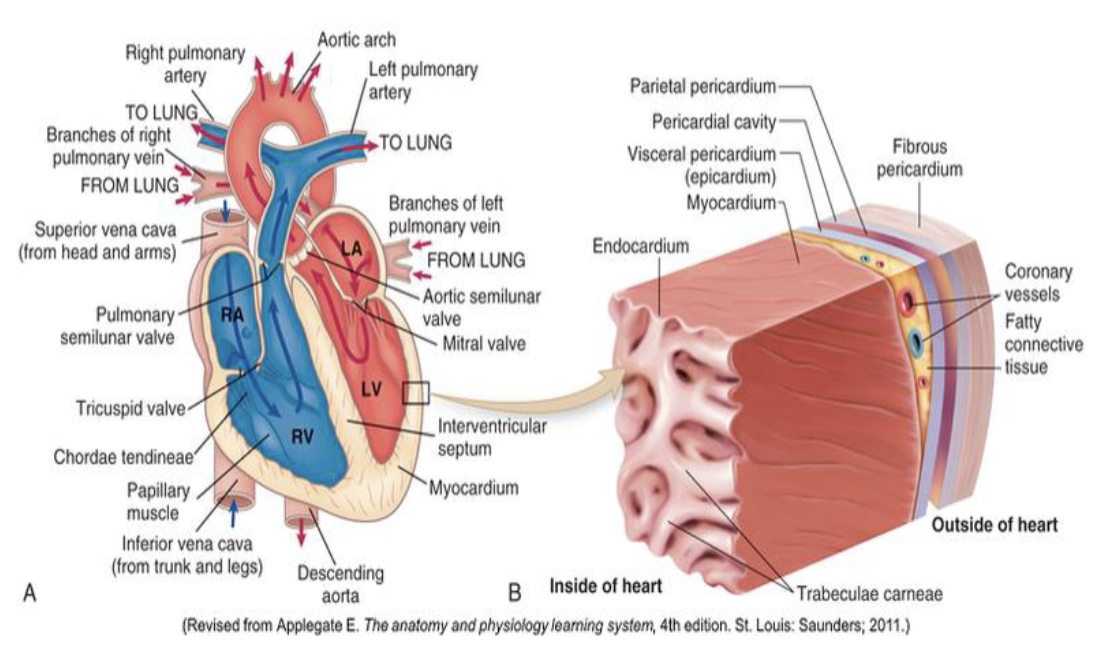
endocardium
innermost layer
lines chambers of heart and covers heart valves
myocardium
middle layer
composed of cardiac muscle tissue
epicardium
outermost layer
also called visceral layer of serous pericardium
right and left coronary arteries branch of:
aorta immediately distal to aortic valve
part of systemic circulation to deliver oxygen-rich blood to myocardium
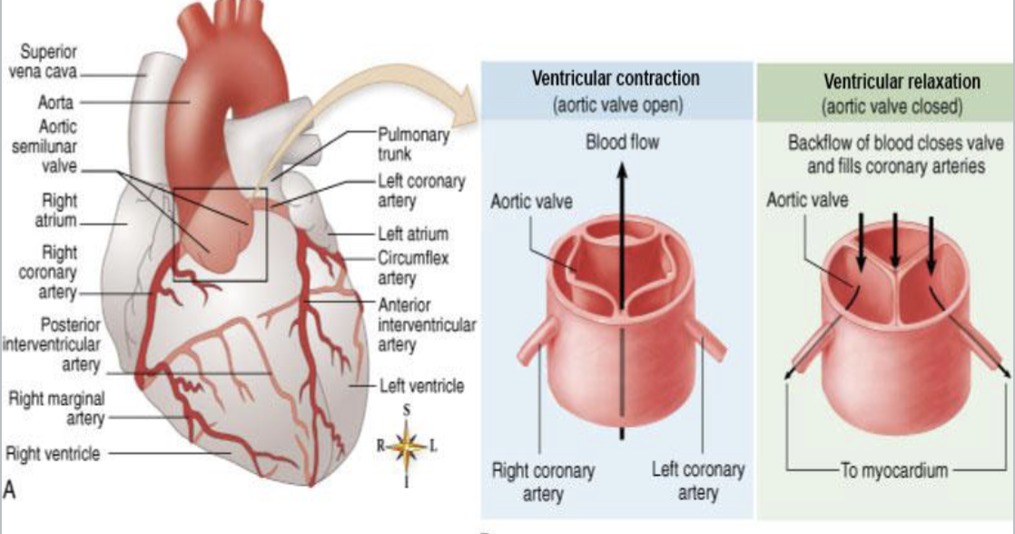
left coronary artery divides into:
left anterior descending or interventricular artery
left circumflex artery
right coronary artery divides into:
right marginal artery
posterior interventricular artery
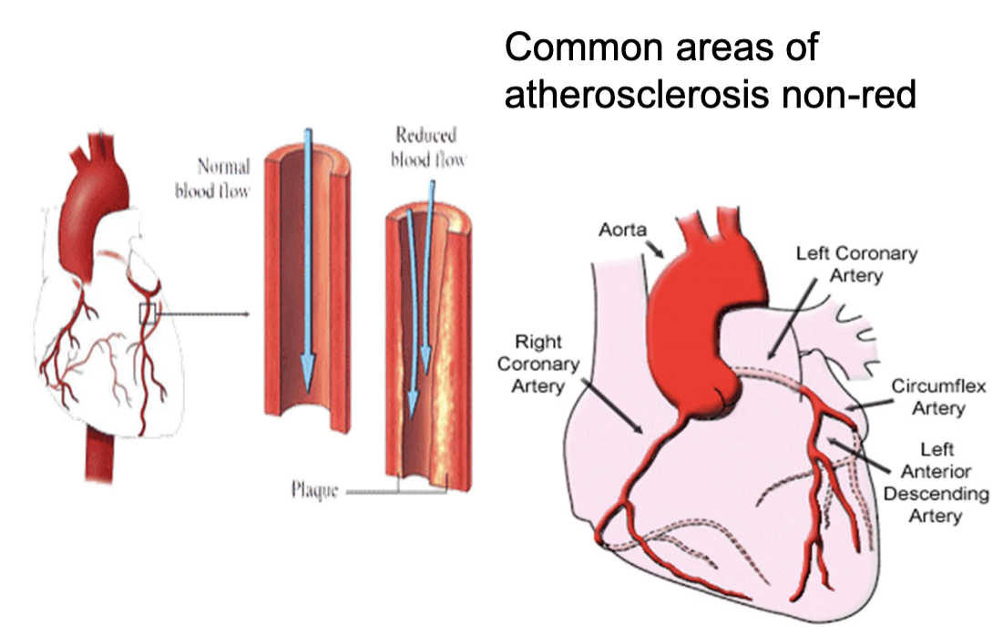
coronary artery disease (CAD)
aka coronary heart disease
occurs when atherosclerotic plaques build up in coronary arteries, narrowing arterial lumen and reducing blood flow to heart muscle
CAD remains silent for many years until blood flow becomes significantly reduced
possible manifestations or complications of CAD
angina attascks
myocardial infarction
cardiac arrhythmias
heart failure
sudden death
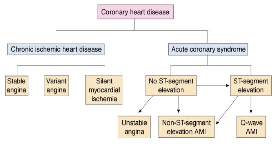
chronic ischemic heart disease (CIHD)
occur when deficit ox oxygen supply to meet myocardial needs
classic symptom: angina - chest pain that may occur in different patterns
recurrent, intermittent brief episodes of substernal chest pain, triggered by physical or emotional stress
angina attacks vary in:
severity and duration but become more frequent and longer as disease progresses
CIHD can be classified into 4 main types:
stable angina
variant angina
unstable angina
silent myocardial ischemia
stable angina
often called classic or exertional
triggered by physical exertion when heart’s O2 demand increases
variant angina
vasospastic (prinzmetal)
cyclical spasms in coronary arteries without increased cardiac workload: pain occurs at rest (early morning or night)
unstable angina
attacks last longer than stable angina and can be less responsive to medication
prolonged pain at rest may precede to acute coronary syndrome
silent myocardial ischemia (angina/type of CIHD)
occurs without pain
reduced blood flow and oxygen supply to heart muscle without noticeable symptoms
areas of angina
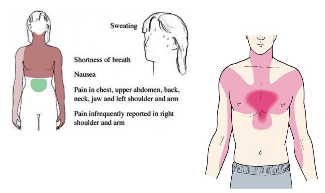
CIHD management screening:
aims to identify asymptomatic stages + reduce risk of complications
stress test: monitors heart activity and coronary perfusion during exercise
treatment of angina attack:
rest, stop activity
sublingual nitroglycerin: dilates coronary arteries, improves blood flow to myocardium
acute coronary syndrome
occurs w/ sudden severe reduction in blood flow to myocardium due to severe narrowing or complete blockage of coronary artery
severe or prolonged lack of blood flow can cause perm dmg to heart muscle
in acute coronary syndrome, atherosclerosis w/ ruputure of plaque is the most:
common cause of ACS
thrombus can form at site of ruptured atherosclerotic plaque and partially or completed block the coronary artery
size and dmg of obstruction determine dmg
manifestations of acute coronary syndrome:
chest pain may occur at rest + may not be relieved by administration of vasodilators
pain described as substernal, crushing, radiating
feeling of pressure, heaviness, or burning in chest
sudden SOB, weakness, fatigue
nausea, indigestion
acute coronary syndrome ECG ischemia:
inadequate blood (O2) supply alters electrical activity of myocardial cell
acute coronary syndrome ECG
assess ST segment
critical part of ECG when ventricles fully depolarized and contracting to eject blood
ECG changes that indicate high probability of MI:
ST-segment elevation (STEMI): likely coronary sthrombus occluding artery
abnormal Q waves: no depolarizing current conducting from necrotic tissue
ECG changes that indicate intermediate or low probability of MI:
NSTEMI: lack of ST segment elevation suggests clot only partly occludes coronary artery
T-wave inversion: repolarization of ventricle altered due to myocardial ischemia or MI
STEMI treatment:
percutaneous coronary intervention (PCI)
balloon angioplasty to widen narrowed arteries
stent placement to keep the artery open
thrombolytics: dissolve blood clots
anticoagulants: prevent clot growth
NSTEMI treatment:
pharm treatment to reduce myocardial workload - beta blockers
anticoagulants - heparin
monitor ECG and serum markers checked
angioplasty if not stable
myocardial infarction
evidence of myocardial necrosis in clinical setting consistent w/ acute myocardia ischemia
time limit: irrev dmg + necrosis may begin within 20-49 mins of severe coronary artery occlusion
cardiac markers: blood lvls of cardiac troponin rise
cardiac markers + types:
substances released into bloodstream when heart muscle is dmgd
troponin I, myoglobin, and CK-MB (creatine kinase-MB)
troponin I (cardiac marker)
preferred marker due to its high sensitivity and specificity, may rise within 3-6 hrs after symptoms onset
myoglobin (cardiac marker)
rise within approx 2 hrs after myocardial injury
not specific to heart since also found in skeletal muscle
CK-MB (creatine kinase-MB) (cardiac marker)
increases within 6-8 hrs after myocardial injury
complications of MI:
sudden death
cardiogenic shock
cardiac tamponade: rupture of necrotic heart tissue w/ blood accumulation in percardium
thromboembolism
ventricular aneurysm: bulging of weakened area of ventricular wall
cardiac cycle
sequence of events that occurs during one complete contraction and relaxation of heart
diastole and systole
diastole (cardiac cycle)
relaxation phase
ventricles relax and their pressure decreases
mitral and tricuspid valves open, blood flows from atria into ventricles
systole (cardiac cycle)
contraction phase
ventricular pressure increases as heart muscle contracts
mitral and tricuspid valves close, aortic, and pulmonary valves open
blood is ejected into aorta and pulmonary artery
cardiac output (CO)
amount of blood pumped by one ventricle in one heart
CO = heart rate x stroke volume
avg CO ~ 5 L/min in healthy adult
stroke volume (SV)
amnt of blood ejected by one ventricle during each contraction
SV calculated by subtracting volume of blood remaining after contraction (ESV) from volume of blood before contraction (EDV)
stroke volume = end-diastolic volume - end-systolic volume
avg SV ~ 70 mL
preload
degree of myocardial stretch before contraction
increased myocardial stretch leads to increased stroke volume
preloasd depends primarily on end-diastolic volume (EDV) and venous return
afterload
resistance ventricles must overcome to eject blood during contraction
primarily determined by arterial pressure + peripheral vascular resistance
pressure in arteries pushing on SL valves is major component of afterload
increased afterload decreases stroke volume (SV)
ejection fraction (EF)
percentage of blood pumpedby ventricle w/ each contraction
norm EF: 55-75%
EF beloe 50% is considered low
cardiac reserve
ability to increase CO to meet body’s increased demands during physical activity or stress
heart failure (HF)
clinical condition in which heart cannot pump out sufficient blood to meet metabolic needs for oxygen and nutrients
HF usually complication of another cardiopulmonary condition
body activates several compensatory mechanisms to maintain blood flow to vital organs when heart’s pumping function declines
some of them often aggravate condition
when heart cannot maintain pumping capability:
cardiac output or stroke volume decreases (EF) below 45%
less blood reaches various organs, cell function and metabolism decreased
mild acidosis develops
backup and congestion develop as:
output from ventricle is less than inflow of blood
congestion in venous circulation draining into affected side of heart
heart failure can be classified accord to:
functional problem: systolic and diastolic
side of heart affected: left and right sided
functional problem (HF)
systolic: decreased contractility
weakness and dilation of ventricle: heart has difficulty contracting and ejecting blood
diastolic: decreased filling
stiffness or hypertrophy of ventricle: heart cannot relax + fill w/ blood
side of heart affected
left sided: left ventricle cannot pump blood effectively into systemic circulation
right sided: right ventricle cannot pump blood effectively into pulmonary circulation
forward effects
consequences of reduced blood flow from heart to body’s tissues, similar in both left and right sided heart failure
reduced oxygen supply: cells receive insufficient oxygen to meet metabolic needsL dyspnea and SOB
impaired cellular function: reduced oxygen availability decreases cellular energy production - fatigue and weakness
compensatory mechanisms are activated to:
maintain BP and blood flow
tachycardia - hypoxia activates SNS
activation of RAAS - due to decreased kidney perfusion
manifestations of HF - back up effects of left sided HF
blood backs up into pulmonary circulation, increasing hydrostatic pressure in pulmonary capillaries resulting in pulmonary congestion
clinical manifestations include:
SOB - dyspnea
difficulty breathing when lying flat - orthopnea
waking up at night w/ sudden breathlessness - paroxysmal nocturnal dyspnea
crackles heard on lung auscultation
cough, sometimes producing pink, frothy sputum in severe pulmonary edema
manifestations of HF - back up effects of right sided HF
blood backs up into systemic circulation increasing hydrostatic pressure in systemic capillaries, forcing fluid into surrounding tissues
clinical manifestations include:
dependent edema in feet and ankles
jugular venous distension: veins in neck to become enlarged
hepatomegaly and splenomegaly
ascites: markely abdominal distention due to fluid
congenital heart diseases
structural defects of heart that develop during first eight weeks of embryonic development
categorized based on structural abnornality + manifestations
valvular defects: abnormality in valves
septal defects: defect in septum
bv defects: abnormality in large arteries
cyanotic: blue tint
acyanotic: absence of blue-ish tint
CHD manifestations based on severity of defect:
mild defects may be asymptomatic
large defects noticed after birth
common manifestations CHD:
pallor or cyanosis: pale of bluish skin
tachycardia: occurs w/ very rapid sleeping pulse and frequent pulse deficit
presence of heart murmur: abnormal heart sound
if untreated, child may dev HF
valvular heart defects (VHD) occur when one more:
heart’s four valves fail to function properly, disrupting norm blood flow through heart
common affected valves: aortic and pulmonary
classification of valvular defects:
stenosis - narrowing
valvular incompetence - regurgitation
valve prolapse - floppy valve
one or more valve leaflets bulge abnormally backward into preceding heart chamber
stenosis (VHD)
narrowed valve not open all way up
harder for blood to flow through
murmur: blood forced through narrowed opening, creating whooshing sound
regurgitation (VHD)
valve will not close completely
valve leaks when it should be closed
murmur: abnorm heart sounds caused by blood leaking backward through valve
tetragoly of fallot
most common cyanotic (R → L shunt) congenital heart condition
cyanosis occurs because shunt bypasses pulmonary circulation
alters pressures in heart and alters blood flow
tetralogy includes four abnormalities:
pulmonary stenosis: narrowing of pulmonary trunk
ventricular septal defect (VSD): opening in ventricular septum
dextroposition of the aorta – aorta is positioned over the VSD
right ventricular hypertrophy – thickened wall of right ventricle due to increased workload.
pericarditis
inflammation of pericardium, double layered membrane that surrounds heart
pericarditis usually secondary to another condition or injury (open heart surgery, MI, rheumatic fever, SLE, cancer)
inflammation that causes fluid to accumulate in pericardial sac is called what?
pericardial effusion!
signs & symptoms of pericarditis
distended neck veins
faint heart sounds: fluid surrounding heart can muffle sounds heard through stethoscope
cardiac tamponade occurs when pressure around heart limits ability to fill w/ blood
chronic pericarditis
long-term inflammation of pericardium results in formation of adhesions btwn pericardial cavity
these adhesions restrict heart’s movmnt during diastole and systole, reducing cardiac output
symptoms result from systemic venous congestion and decreased cardiac output
manifestations include: SOB, fatigue, peripheral edema, elevated jugular venous pressure, pleural effusion
rheumatic fever
acute systemic inflammatory condition result from abnormal immune reaction after untreated infection (group A B-hemolytic streptococcus)
usually follows strep throat or another group A strep infection
most common in children ages 5-15
long term effects of rheumatic fever
rheumatic fever can cause inflammation + permanent scarring of heart valves
mitral valve is commonly affected
leading cause of adult mitral valve replacement in US
during acute stage of RA, heart may become
inflamed!
for rheumatic fever’s acute stage, inflammation can affect all 3 layers of heart, which are:
pericarditis: inflam of pericardium (membrane surrounding heart)
myocarditis: inflam of myocardium (heart muscle)
endocarditis: inflam of endocardium (inner lining of heart, which may dmg heart valves)
other areas affected by inflammation from rheumatic fever
large joints: arthritis, causing joint paint, swelling, tenderness
skin: vasculitis, associated w/ erythema marginatum (skin rash)
signs & symptoms of rheumatic fever:
low-grade fever, malaise
leukocytosis
tachycardia
heart murmurs
erythema marginatum
diagnostic tests of rheumatic fever
antistreptolysin O (ASO) titer: blood test that measures antibodies against group A streptococcus
heart function test
infective endocarditis
infection of endocardium (inner lining of heart and heart valves)
usually caused by microorganisms, such as bacteria entering bloodstream and attaching to heart valves
predisposing factors for infective endocarditis:
presence of abnormal valves in heart: dmgd valves make it easier for microorganisms to attach and grow
bacteremia: bacteria in bloodstream
reduced host defenses
infective endocarditis - abnormal or prosthetic heart valves:
provide an easy surface for bacteria to attach
infective endocarditis - bacteria, platelets, and fibrin accumulate on:
valves forming masses called vegetations
infective endocarditis - complication
pieces of vegetations may break off and travel through bloodstream - thromboembolism
two common types of infective endocarditis
acute endocarditis
subacute endocarditis
acute endocarditis
rapidly developing, severe, and potentially life-threatening infection of the inner lining of the heart chambers and valves (the endocardium)
common symptoms: high spiking fever, and chills
subacute endocarditis
has gradual insidious onset and progresses slowly (over days to weeks)
common symptoms: increasing fatigue, loss of appetite (anorexia), and SOB
cardiomyopathies
heterogeneous group of diseases of myocardium associated w/ mechanical and/or electrical dysfunction
usually (but not invariably) exhibit inappropriate ventricular hypertrophy or dilation and that are due to variety of causes that frequently are genetic
cardiomyopathies either are confined to heart or:
part of generalized systemic disorders, often leading to cardiovascular death or progressive heart failure-related disability
cardiomyopathy classified into 2 main types:
primary CMP
secondary CMP
primary cardiomyopathy
affects heart muscle without any underlying cause
common types of primary CMP;
genetic: caused by inherited gene mutations
acquired: idiopathic - exact cause unknown
mixed: has both genetic and acquired causes
secondary cardiomyopathy
heart muscle is affected as a result of underlying systemic disease
causes and examples of secondary CMP:
metabolic disorders: diabetes mellitus or thyroid disorders
nutritional deficiencies: thiamine deficiency
toxins: alcohol or certain chemotherapy drugs
cardiomyopathy classified into 2 main types based on type of cardiac dysfunction:
dilated cardiomyopathy (DCM)
hypertrophic cardiomyopathy (HCM)
dilated cardiomyopathy (DCM):
ventricular dilation and enlargement lead to decreased myocardial contractility
systolic function
hypertrophic cardiomyopathy (HCM):
ventricular hypertrophy (thickening of ventricular walls and septum) causes reduced ventricular filling
diastolic function
HCM may lead to sudden death during exertion due to mitral valve regurgitation and atrial thrombus formation
shock
acute failure of circulatory system to deliver adequate oxygen to body tissues, resulting in cellular dysfunction and, if untreated, organ failure
classified into 3 main types
hypovolemic
cardiogenic
distributive
hypovolemic (shock)
loss of circulating blood volume
cardiogenic (shock)
heart fails to pump enough blood to maintain adequate circulation
distributive (shock)
vasodilation causes abnormal distribution of blood
distributive shock incldue4s three subtypes:
neurogenic shock
septic shock
anaphylactic shock
neurogenic shock
caused dmg to CNS, especially spinal cord, leading to loss of sympathetic vascular tone and vasodilation
septic shock
caused by severe, systemic infection, leading to vasodilation, capillary leakage, and circulatory dysfunction
anaphylactic shock
caused by severe allergic reaction (type I) that releases inflammatory mediators, leading to vasodilation and increased permeability
SNS and adrenal medulla stimulation releases:
epinephrine and norepinephrine
increased heart rate
systemic vasoconstriction
increased renin secretion:
happens when the kidneys detect low blood pressure, low blood volume, or low sodium levels
angiotensin II causes vasoconstriction
aldosterone increases sodium and water retention